Radio Ion Chromatography
Metrohm
Positron emission tomography (PET) is one of the most powerful non-invasive diagnostic tools for tracing organ functioning. Quality control of the short-lived radiopharmaceuticals is challenging, not least because of the tough time limits, the radiation issue and the near nanomole radiotracer quantities.
This article presents a likewise rugged and versatile multichannel radio IC system that controls the production of the radionuclide [18F]fluoride (precursor) and the two radiotracers synthesized from it, [18F]fluorodeoxyglucose and [18F]fluorocholine, in accordance with pharmacopoeial regulations.
Principles of Positron Emission Tomography (PET)
Radiopharmaceuticals are radioactive substances used in nuclear medicine to diagnose, treat or prevent disease. They contain a radioactive isotope, a so-called radionuclide, attached to a biologically inert or active molecule.
Radionuclides are unstable isotopes that have an excess of either neutrons or protons and, therefore, radioactively decay, resulting in the emission of gamma rays or subatomic particles. In proton-rich nuclides, a proton changes to a neutron, whereby a positron (antiparticle of the electron or an electron with a positive charge, also called B+ particle) is emitted together with a neutrino (ν) according to proton (p) → neutron (n) + positron (B+) + neutrino (ν).
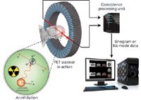
While travelling in the surrounding media, the released positron loses its kinetic energy and then combines with an electron. The encounter annihilates both positron and electron and results in two photons (gamma rays) each with an energy of 0.511 MeV that are emitted in opposite directions (Figure 1).

Figure 1: Principle of positron emission tomography.
me+ + me– → 2γ
Sophisticated scanners (detector) can detect such pairs of photons by coincidence detection. From the data collected, three-dimensional images of tissue structures are then calculated. The most commonly used short-lived, cyclotron-produced radionuclides in radiopharmacy are 11C, 13N, 15O and 18F. Their respective half-lives are 20.38, 9.96, 2.03 and 109.7 min.
Radiopharmaceuticals
To administer the radionuclide to a living human or animal, it is either incorporated in a biologically inert molecule (e.g., the blood flow tracers [15O]water or [15O]butanol) or in a biologically active molecule that is absorbed by the organ of interest.
After the radiopharmaceutical is concentrated in the tissue of interest, the patient is placed in the PET scanner. By tracking the photons, computers with sophisticated software generate three-dimensional images of the source of the photons. This allows the study of physiological, biochemical and pharmacological functions at a molecular level. Illnesses such as cancer, cardiovascular disease and even neurological disorders can be detected long before symptoms appear.
Production of PET Radiopharmaceuticals
Radionuclides used in PET experiments such as 11C, 13N, 15O and 18F are artificially produced in a cyclotron, where a beam of accelerated charged particles irradiates a prepared target. Subsequently, the resulting radionuclides are isolated and synthetically incorporated into the radiotracer. Since the fluorine atom is similar in size to the hydrogen atom, it acts as a pseudohydrogen and is therefore ideally suited for replacing hydrogen atoms in organic molecules. The positron emitter 18F is thus one of the most important imaging radionuclides in diagnostic nuclear medicine. It is produced by proton bombardment of an 18O-enriched water target. In an 18O(p, n)18F reaction, highly accelerated protons (p) react with the 18O atomic nucleus to emit a neutron (n) and 18F, which immediately decays by positron emission with a half-life of 109.7 min. The product of the 18F decay is the stable isotope 18O. After isolation of 18F from the target water, the radionuclide is incorporated into the chemical compound required. To this end, the preparation of reactive fluorine radionuclides in organic solvents is an important prerequisite for the synthesis of aliphatic carbon-fluorine bonds.
a) [18F]Fluorodeoxyglucose
[18F]Fluorodeoxyglucose, commonly abbreviated [18F]FDG, or simply FDG, is a glucose analogue in which the hydroxyl group at the 2' position of the glucose molecule is substituted by [18F]fluorine (Figure 2a). It throws light on the use and metabolism of glucose in heart, lungs and brain. Additionally, it is used in oncology to determine abnormal glucose metabolism to characterize different tumour types. After administering [18F]FDG to the patient, it is incorporated into the cells by the same transport mechanism as the normal glucose, but unlike this, once inside the cell, it is not metabolized and thus remains in the cell allowing PET tomographic imaging. Not least because of its many diagnostic uses, the high number of existing labelling procedures and its advantageous half-life of approximately two hours, which allows the transport to sites that have no cyclotron, [18F]FDG is actually the most frequently used organic PET radiopharmaceutical.

Figure 2: Chemical structures of two PET radiopharmaceuticals. (a) In [18F]FDG, the hydroxyl group at the 2’ position of normal glucose is substituted by 18F. (b) In [18F]fluorocholine, a [18F]fluoroalkyl group is attached to the nitrogen atom of N,N-dimethylaminoethanol (DMAE).
b) [18F]Fluorocholine
In cells, choline is used as a precursor for the biosynthesis of phospholipids. As the latter are essential cell membrane components and because tumours reveal increased metabolism of cell membrane constituents and increased choline uptake, radiolabelled choline tracers are invaluable diagnostic tools for cancer detection. [18F]Fluorocholine (Figure 2b) is a recently developed PET radiotracer that allows choline metabolism to be imaged in vivo. It is based on the tumour-detecting radiotracer [11C]choline. The driving force of the production of the 18F-labelled derivative was the substantially longer half-life, which allows the distribution of this tracer to PET institutions without on-site cyclotron.
Radio Ion Chromatography (IC)
Radio IC is a powerful, fast and sensitive tool for the quality control of a wide range of PET pharmaceuticals. It aims at determining the radiochemical purity of radiopharmaceuticals after radiosynthesis with cyclotron-produced nuclides. To this end, Radio IC uses a flow-through mass and radioactivity detector.
Besides the accuracy and reproducibility of the analytical results, high throughput is a must. One and the same multichannel radio IC takes over the quality control of three production lines. The analytical unit provides the following benefits:
a) Flexibility of the System
The ion chromatography system installed at the ITP (Instituto Tecnológico PET) in Spain combines three quality control systems for PET pharmaceuticals in one. From the very same injection system, the flow can be automatically directed to the three channels. By selecting between an array of different columns, mobile phases and detectors, [18F]fluoride, [18F]FDG and [18F]fluorocholine can be separately determined (Table 1). All aspects of system operation and data acquisition are controlled by MagIC Net™ software.

Table 1: Chromatographic conditions for the quality control of [18F]fluoride, [18F]FDG and [18F]fluorocholine.
b) Safety
The automated sample injection with Metrohm's patented Dosino technology allows the aspiration of very low sample volumes with accuracy and precision. By using the MagIC Net™ software, liquid handling as well as dosing and rinsing tasks are completely automated without any carryover. The system's modular design supports the installation of the required lead shielding and thus guarantees user safety. The injection valve is placed inside a 5 cm thick tailor-made lead housing, while radiation from the radiotracers in the separation columns is attenuated to a safe level by a sufficiently thick lead shielding. In addition, a lead sample holder avoids user exposure to gamma radiation.
c) Comprehensive Quality Control Using Multichannel Radio IC
Radiopharmaceuticals have unique characteristics and require special tests described in numerous pharmacopoeias. The quality control includes testing for both chemical and radiochemical purity before the radiotracer is administered to the patient. The radiochemical purity of a radiotracer – the ratio of the radionuclide in bound form (e.g., [18F]FDG) to the radionuclide in unbound form (e.g., [18F]fluoride) – guarantees the image quality of the PET scan and protects the patient from unnecessary radiation.
In the first instance, the concentration of the cyclotron-produced radionuclide, the 18F, which is used as a precursor in the subsequent radiosynthesis, has to be determined (Figure 3).

Figure 3: (a) Conductivity and (b) radioactivity chromatogram of cyclotron-produced [18F]fluoride. In the subsequent radiosynthesis (nucleophilic fluorination), tracer (i.e., very low) quantities of [18F]fluoride ions are used to form carbon-fluorine bonds. The IC software converts the radiation units, counts per second (cps), to mV. Chromatographic conditions are shown in Table 1.
Subsequently, the appropriate chemical purity – as for any other pharmaceutical preparation – and the concentration of the synthesized radiopharmaceutical, the 18F, which mostly is in the nanomolar range, have to be determined. In doing so, excessive precursors and radiolabelling-derived impurities have to be quantified also. Figure 4 shows the chromatogram with the glucose precursor, the carrier-free [18F]FDG and the chlorodeoxyglucose impurity. The analysis is completed in less than 10 min.

Figure 4: (a) IC-PAD chromatogram with the glucose precursor, the carrier-free [18F]FDG and the impurity chlorodeoxyglucose. (b) Radioactivity chromatogram of the [18F]FDG. The IC software converts the radiation units, counts per second (cps), to mV. Chromatographic conditions are shown in Table 1.
Figure 5 shows the chromatogram of the reaction mixture of [18F]fluorocholine synthesis. Besides nanomole quantities of the [18F]fluorocholine radiotracer, considerable amounts of the reactant N,N-dimethylaminoethanol and trace-levels of calcium impurities were detected in the reaction mixture.

Figure 5: (a) Conductivity and (b) radioactivity chromatogram of the [18F]fluorocholine reaction mixture. [18F]Fluorocholine is synthesized by 18F-fluoroalkylation of N,N-dimethylaminoethanol (DMAE) using gaseous 18F-fluorobromomethane. This labelling reaction results in high levels of residual DMAE. Other potential byproducts such as bromocholine (not detected) can additionally be determined. The IC software converts the radiation units, counts per second (cps), to mV. Chromatographic conditions are shown in Table 1.
d) Analysis Time
As most positron-emitting radiopharmaceuticals are characterized by short half-lives, there is a strong drive to reduce the time spent on quality control. Fast and precise analyses are guaranteed by optimally harmonized and computer-controlled determination and rinsing sequences for detector pathways and the sample injection circuit.
Conclusion
Metrohm's highly customizable chromatography system copes with the tough requirements of the radiopharmaceutical industry and pharmacopoeial regulations. One single multichannel radio IC meets the quality control requirements of various production lines. Besides the high quality, the Metrohm IC presented ensures user safety, low maintenance costs and outstanding ruggedness.
Acknowledgements
The authors thank the entire group of the Ion Chromatography Department of Gomensoro for their outstanding support and fruitful discussions during this project.
References
(1) D. Slaets, S. De Bruyne, C. Dumolyn, L. Moerman, K. Mertens and F. De Vos, Reduced dimethylaminoethanol in [18F]fluoromethylcholine: an important step towards enhanced tumour visualization, Europ. J. Nucl. Med. Mol. Imaging 37(11), 2136–2145 (2010).
(2) D. Kryza, V. Tadino, M. Azuzurra Filannino, G. Villeret and L. Lemoucheux, Fully automated [18F]fluorocholine synthesis in the TracerLab MXFDG Coincidence synthesizer, Nucl. Med. Biol. 35, 255–260 (2008).
(3) J. Passchier, Fast high-performance liquid chromatography in PET quality control and metabolite analysis, Q. J. Nucl. Med. Mol. Imaging 53, 411–416 (2009).
Metrohm International Headquarters
Ionenstrasse, 9101 Herisau, Switzerland
tel. +41 71 353 85 04
Website: www.metrohm.com


