UHPLC Analysis of Immunoglobulins with TSKgel® UP-SW3000 SEC Columns
The Application Notebook
Antibody therapeutics are enjoying high growth rates, the major areas of therapeutic application being cancer and immune/inflammation-related disorders, including arthritis and multiple sclerosis. In 2013, six of the top ten best-selling global drug brands were monoclonal antibodies (mAbs) and more than 400 monoclonals were in clinical trials. The characterization of these complex biomolecules is a major challenge in process monitoring and quality control. The main product characteristics that need to be monitored are aggregate and fragment content, glycosylation pattern, and charged isoforms.
The standard method used in biopharmaceutical QC for mAb aggregate and fragment analysis is size-exclusion chromatography (SEC). A new series of 2 µm silica-based ultrahigh-pressure liquid chromatography (UHPLC) columns with 25 nm (250 Å) pore size can be applied to either increase speed or improve resolution of the separation of antibody fragments, monomers, and dimers.
Experimental
TSKgel UP-SW3000 (P/N 0023449), 2 µm Competitor Protein SEC Column, 1.7 µm
Results
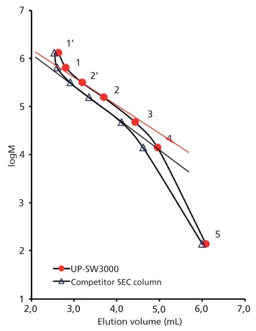
Figure 1 shows the calibration curves of the new TSKgel UPâSW3000 2 µm column and a commercially available 1.7 μm UHPLC column. The calibration of TSKgel UP-SW3000 shows a shallower slope in the region of the molecular weight of γ-globulin. These differences in the separation range and steepness of the curves are related to a slight difference in pore size (25 nm for TSKgel versus 20 nm for the 1.7 µm material).
Figure 1: Calibration curves.

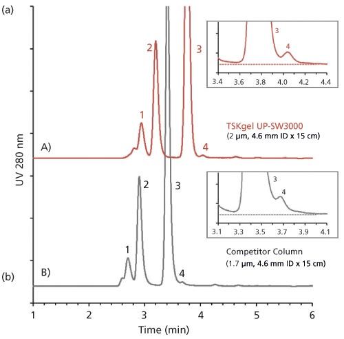
Figure 2: Comparison of antibody analysis results: mouse-human chimeric mAb. 1: trimer; 2: dimer; 3: monomer ; 4: fragment.

The separation of an antibody sample on the new 2 µm packing compared to the competitor UHPLC column is depicted in Figure 2. The difference in pore sizes results in a better separation in the molecular weight range of antibodies, fragments, and aggregates. Based on the wider separation window the resolution between monomer and dimer as well as dimer and trimer is slightly higher with the TSKgel UP-SW3000 column, although particle size is slightly larger than in the competitor column. Moreover, the fragment peak is more clearly separated from the monomer peak.

Conclusion
TSKgel UP-SW3000 is ideally suited for the analysis of the aggregate and fragment contents of antibody preparations. It features the same pore size as the renowned TSKgel G3000SWXL and TSKgel Super mAb columns while improving resolution through a smaller particle size. Based on the optimized pore size and the high degree of porosity the resolution in the molecular weight range of immunoglobulins is superior to a competitive UHPLC column with slightly smaller particle and pore size.

Tosoh Bioscience GmbH
Im Leuschnerpark 4 64347 Griesheim, Darmstadt, Germany
Tel: +49 6155 7043700 fax: +49 6155 8357900
E-mail: info.tbg@tosoh.com
Website: www.tosohbioscience.de

SEC-MALS of Antibody Therapeutics—A Robust Method for In-Depth Sample Characterization
June 1st 2022Monoclonal antibodies (mAbs) are effective therapeutics for cancers, auto-immune diseases, viral infections, and other diseases. Recent developments in antibody therapeutics aim to add more specific binding regions (bi- and multi-specificity) to increase their effectiveness and/or to downsize the molecule to the specific binding regions (for example, scFv or Fab fragment) to achieve better penetration of the tissue. As the molecule gets more complex, the possible high and low molecular weight (H/LMW) impurities become more complex, too. In order to accurately analyze the various species, more advanced detection than ultraviolet (UV) is required to characterize a mAb sample.

.png&w=3840&q=75)

.png&w=3840&q=75)


.png&w=3840&q=75)


.png&w=3840&q=75)


