Electrophoretic Concentration—A Simple and Green Approach for Sample Preparation
LCGC North America
We explain the principles of EC and demonstrate its use to enrich anionic pollutants and cationic drugs in environmental samples.
Electrophoretic concentration (EC) is an electric field-driven and environmentally friendly off-line sample preparation for charged analytes. EC was demonstrated for the enrichment of either six anionic pollutants or five cationic drugs from purified, drinking, river, or waste-water samples. EC provided analyte enrichment in 15–50 min with concentration factors of 30–249 and 12–243 for the negatively and positively charged analytes, respectively. A modification of the EC device enabled simultaneous EC and separation (SECS) of six cationic and anionic herbicides with concentration factors of 18–337 in 30 min. The potential of SECS has also been evaluated for the determination of high mobility ions in urine and the results obtained have been compared to common acetonitrile treatment of urine. SECS provided an enrichment of high mobility ions and revealed more peaks compared to the acetonitrile treatment.
State-of-the-art analytical separation and detection instrumentation enables rapid chemical analysis. However, the total time of an analytical workflow can be long because of the need to transform the analyte into a suitable state for separation and detection. This transformation is the domain of sample preparation and can involve substantial resources, time, and labor. A general strategy to address these disadvantages is to accelerate the transfer of analyte from the sample into a suitable phase prior to analysis (that is, an acceptor phase). Accelerated mass transfer between the sample and acceptor phases has been achieved by reducing the volume of acceptor phase or by using auxiliary forces. For the latter approach, common examples include pressure (1), heat, sonication (2), microwave-irradiation (3), and the application of electric fields. The use of smaller volumes of acceptor phase has also led to the introduction of organic solvent-free or virtually solvent-free sample preparation. Examples of these approaches are dispersive liquid-liquid extraction (DLLE) (4), liquid-phase microextraction (LPME) (5) or single-drop microextraction (SDME) (6), stir-bar sorptive extraction (SBSE) (7), or solid-phase microextraction (SPME) (8).
Electric field-driven sample preparation has attracted considerable research interest, but has not yet found wide use in the analytical routine laboratory. The applied electric field causes migration of charged analytes based on their electrophoretic mobility (9). In practice, the electric field is imposed between the sample and acceptor phases. These two phases can be separated by a solid or liquid phase in electromembrane extraction (10), electrodialysis (11), and three-phase liquid electroextraction (12). When the sample and acceptor phase are in contact with each other, the electrodriven techniques are referred to as electric field-assisted solid-phase extraction (SPE) and solid-phase microextraction (13-15), electroextraction (16), and more recently electrophoretic concentration (EC).
In this article, the principles of EC are discussed and highlighted using application-based examples. EC is described for sample cleanup and analyte enrichment of pollutants, drugs, and herbicides from water samples. As a short demonstration, simultaneous EC and separation (SECS) is evaluated on urine samples as an alternative to acetonitrile precipitation.
Electrophoretic Concentration
Principle: In Figure 1a, a schematic single EC setup for the concentration of negatively charged analytes is shown. Voltage with the positive sign or anodic voltage is applied at the micropipette. The same setup can be used for cationic analytes except that the cathode is placed at the micropipette. The electric field causes the migration of the anionic analytes (pink dots) from an aqueous sample into an aqueous acceptor electrolyte immobilized in the lumen of a glass micropipette. The analyte enrichment that occurs is based on field-enhanced sample injection (FESI), which is an on-line sample concentration technique used widely in capillary electrophoresis (CE) (17,18). In FESI, electrokinetic injection is performed from a low conductivity sample into a high conductivity separation electrolyte inside a CE capillary. Upon application of an electric field, the rate of analyte migration in the sample is faster than in the separation electrolyte (because the applied electric field strength is higher in the low conductivity sample), which results in an analyte enrichment or stacking at the boundary of sample and separation electrolyte. The analyte enrichment factor achieved using FESI is proportional to the conductivity ratio between the sample and the separation electrolyte. Under ideal FESI conditions, improvements of analyte detection sensitivity of two to three orders of magnitude compared to a typical direct injection can be achieved.
Figure 1: Schematic setup for (a) electrophoretic concentration (EC) of anions and (b) simultaneous EC and separation (SECS) of anions and cations. The sample is at the bottom of the setup and Pt-electrodes and micropipettes are dipped into the sample. The micropipettes contain the acceptor electrolyte for the analytes. The Pt-electrodes are connected to a high voltage (HV) supply to close the electrical circuit. Upon voltage application, the charged analytes migrate to the opposite sign of the electrode situated in the micropipette. Enrichment of the analytes is based on field-enhanced sample injection, a concentration technique used in capillary electrophoresis (CE).
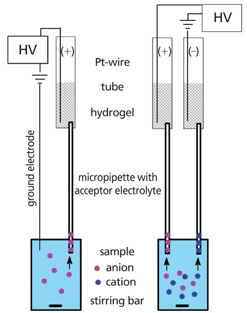
The experimental setup for EC was realized using an eight-channel high voltage (HV) device, which allowed eight samples to be prepared simultaneously. In Figure 1a, the aqueous sample is at the bottom. The ground electrode and a glass micropipette containing 20 µL of a high conductivity acceptor electrolyte were dipped into the sample. The micropipettes used were common capillary pipettes used for spotting applications such as in thin-layer chromatography (TLC). The other end of the micropipette was inserted into an electrolyte-fortified polyacrylamide hydrogel. The hydrogel was prepared inside the barrel of a disposable syringe by thermal polymerization (10 min at 60 °C) of a mixture made of separation electrolyte, acrylamide (monomer), and potassium persulfate (initiator). The hydrogel was conductive and prevented the flow of acceptor electrolyte out of the pipette because of gravity. During EC, the hydrogel also caused a zero net-flow (that is, the sum of electroosmotic flow [EOF] and hydrodynamic fluid flows) inside the pipette (19). A zero net-flow was important because the presence of EOF has an adverse effect on analyte enrichment. A Pt-wire was attached to the hydrogel and connected to the HV power supply. The sample was stirred during voltage application. After EC, the acceptor electrolyte was transferred to a suitable vial for separation and detection using liquid chromatography (LC) or CE.
In EC, the method can be optimized by studying the applied voltage, voltage application time, and the type and concentration of the acceptor electrolyte. The applied voltage is generally maximized using the maximum observed current that does not cause bubble formation inside the micropipette. Typically, the applied voltage is selected when the observed current is <300 µA after 1 min. The optimized values for voltage application time, type, and concentration of acceptor electrolyte are then evaluated by plotting the analyte concentration factor (CF) versus the investigated parameter. The CF is calculated by dividing the peak areas of the analyte obtained after analysis of the acceptor electrolyte after EC with the peak area of the analyte obtained after analysis of the sample before EC. The value of each parameter that provides the highest CF for an analyte, or the highest averaged CF in the case of multiple analytes, is then selected.
EC of Cationic or Anionic Analytes: The EC setup in Figure 1a has been applied to the determination of anionic pollutants in purified, drinking, and river water (20), as well as to cationic drugs in purified and wastewater (21). Nine anionic species, including inorganic ions, a dye, and benzenesulfonate derivatives, were concentrated from 10 mL of sample into an acceptor electrolyte of 50 mM ammonium acetate at pH 8.3. The optimized values for applied voltage and voltage application time were 1.3 kV, 0.6 kV, and 0.4 kV and 15 min, 20 min, and 20 min for purified, drinking, and river water, respectively. The corresponding CFs were 48–249, 43–86, and 30–81. These values facilitated sensitive analysis, resulting in method detection limits (MDL) as low as 1 ng/mL by CE with UV detection. The developed method of EC–CZE–UV showed a linear concentration range of a factor of 50 to 100 for all analytes.
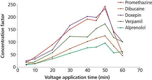
Sensitive determination of five cationic drugs including promethazine, dibucaine, doxepin, verapamil, and alprenolol was achieved by the combination of off-line and on-line sample concentration by EC and sweeping. This combination was chosen because the EC concentrate was free of micelles and thus suitable for further enrichment using sweeping in micellar electrokinetic chromatography (MEKC)–UV. The sample volume was 20 mL and the EC acceptor electrolyte was 50 mM ammonium acetate at pH 5. An optimized voltage and voltage application time of 1.0 kV for 50 min and 0.5 kV and 15 min for purified and 10-fold diluted wastewater provided the highest average CF (that is, the CF obtained by EC only-without the contribution of sweeping) for all five analytes. Figure 2 shows the dependence of the CF on the voltage application time for the cationic analytes spiked in purified water. The CF values increased from 11–26 to 97–243 from 5 min to 50 min. After 50 min, the analytes started to migrate out of the micropipette, which resulted in a significant decrease in the analyte enrichment. At 55 min, the analyte concentration in the acceptor electrolyte dropped to 57–102, which was less than half the values compared to 50 min. The steep decrease in CF was in agreement with moving boundary electrophoresis where the analytes migrate as concentrated boundaries and according to their electrophoretic mobility.
Figure 2: Effect of the voltage application time on the concentration factor of cationic drugs. EC was performed with an applied voltage of 1 kV. The acceptor electrolyte in the micropipette was 50 mmol/L ammonium acetate at pH 5. The sample was 20 mL purified water with an analyte concentration of 100 ng/mL. The analytes enriched steadily inside the lumen of the micropipette from 5 to 50 min. At 55 min, a decrease in concentration factors was obtained as a result of the migration of the analytes out of the micropipette. Adapted with permission from reference 21.
The CFs for EC used in combination with sweeping reached very high values of up to almost 45,000 for purified water and 6500 for diluted wastewater. This analytical procedure exhibited a linear concentration range of two orders of magnitude and gave an MDL as low as 0.04 ng/mL and 1.2 ng/mL in purified water and undiluted wastewater, respectively. It was noted that the CFs by EC (without sweeping) for the undiluted wastewater were 1–3 because of the relatively high conductivity of the sample, thus EC served primarily as a rapid and simple sample cleanup for this sample.
Simultaneous EC of Cationic and Anionic Analytes: Figure 1b shows the setup of SECS for the simultaneous enrichment and sample cleanup of both positively and negatively charged analytes and is based on the EC setup shown in Figure 1a with some modifications. These modifications included the use of an additional assembly of micropipette and hydrogel. This assembly was also connected to the HV power supply and replaced the ground electrode. In SECS, the cathodic and anodic micropipettes contain the acceptor electrolyte for the cationic and anionic analytes, respectively.
SECS was applied to the simultaneous enrichment of cationic (paraquat, diquat, and difenzoquat) and anionic (mecoprop, dichlorprop, and fenoprop) herbicides in drinking and river water (22). The acceptor electrolytes for the cations and anions were 50 mM ammonium acetate at pH 5 and pH 9, respectively. The optimized SECS was performed for 30 min at 2.0 kV, 1.0 kV, and 0.5 kV for purified, drinking, and river water samples, respectively. The cationic and anionic acceptor electrolytes were analyzed by CZE–UV and LC–UV, respectively. The CF values obtained were 18–337, 32–131, and 31–83, respectively. The linear range for all water samples encompassed two orders of magnitude and provided MDLs for purified water of 0.5-5.0 ng/mL and for both drinking and river water of 5.0 ng/mL.
The optimized SECS was then compared to two commercial SPE procedures using polymeric weak anion-exchanger and cation-exchanger sorbent for the anionic and cationic herbicides, respectively. For SECS, the sample preparation time was less than 45 min and involved four simple steps: filling of the micropipettes with the acceptor electrolyte; attaching the micropipettes to the hydrogel and sample; voltage application; and transfer of the acceptor electrolyte for analysis. In SPE, the time required was more than 12 h, even when enrichment of the anions and cations was performed in parallel, and included seven steps: conditioning of the sorbent; equilibrating of the sorbent; loading of the sample; washing of the sorbent; elution of the analyte from the sorbent; evaporation of the elution solvent; and reconstitution of the analyte before analysis. Step 6 (evaporation of the elution solvent) accounted for most of the preparation time because it was performed overnight. It is noted that a typical SPE procedure can be performed in less than 60 min, for instance when drying of the elution solvent is performed at elevated temperature and reduced pressure.
In a green sample preparation workflow, the use of solvents and toxic reagents should be reduced or eliminated and the demand in energy and labor should be minimized (23). The sample preparation time and labor were significantly reduced by SECS (<45 min compared to >12 h in SPE). The consumption of reagents was also substantially reduced. The volume of reagents in SECS was ~2.4 mL (that is, two hydrogels and aqueous acceptor electrolytes) and SECS did not involve any organic solvents. In SPE, the volume of consumables was ~200 mL, with ~54% organic solvents. SECS can therefore be considered to be a simple and environmentally friendly sample preparation technique for polar and highly water-soluble herbicides.
Preliminary Studies of SECS for Biological Samples: We were interested in the use of SECS for the enrichment and sample cleanup of high mobility ions present in urine. In a first attempt, the SECS setup was applied for sample preparation of undiluted urine samples (using the same acceptor electrolytes as described in the previous section). The conductivity values of the urine and pH 5 and pH 9 acceptor electrolytes were 23.6 mS/cm, 4.3 mS/cm, and 4.1 mS/cm, respectively. These values do not fulfil the major criterion for analyte enrichment by FESI since FESI requires a minimum conductivity ratio of separation electrolyte and sample of 10. Enrichment of high mobility ions can still be achieved by electrokinetic injection (EKI) (24). In EKI, the applied electric field causes the migration of ions according to their electrophoretic mobility and thus there is bias toward high mobility ions. The SECS setup was used to perform EKI for 15 min at 200 V on 20 mL of urine. The sample obtained after SECS was compared to urine subjected to sample cleanup using protein precipitation by the addition of acetonitrile. The protein precipitation of 400 µL of urine was performed by adding 600 µL of acetonitrile followed by 2 min of vigorous shaking and 2 min of centrifugation. The acceptor electrolytes from SECS and an aliquot of the supernatant obtained from the protein precipitated urine were analyzed by CE in a fused-silica capillary (internal diameter of 50 µm, effective and total length of 39 cm and 50 cm, respectively) with contactless conductivity detection. The separation electrolyte was 50 mM L-histidine/50 mM 2-(N-morpholino)ethanesulfonic acid at pH 6. The sample was injected at 50 mbar for 6 s. The separation voltage was +15 kV and -15 kV for analysis of the cationic and anionic species in the sample, respectively.
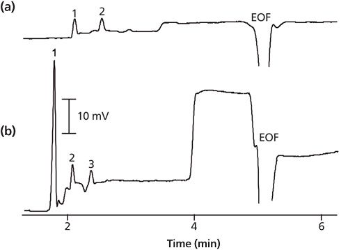
Figure 3 shows the electropherograms obtained for the anions of the urine sample after treatment by protein precipitation (Figure 3a) and EKI (Figure 3b). The detection of the anions was in counter-EOF mode, and thus only anions with migration velocity values higher than the EOF could be detected. Common anions in urine with high mobility values are inorganic compounds such as chloride, nitrate, sulfate, and phosphate. In a comparison of protein precipitation and EKI, EKI provided an enrichment of the first three peaks (peak 1–3) and also revealed more peaks (peaks 4–6). As a preliminary study, the identification of the peaks was not attempted but will be considered in the future. Peaks 1–3 were probably chloride, nitrate, and sulfate, however, according to a similar method (25,26). Figure 4 shows the electropherograms from the cationic analysis of the same urine sample as in Figure 3. The detection of the cations was in co-EOF mode and so positively, neutral, and negatively charged analytes could be detected if the apparent velocity was toward the cathode. The cations were detected before the EOF (<5 min). In a comparison of protein precipitation and EKI, EKI provided a significant enrichment of the first peak (peak 1) and revealed three peaks in total, while protein precipitation only showed two peaks. Peaks 1–3 were presumably sodium, calcium, ammonium, and magnesium (25,26). In EKI, an additional large and flat peak was observed before the EOF, which could be attributed to unresolved cationic compounds with lower electrophoretic mobility values. EKI applied to urine samples therefore provided an alternative to acetonitrile precipitation with the advantage of selective enrichment of high mobility analytes.
Figure 3: Anionic electropherograms of urine sample obtained after sample preparation and analysis by counter-EOF capillary electrophoresis with contactless conductivity detection. The sample preparations were (a) protein precipitation and (b) SECS for 15 min at 200 V, respectively. In (b), SECS provided a distinct concentration of peaks 1–3 and revealed more peaks (peaks 4–6) in comparison to (a) protein precipitation.
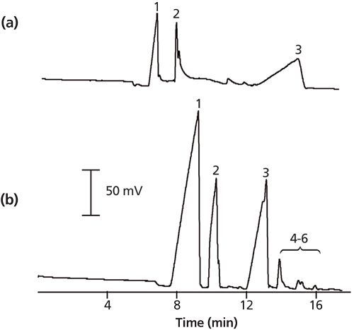
Figure 4: Cationic electropherograms of urine sample obtained after sample preparation and analysis by co-EOF capillary electrophoresis with contactless conductivity detection. The sample preparation were (a) protein precipitation and (b) SECS for 15 min at 200 V, respectively. In (b), SECS provided a significant concentration of peak 1 and revealed three peaks in total compared to two peaks obtained in (a) by protein precipitation.
Conclusions
EC and SECS are purely aqueous and electric field-driven non-exhaustive sample preparations. The principle of these approaches is derived from the on-line sample concentration technique of FESI used in CE. The implementation of FESI for off-line sample preparation is simple and uses inexpensive glass micropipettes and hydrogels. EC and SECS have been shown to allow the enrichment of analytes from a low conductivity aqueous sample into a higher conductivity acceptor electrolyte. EC and SECS have been demonstrated successfully for various water samples and have provided analyte enrichments of up to 337 in less than 50 min. In addition, preliminary studies on the use of the SECS setup for the extraction of high mobility ions in undiluted urine gave improved results when compared to the commonly used acetonitrile precipitation process for removal of proteins from the sample. The acceptor electrolytes obtained after EC and SECS were directly transferred for CE, MEKC, and LC analysis, without the requirement for subsequent complicated steps, such as evaporation and reconstitution.
In a comparison of SECS with commercial SPE, the sample preparation time for SECS (<45 min) was shorter than for SPE (>12 h) and SECS significantly reduced the consumption of reagents. These green features strongly support the criteria for green analytical chemistry. EC and SECS might serve as an alternative to traditional liquid–liquid or solid–liquid-based extraction in environmental and food analysis for hydrophilic analytes (27). Future investigations will be geared toward extending the breadth of SECS for a wider range of samples (environmental waters and biological samples) and to increasing the concentration efficiency (larger volumes and the use of a continuous flow of sample).
References
- L. Ramos, E.M. Kristenson, and U.T. Brinkman, J. Chromatogr. A975, 3–29 (2002).
- S. Li, S. Cai, W. Hu, H. Chen, and H. Liu, Spectrochim. Acta Part B At. Spectrosc.64, 666–671 (2009).
- C. Sparr Eskilsson and E. Björklund, J. Chromatogr. A902, 227–250 (2000).
- M. Rezaee, Y. Assadi, M.-R. Milani Hosseini, E. Aghaee, F. Ahmadi, and S. Berijani, J. Chromatogr. A1116, 1–9 (2006).
- M.A. Jeannot and F.F. Cantwell, Anal. Chem.68, 2236–2240 (1996).
- Y. He and H.K. Lee, Anal. Chem.69, 4634–4640 (1997).
- E. Baltussen, P. Sandra, F. David, and C. Cramers, J. Microcolumn Sep.11, 737–747 (1999).
- R.P. Belardi and J.B. Pawliszyn, Water Pollut. Res. J. Canada 24, (1989).
- A. Wuethrich, P.R. Haddad, and J.P. Quirino, TrAC Trends Anal. Chem.80, 604–611 (2016).
- S. Pedersen-Bjergaard and K.E. Rasmussen, J. Chromatogr. A1109, 183–190 (2006).
- P. Kubán, A. Slampová, and P. Bocek, Electrophoresis31, 768–785 (2010).
- R.J. Raterink, P.W. Lindenburg, R.J. Vreeken, and T. Hankemeier, Anal. Chem. 85, 7762–7768 (2013).
- R.M. Orlando, J.J.R. Rohwedder, and S. Rath, Chromatographia77, 133–143 (2013).
- J. Wu, W.M. Mullett, and J. Pawliszyn, Anal. Chem.74, 4855–4859 (2002).
- R. Xu and H.K. Lee, J. Chromatogr. A1350, 15–22 (2014).
- J. Stichlmair, J. Schmidt, and R. Proplesch, Chem. Eng. Sci. 47, 3015–3022 (1992).
- R.-L. Chien and D.S. Burgi, J. Chromatogr. 559, 141–152 (1991).
- R.-L. Chien and D.S. Burgi, J. Chromatogr. 559, 153–161 (1991).
- A. Wuethrich, P.R. Haddad, and J.P. Quirino, Analyst 139, 3722–3726 (2014).
- A. Wuethrich, P.R. Haddad, and J.P. Quirino, J. Chromatogr. A1369, 186–190 (2014).
- A. Wuethrich, P.R. Haddad, and J.P. Quirino, J. Chromatogr. A 1401, 84–88 (2015).
- A. Wuethrich, P.R. Haddad, and J.P. Quirino, Anal. Chem.87, 4117–4123 (2015).
- A. GaÅuszka, Z. Migaszewski, and J. NamieÅnik, TrAC Trends Anal. Chem.50, 78–84 (2013).
- X. Huang, M.J. Gordon, and R.N. Zare, Anal. Chem.60, 375–377 (1988).
- Q.J. Wan, P. Kubán, J. Tanyanyiwa, A. Rainelli, and P.C. Hauser, Anal. Chim. Acta 525, 11–16 (2004).
- M. Greguš, F. Foret, and P. Kubán, Electrophoresis 36, 526–533 (2015).
- A. Wuethrich, P.R. Haddad, and J.P. Quirino, Electrophoresis 37, 1122–1128 (2016).
Joselito P. Quirinois an associate professor at ACROSS, University of Tasmania, Australia. He graduated with a B.Sc. in industrial pharmacy from the University of the Philippines in Manila, Philippines, in 1992. He finished his M.Sc. and PhD in analytical chemistry with Professor Shigeru Terabe at the Himeji Institute of Technology, Japan, in 1998-1999. He was then a postdoctoral researcher in Professor Richard Zare’s laboratory at Stanford University until 2001. This was followed by an industrial stint in pharmaceutical companies in California until 2007. He has been awarded an Australian Research Council Future Fellowship and Japanese Society for the Promotion of Science Fellowships. He is interested in analytical method development for chemical, pharmaceutical, biological, environmental, and food sciences.
Paul R. Haddadis an Emeritus Distinguished Professor at ACROSS, University of Tasmania. He has a D.Sc. from the University of NSW and is a Fellow of both the Australian Academy of Science and the Academy of Technological Sciences and Engineering. He was an ARC Federation Fellow from 2006 to 2011. He is the recipient of a number of national and international awards, including the American Chemical Society Chromatography Award. His research is in the field of separation science and covers ion chromatography, capillary zone electrophoresis, capillary electrochromatography, and high performance liquid chromatography, used especially for the separation of inorganic ions, metal complexes, and low-molecular-weight organic species.
Alain Wuethrichis a postdoctoral fellow at the Australian Institute for Bioengineering and Nanotechnology, University of Queensland, Australia. He graduated with an M.Sc. in life sciences from the University of Applied Sciences Northwestern Switzerland in 2012. He obtained his Ph.D. at ACROSS, University of Tasmania, in 2016. Alain has been awarded an Early Postdoc Mobility Fellowship from the Swiss National Science Foundation to conduct research on microfluidic approaches for early cancer detection. His research interests are in microfluidic systems, sample preparation, chromatography, electrophoresis, and mass spectrometry.

Thermodynamic Insights into Organic Solvent Extraction for Chemical Analysis of Medical Devices
April 16th 2025A new study, published by a researcher from Chemical Characterization Solutions in Minnesota, explored a new approach for sample preparation for the chemical characterization of medical devices.
Study Explores Thin-Film Extraction of Biogenic Amines via HPLC-MS/MS
March 27th 2025Scientists from Tabriz University and the University of Tabriz explored cellulose acetate-UiO-66-COOH as an affordable coating sorbent for thin film extraction of biogenic amines from cheese and alcohol-free beverages using HPLC-MS/MS.
Multi-Step Preparative LC–MS Workflow for Peptide Purification
March 21st 2025This article introduces a multi-step preparative purification workflow for synthetic peptides using liquid chromatography–mass spectrometry (LC–MS). The process involves optimizing separation conditions, scaling-up, fractionating, and confirming purity and recovery, using a single LC–MS system. High purity and recovery rates for synthetic peptides such as parathormone (PTH) are achieved. The method allows efficient purification and accurate confirmation of peptide synthesis and is suitable for handling complex preparative purification tasks.


