Membrane-Assisted Liquid–Liquid Extraction Trace Analysis of Pharmaceutical Compounds in Aquatic Environments
LCGC Asia Pacific
The concept of membrane-controlled processes is widespread in nature. Nearly all biological mechanisms concerning mass transport and exchange are regulated by membrane barriers and a variety of technical and biotechnological applications have been devised based on this mechanism. Membrane applications in analytical chemistry are geared towards the enrichment of target substances from an aqueous solution or the separation of compounds from a complex matrix. This article describes membrane-assisted extraction processes to separate traces of polar pharmaceutical substances the so called emerging micropollutants from aqueous samples. Basic prospects and examples of membrane-supported extractions are presented.
The concept of membrane-controlled processes is widespread in nature. Nearly all biological mechanisms concerning mass transport and exchange are regulated by membrane barriers and a variety of technical and biotechnological applications have been devised based on this mechanism. Membrane applications in analytical chemistry are geared towards the enrichment of target substances from an aqueous solution or the separation of compounds from a complex matrix. This article describes membrane-assisted extraction processes to separate traces of polar pharmaceutical substances the so called emerging micropollutants from aqueous samples. Basic prospects and examples of membrane-supported extractions are presented.
Since the introduction of dialysis 30 years ago, membranes have proven to be useful tools for the separation of polar and ionic compounds in a variety of applications, including drugs from biofluids and pollutants from aqueous environmental samples.
Nano- to micromolar concentrations of drugs present in plasma or urine can be determined, even when a complex biological matrix is present. When drugs have been administered, about 10–30% of the initially applied amount is excreted unchanged. Together with conjugated and metabolized drug portions they enter the environment via wastewater because conventional wastewater treatment cannot completely eliminate these mostly polar and persistent substances.
Common drug concentrations in wastewater range from mean ng/L to some μg/L. Much lower concentrations occur in surface and ground water (<20 ng/L).1
Let's assume that these compounds are suspected to present certain risks related to reproduction cycles and the health of organisms. Permanent monitoring is recommended but the low concentration of drugs within complex matrices makes their determination challenging. Simple and economic protocols particularly for preconcentration and matrix separation have to be established.
Can membrane-assisted extraction be a possible alternative to solid-phase extraction — the current standard procedure for the trace analysis of bioactive compounds?
In recent times, membrane-assisted microextraction techniques are among the most promising developments in sample enrichment and separation. The membrane probe design is flexible and easy to adapt to quite different analytical instruments. The high potential for automation makes membrane-assisted microextraction interesting for high-throughput analysis.
Membrane-assisted extraction processes become apparent as an attractive alternative to time- and solvent-consuming liquid–liquid extraction processes. Particularly, the opportunity to miniaturize a membrane-assisted extraction process popularized this sample preparation technique in the last decade.
In addition to the demands in environmental monitoring, drug design and admission procedures, medical therapies require analytical methods that are able to manage little sample volumes to detect traces (μg/L and below) of polar pharmaceutical compounds in an aqueous, complex matrix that includes proteins, carbohydrates, fatty acids etc.
Despite high sensitive instruments such as LC–MS–MS an enrichment of drugs prior to analysis is absolutely necessary. Current procedures in environmental drug analysis use solid-phase extraction (SPE) of high sample volumes (1 L and more), solvent amounts between 5 and 12 mL for elution, and further portions for subsequent clean-up are required mostly when wastewater has to be analysed.
The multiple-step sample preparation is time- and labour-consuming, and difficult to automate (Figure 1 — green blocks). Analyte loss during sample transfer, evaporation and redissolution is possible and can reduce the recovery of the analytes.

Figure 1
Among the well-established solid-phase microextraction techniques such as SPME,2 in-tube extractions3 and stir bar sorptive extraction (SBSE),4 membrane-assisted extractions are included increasingly in drug analysis within pharmaceutical,5,6 medical and environmental studies.7,8 Because of its compact and small design, particularly, micro hollow-fibre membranes can clearly save time and solvent for sample preparation when compared with standard SPE (Figure 1).
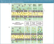
When drugs have to be concentrated from aqueous solution two major versions of membranes are generally used [Figures 2(a)–2(b)]. Porous and non-porous polymer materials can be distinguished that define the mode of extraction.

Figures 2
Liquid-Phase Microextraction (LPME)
LPME uses a porous polymer membrane. The pores are filled with an inert, viscous liquid that serves as a liquid membrane the analytes permeate through. As shown in Figure 2(a), the lumen of the hollow fibre can be filled with an aqueous acceptor solution. The driving force for the mass transport is a pH gradient between donor and acceptor phases. The pH in both phases has to be adjusted so that acidic or basic compounds keep neutral in the donor phase.
After passage through the membrane they become dissociated in the acceptor side. Thus, the selectivity of analyte enrichment can be easily controlled.
Comprehensive reviews on theory and application of LPME were presented by Jönsson and Matthiason,9 Rasmussen and Pederson-Bjergard6 and Majors.10
A special application of LPME uses electrokinetic migration through a porous polypropylene hollow fibre impregnated with 2-nitro octylether as liquid membrane to enrich the basic drugs pethidine, nortriptyline, metadone, haloperidol, and loperamide.11 The donor and acceptor solution consists of 10 mM HCl. The transport of the dissociated amino drugs through the liquid membrane was forced by an electric field of 300 V (d.c). Already, 5 min of extraction resulted in enrichment factors of about 8. It was found that the efficiency of the electrokinetic migration of the drugs was strongly dependent on the kind of liquid membrane used and the electric potential applied. Too high a voltage can heat up the sample or initiate electrochemical reactions that cause adverse changes of the analyte structures.
Otherwise, specific oxidation reactions could be useful when, for instance, additional polar groups should be introduced in the analyte molecule to enable separation by capillary electrophoreses or detection by electrospray-ionization mass spectrometry (EIMS).12
Membrane-Assisted Solvent Extraction
When an organic solvent is used instead of an aqueous acceptor phase then analytes will permeate favourably in their neutral form [Figure 2(b)] into the organic solvent until the partition equilibrium is achieved. Thus, for the extraction of drugs containing carboxylic groups such as ibuprofen, naproxen, gemfibrozil, and clofibric acid, a pH value of 2 should be preferred.
However, when a cocktail of substances with quite different properties has to be determined then the optimal extraction conditions for all analytes must be found. Thus, Mueller et al.8 extracted phenolic compounds known as endocrine disrupters (technical nonylphenol, bisphenol A, 17α–ethinylestradiol) at pH 7. Simultaneously, acidic drugs (ibuprofen), amides (phenazone, carbamzepine) and polycyclic musk compounds (tonalide, galaxolide) used as additives in several personal care products were enriched using a porous polypropylene hollow-fibre loop filled with n-octanol as the extraction solvent.
The membrane probe uses conventional autosampler vials thus automated analysis by GC–MS is possible. Enrichment factors between 7 (phenazone) and 187 (ibuprofen) resulted in limits of detection between 100 and 20 ng/L. Analysis disturbing matrix compounds such as fatty acids and humic matter were completely separated. Monitoring of wastewater is a promising application area for membrane extractions that was successfully demonstrated also for the determination of priority pollutants such as phenols, PCB and pesticides.13
Important Parameters for Membrane Probe Design
Among the properties of the analytes, the configuration of the membrane device determines the result of membrane-assisted extraction. Several types of polymers and membrane formats are currently used whereas the hollow fibres or membrane bags are preferred against flat membrane assemblies. Single-film tubing, hollow-fibre loop or knotted arrangements are known.
Film tubing material is commercially available and it is often modified and tailored for special purposes. Photochemically grafted surfaces or immobilized enzymes can prepare the character of membranes for extra functions beyond analyte enrichment and matrix separation.14,15 Maximum enrichment of analytes is the principle objective of every probe design.
The following features are important for successful analyte enrichment using a membrane-assisted solvent extraction device:
- Membrane material: The demand that users face in terms of membrane selection is to reduce interactions of analytes and membrane material. Highest permeability and lowest sorption of the analytes are the objectives. Polypropylene and -ethylene polymers seem best suited for membrane-assisted extraction of polar and semi-polar bioactive compounds. Silicon or polyethersulphone materials can reduce the recovery of analytes to 76%.10,16 Miniaturization can help to diminish the contact to glass and membrane surfaces thus, ad-/absorption effects are minimized. Furthermore, the thinner the membrane the faster the analyte permeation and the less the absorption of analytes.
- Type of acceptor solvent and extraction time: These parameters control the partition equilibriums of analytes. The analytes should exhibit high affinity to the acceptor solvent thus an effective concentration in the organic phase becomes possible. An appropriate long extraction time allows maximum extraction yield when the partition equilibrium for the analyte is achieved.
- Adjusting pH: The dissociation of analytes can be influenced to enhance the selectivity of extraction.
- Mixing and extraction temperature: These influence the diffusion of analytes. Mostly fast stirring of the slightly heated sample (30–50 °C) is recommended for non-volatile, polar pharmaceutical compounds and polycyclic musk compounds.16
- Ion strength of the donor solution: Salting-out effect. Similar to liquid–liquid-extraction the addition of salt (e.g., NaCl) to the aqueous sample solution can enhance the extraction yields especially of polar analytes like acidic drugs.
- Membrane modification: Liquid-phase microextraction (LPME) operates with liquid membranes within the pores, which is the simplest way to modify a membrane. Permanent membrane modifications can be accomplished by photochemical or chemical treatment to introduce functional groups changing the properties of the membrane.
Previous comprehensive reviews of Rasmussen et al.6 and Jönsson et al.9,17,18 described these important parameters in more detail.
Good compatibility of a membrane probe with downstream analytical instruments is another way of enhancing automation possibilities. When GC–MS is used for analysis then only organic GC-compatible solvents are implementable as acceptor solutions. Conversely, a combination with LC–MS requires more polar acceptor solvents such as methanol or acetonitrile. When LPME is used together with LC–MS, only volatile buffers are allowed to avoid trouble with the instrument.
When, for instance, acidic drugs such as ibuprofen, naproxen or gemfibrozil (analgesics) have to be determined by GC–MS then derivatization reactions must be included in the membrane extraction process because most of the polar drugs are thermally labile and cannot be injected into GC–MS without decomposition. Additionally, the blocking of polar functional groups by derivatization improves the chromatographic behaviour and mass spectrometric response of the analytes too. In this context, some special versions of membrane-assisted extraction should be mentioned that include chemical reactions to change the properties of the target analytes. Two principle modes of derivatization are possible, in-fibre and in-sample derivatization [Figure 2(b)].
Derivatization in Membrane-Assisted Extraction
Leinonen et al.19 determined anabolic steroids in urine in accordance with the LPME-method of Pedersen-Bjergard et al.20 A two-phase extraction module based on a porous polypropylene hollow fibre was prepared. The urine samples buffered with K2CO3/KHCO3 phosphate (pH 7) were extracted by a hollow fibre preconditioned with dihexylether and then filled with a mixture of N-methyl-N-(trimethylsilyl)trifluoroacetamide (MSTFA), ammonium iodide and dithioerythritol as acceptor and reaction phase for derivatization. The extract/reagent mixture is directly injectable to GC. The ketosteroids were transformed into the corresponding trimethylsilylether, which shows good GC behaviour and high specific mass spectrometric features. Because moisture sensitive reagents such as MSTFA are used for derivatization the aqueous sample solution must be kept away from the solution inside the hollow fibre. The dihexylether inside the membranes pores takes the function of a tight barrier to the urine sample. The limit of detection (LOD) of 2 ng/mL for methyltestosterone and an excellent precision of 3% (intra) and 8% (inter-day) emphasized the feasibility of the technique.
The analytical performance parameters were comparable with those of a conventional liquid–liquid extraction (LLE) but LPME offers some benefits over the classic LLE, such as a simple and low-cost assembly with microlitres of solvent consumption, easy handling and in total, short analysis times. Disposable materials avoid cross contamination and memory effects. All sample preparation steps were performed in the same volume that minimizes methodical errors.
When extraction and derivatization are simultaneously performed the partition equilibrium of the analyte is changed. A fast derivatization permanently renews the concentration gradient in the extraction system and the extraction yield increases. Elevated temperature is primarily required for fast derivatization but improves the diffusion of analytes as well. In sum, a proper in-fibre derivatization can enhance the extraction efficiency of polar compounds; can improve their chromatographic behaviour and the selectivity of detection. Alternatively derivatization prior to extraction should be chosen when good water soluble analytes can hardly be extracted. Acetic anhydride and butylchloroformate (BuCF) allow in situ derivatization under aqueous conditions then, the resulting less polar derivatives of the analytes can be extracted more efficiently by an organic acceptor phase.
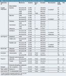
Table 1 contains analytical procedures including several types of membrane-assisted extraction that are focused on the determination of environmentally relevant drugs.

Table 1: Membrane-assisted extraction methods reported for drug detection in water samples.
Beyond chemical conversion of analytes ion-pairing or chelating reagents can be added to ionic analytes to enable their extraction into an organic acceptor phase [Figure 2(b)]. Reubsaet et al.21 presented an ion-pair mediated LPME of angiotensin, neurotensin and their metabolites. Under acidic conditions the analyte transport across n-octanol as liquid membrane was mediated by heptane-1-sulphonic acid. The alkyl sulphonic acid formed with the charged peptides ion-pairs that migrated through an n-octanol layer present in the pores of the polypropylenene membrane. The ion-pair transport was driven by a pH gradient from pH 3 in the donor phase to pH 1 in the acceptor solution of the LPME hollow fibre lumen.
Recoveries up to 82% could be achieved. A comparable strategy was pursued by Ho et al.22 to enrich hydrophilic drugs such as amphetamine, morphine and practolol, an adrenergetic beta-receptor antagonist (β-blocker). Ion pairs were formed with sodium octanoate at pH 7 and the neutral complexes can be extracted into n-octanol, the liquid membrane within the pores. Protons delivered from the acidic acceptor phase support the release of the amino drugs at the octanol and neutralize the counter ion. Capillary electrophoresis was used for the detection of the analytes.
The use of heptanoic acid as carrier for membrane-assisted extraction of quaternary ammonium compounds was described by Norberg et al.23
Alkyldimethylbenzylammonium salts widely used as fabric softener and antiseptic additives are known for their toxicity towards aquatic organisms. Determination of the cationic surfactants can be realized by continuous membrane-assisted extraction combined with normal phase HPLC. The ion complexes formed with heptanoic acid can permeate through a microporous membrane into chlorobutane as acceptor phase. Enrichment factors of about 20 were obtained resulting in limits of detection between 0.7 and 5 μg/L (for the three most abundant homologues).
Counter ions can also be incorporated in the liquid membrane (tri-2-ethylhexyl phosphate or di-n-hexyl ether) as demonstrated by Romero et al. for the extraction of biogenic amines from wine.24 The HPLC/UV detection of the extracted amines was enhanced by their conversion into the dabsyl derivatives.
When atmospheric pressure ionization mass spectrometry (APIMS) is selected for detection, the ion-pair reagents should be carefully selected because excess of ionic substances can suppress the ionization of the target analytes.
An excellent overview on currently applied microextraction techniques for the determination of polar pollutants in water is presented by Quintana and Rodriguez.25 They summarized that most of the currently used (micro) extraction techniques are based on SPE and SPME whereas the last technique is more frequently applied for the extraction of hydrophobic and semi polar analytes. The review highlighted also the main advantages of LPME, such as the time- and solvent saving mode, as well as the easy separation of matrix compounds.
Furthermore, only small sample volumes (10–50 mL) are needed for a sensitive analysis at the ng/L to μg/L range and the formation of inconvenient emulsions that can occur in liquid–liquid extractions is completely avoided. Actually, most of the micromembrane devices used for analytical purposes are custom-made.
A wider acceptance of the elegant technique for routine analysis could be forced when membrane probes could be commercialized. Currently, only the Gerstel cooperation offers a membrane extraction device that allows fully automated analyte enrichment coupled to large volume injection and GC–MS analysis.26
This short review demonstrates that the high potential of membrane-assisted extraction is not exhausted yet. Particularly, additional membrane modifications and specific chemical or biochemical reactions can expand the application field of membranes for analytical and biomedical purposes.
Perspectives
In 2001 Torto and Mwatseteza realized in a review that the application of membrane-assisted extraction processes, here called microdialysis, were focused on limited fields and hardly any samples other than biofluids had been investigated.27 The last five years have shown great advances in perfecting the technical design of membrane supported assemblies.
The membrane permeation processes can be controlled and invented by specific tailoring membrane units and extraction conditions. Furthermore, miniaturization will allow the combination of sample preparation and microcapillary electrophoresis or HPLC, a promising step to operate at a lab-on-a-chip scale28 that would be interesting for microsensor development in neurosciences, medical and biotechnological applications.29,30
Monika Moeder is senior fellow at the Helmholtz Centre for Environmental Research — UFZ (Leipzig, Germany) and works in the field of analytical method development for environmental monitoring.
Franziska Lange is PhD student at the Martin-Luther-University of Halle-Wittenberg (Halle/Saale, Germany). Her thesis concerns the development of membrane-assisted micro-extraction methods.
References
1. T. Heberer, Tox. Lett., 131, 5–17(2002).
2. J. Pawliszyn, Anal. Chem., 75, 2543–2558 (2003).
3. R. Eisert and J. Pawliszyn, Anal. Chem., 69, 3140–3147 (1997).
4. E. Baltussen et al., Microcolumn Sep., 11, 737–747 (1999).
5. H.G. Ugland, M. Krogh and K. E. Rasmussen, J. Chromatogr. B, 749, 85–92 (2000).
6. K.E. Rasmussen and S. Pedersen-Bjergaard, Trends in Anal. Chem., 23, 1–10 (2004).
7. J.B. Quintana, R. Rodil and T. Reemtsma, J. Chromatogr. A, 1061, 19–26 (2004).
8. S. Mueller et al., J. Chromatogr. A, 985, 99–106 (2003).
9. J.A. Jönsson and L. Mathiasson, J. Chromatogr. A, 902, 205–225(2000).
10. R. E. Majors, LCGC Eur., 5, 284–292 (2006).
11. S. Pedersen-Bjergaard and K. E. Rasmussen, J. Chromatogr. A, 1109, 183–190 (2006).
12. U. Karst, Angew. Chem.-Int., 43, 2476–2478 (2004).
13. B. Hauser, M. Schellin and P. Popp, Anal. Chem., 76, 6029–6038 (2004).
14. M. Ulbricht, M. Riedel and U. Marx, J. Membr. Sci., 120, 239–259 (1996).
15. M. Moeder, C. Martin and G. Koeller, J. Membr. Sci., 245, 183–190 (2004).
16. T. Einsle et al., J.Chromatogr. A, 1124, 196–204 (2004).
17. J. A. Jönsson and L. Mathiasson, Trends in Anal. Chem., 18, 318–325, 325–334 (1999).
18. J.A. Jönsson and L. Mathiasson, J. Sep. Sci., 24, 495–507 (2001).
19. A. Leinonen et al., Anal. Chim. Acta, 559, 166–172 (2006).
20. S. Pedersen-Bjergaard and K. E. Rasmussen, Anal. Chem., 71, 2650–2656 (1999).
21. J.L.E. Reubsaet, H. Loftheim and A. Gjelstad, J. Sep. Sci., 28, 1204–1210 (2005).
22. T.S. Ho et al., J. Chromatogr. A, 998, 61–72 (2003).
23. J. Norberg et al., J. Chromatogr. A, 869, 523–529 (2000).
24. R. Romero et al., J. Sep. Sci., 25, 584–592 (2002).
25. J.B. Quintana and I. Rodriguez, Anal. Bioanal. Chem., 384, 1447–1461 (2006)
26. B. Hauser, P. Popp and E. Kleine-Benne, Gerstel Application Note 12/2002
27. N. Torto, J. Mwatseteza and T. Laurell, LCGC Eur., 9, 536–546 (2001).
28. X. Wang, C. Saridara and S. Mitra, Anal. Chim. Acta, 543, 92–98 (2005).
29. M.A.F. Elmosallamy et al., Microchimica Acta, 151, 109–113 (2005).
30. G.A.E. Mostafa, J. Pharm. Biochem. Anal., 31, 515–521 (2003).

Common Challenges in Nitrosamine Analysis: An LCGC International Peer Exchange
April 15th 2025A recent roundtable discussion featuring Aloka Srinivasan of Raaha, Mayank Bhanti of the United States Pharmacopeia (USP), and Amber Burch of Purisys discussed the challenges surrounding nitrosamine analysis in pharmaceuticals.
Extracting Estrogenic Hormones Using Rotating Disk and Modified Clays
April 14th 2025University of Caldas and University of Chile researchers extracted estrogenic hormones from wastewater samples using rotating disk sorption extraction. After extraction, the concentrated analytes were measured using liquid chromatography coupled with photodiode array detection (HPLC-PDA).


