Liposome Characterizatio by FFF–MALS–QELS
The Application Notebook
Liposomes are made of lipid bilayers and are often used in drug delivery by encapsulating the core with therapeutic drugs. During liposome research, formulation, manufacturing, and quality control, it is of great importance to monitor liposome size and encapsulation.
Liposomes are made of lipid bilayers and are often used in drug delivery by encapsulating the core with therapeutic drugs. During liposome research, formulation, manufacturing, and quality control, it is of great importance to monitor liposome size and encapsulation. Field flow fractionation (FFF) with the concomitant use of multi-angle light scattering (MALS) and quasi-elastic light scattering (QELS, aka dynamic light scattering) is an ideal tool for such characterization.
Here, we report the analytical results of two liposome samples, one empty, and one filled. Using the Eclipse FFF system followed by a DAWN HELEOS (with embedded WyattQELS instrumentation), the FFF method was optimized with the aid of Wyatt ISIS FFF simulation software. The online QELS directly measures the hydrodynamic radius, Rh, whereas the HELEOS measures the root-mean square radius, Rg.
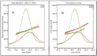
The WyattQELS detector was placed at approximately 143° in order to extend the Rh measurement up to 300 nm. Both Rg and Rh are plotted against elution time in Figure 1. The results from duplicate runs demonstrate excellent reproducibility of the FFF–MALS–QELS method. Figure 1 also shows that the Rh values for both empty and filled liposomes are well overlaid, suggesting the separation is based on hydrodynamic radius as expected from an FFF separation. However, Rg values for these two liposomes do not overlay, which indicates these two liposomes have different degrees of encapsulation.

Figure 1
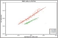
Root-mean square radii, Rg, were then plotted against hydrodynamic radii, Rh, for these two liposomes. The slope of Rg vs. Rh plot yields the internal structure of the liposomes. The empty liposome sample has a slope of 1.0, consistent with a spherical shell structure. The filled liposome sample, on the other hand, has a slope of 0.75, in good agreement with that of a solid sphere structure of uniform density.

Figure 2
For liposomes or other nanoparticles, FFF–MALS–QELS provides an easily adaptable yet powerful characterization tool to obtain information on particle size, size distribution, particle count, as well as structure — all without making assumptions about the particles or their composition.
DAWN®, miniDAWN®, ASTRA®, Optilab® and the Wyatt Technology logo are registered trademarks of Wyatt Technology Corporation. ©2011 Wyatt Technology Corporation.
Wyatt Technology Corporation
6300 Hollister Avenue, Santa Barbara, CA 93117
tel. (805) 681-9009, fax (805) 681-0123
Email: info@wyatt.com, Website: www.wyatt.com

Separating Impurities from Oligonucleotides Using Supercritical Fluid Chromatography
February 21st 2025Supercritical fluid chromatography (SFC) has been optimized for the analysis of 5-, 10-, 15-, and 18-mer oligonucleotides (ONs) and evaluated for its effectiveness in separating impurities from ONs.


