UHPLC Separations Using HPLC Methods and TSKgel Columns
The use of UHPLC systems for small molecule analysis has gained widespread acceptance among researchers in recent years.
The use of UHPLC systems for small molecule analysis has gained widespread acceptance among researchers in recent years. A UHPLC system is a HPLC system optimized with regards to dead volume, injection performance, and detector sampling rate and is able to tolerate application pressures exceeding 1000 bar. It is therefore advantageous to use UHPLC instrumentation for methods developed on conventional HPLC systems with HPLC columns. The use of HPLC columns with UHPLC systems offers the advantages of cost and time savings over having to purchase and develop new methods with UHPLC columns. This application note demonstrates the excellent performance of conventional TSKgel HPLC columns on a UHPLC system.
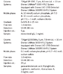
Experimental Conditions

Results and Discussion
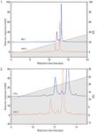
Figure 1 shows the analysis of a monoclonal lgG using a TSKgel SP-STAT cation exchange column. The same column, sample, and method were used to verify the column-system compatibility on both the HPLC (blue chromatogram) and UHPLC (red chromatogram) systems. The column performs better in the UHPLC system than it does when connected to the HPLC system. The number of theoretical plates is 6% higher for the UHPLC setup.

Figures 1 and 2: Analysis of a monoclonal lgG using a TSKgel SP-STAT column and a Zoomed Elution Profile of the mAb charge variants on the TSKgel SP-STAT column.
Figure 2 shows a zoomed elution profile of the mAb charge variants on the TSKgel SP-STAT column, which provides better insight into what extent the elution profile benefits from using a UHPLC system. The peak width is smaller for the UHPLC chromatogram. The decreased system dead volume resulted in the peak elution to occur earlier than it did on the HPLC system.
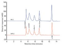
Figure 3 shows a standard protein mixture analyzed using a TSKgel G2000SWXL size exclusion column. The number of theoretical plates exceeds 32,000 for para-aminobenzoic acid when connected to the UHPLC system (blue chromatogram), while the column connected to the HPLC system (red chromatogram) only reaches 29,000 for the same component.

Figure 3: Separation of a standard protein mixture using a TSKgel G2000SWXL column.
Of course, resolution is an important factor when considering chromatographic performance. The HPLC data shows a resolution of 2.1 for the separation of γ-globulin and ovalbumin, and 10.2 for ribonuclease A and para-aminobenzoic acid. For the UHPLC data, the resolution factors increase to 2.2 and 10.9 for the respective peak pairs.
Conclusions
The results of both analyses using a TSKgel SP-STAT ion exchange column and a TSKgel G2000SWXL size exclusion column indicate excellent separation with high resolution when used on a UHPLC system. As a UHPLC system is simply an optimized HPLC system, bioseparation method transfer from HPLC to UHPLC is hardly more complicated than from one HPLC instrument to another. The applied pressure in these applications was far from exceeding the limit of a conventional HPLC system. This is the case in most bioseparation applications, since many biological sample molecules themselves cannot withstand very high pressure. Transferring established HPLC methods using HPLC columns to a UHPLC system is an economical and time-saving way to take advantage of this latest wave of development in HPLC technology.
Tosoh Bioscience and TSKgel are registered trademarks of Tosoh Corporation.
UltiMate is a registered trademark of the Dionex Corporation.

Tosoh Bioscience LLC
3604 Horizon Drive, Suite 100, King of Prussia, PA 19406
tel. (484) 805-1219, fax (610) 272-3028
Website: www.tosohbioscience.com

SEC-MALS of Antibody Therapeutics—A Robust Method for In-Depth Sample Characterization
June 1st 2022Monoclonal antibodies (mAbs) are effective therapeutics for cancers, auto-immune diseases, viral infections, and other diseases. Recent developments in antibody therapeutics aim to add more specific binding regions (bi- and multi-specificity) to increase their effectiveness and/or to downsize the molecule to the specific binding regions (for example, scFv or Fab fragment) to achieve better penetration of the tissue. As the molecule gets more complex, the possible high and low molecular weight (H/LMW) impurities become more complex, too. In order to accurately analyze the various species, more advanced detection than ultraviolet (UV) is required to characterize a mAb sample.


