TSKgel Amide-80, 2 m Columns for the Analysis of Glycans
The Application Notebook
Most proteins, particularly soluble and membrane-bound proteins expressed in the endoplasmic reticulum, are glycosylated. The extent and result of glycosylation varies.
Most proteins, particularly soluble and membrane-bound proteins expressed in the endoplasmic reticulum, are glycosylated. The extent and result of glycosylation varies. For example, some proteins do not fold correctly unless they are glycosylated. The polysaccharide side chains (glycans) play critical roles in physiological and pathological reactions ranging from immunity to cell signaling, development, and death. The efficacy of biotherapeutic drugs is affected by glycosylation and therefore the profiling of the glycan pool released from a glycoprotein must be monitored to control product consistency.
Hydrophilic interaction chromatography (HILIC) with an amide-based stationary phase, used in conjunction with fluorescent-
labeled oligosaccharides, is a well-established, robust technique used by many laboratories to obtain high resolution separation of isolated glycans. TSKgel Amide-80 column chemistry is ideally suited for the separation of charged and neutral fractions of glycan pools in a single run. The application of a TSKgel Amide-80, 2 μm column for the analysis of glycans from monoclonal antibodies is demonstrated in this note, including a comparison to a 3 μm TSKgel Amide-80 column.
Materials and Methods

Results and Discussion
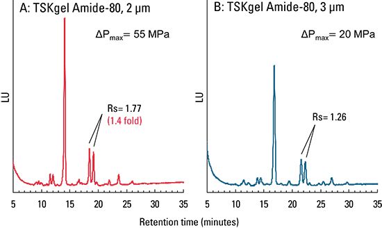
A comparison was done on the separation of pyridylaminated oligosaccharides released from mouse IgG on TSKgel Amide-80, 2.0 mm ID × 15 cm columns differing in particle size. As seen in Figure 1, the TSKgel Amide-80, 2 μm showed a 1.4-fold higher resolution of PA-glycan peaks compared to the TSKgel Amide-80, 3 μm column. With similar selectivity, method transfer from a 3 μm to a 2 μm column is easily accomplished. Note that the TSKgel Amide-80, 2 μm column performed at a pressure of approximately 55 MPa during the gradient elution when run at the flow rate of 0.5 mL/min. A UHPLC system or a conventional HPLC system with a maximum pressure rating of greater than 40 MPa is required if the separation is run at this higher flow rate.

Figure 1: Separation of PA-glycans from mouse monoclonal antibody.
Pyridylamination is a fluorescence-tagging method for oligosaccharides that enables measurement and structural analyses of glycans. Glycans do not contain fluorophores and thus must be labeled with fluorescent tags prior to analysis. 2-Aminobenzamide (2-AB) is one of the most common labels used.
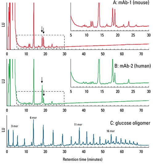
Several peaks of glycans were separated from both mouse IgG and human IgG (Figure 2). These peaks were similar in elution time to 6-8 mer glucose. When comparing chromatograms A and B, different ratios of glycan attached to each mAb is implied.

Figure 2: Comparison of glycan peak profile of monoclonal antibodies.
Conclusions
A TSKgel Amide-80, 2 μm column has similar selectivity as an Amide-80, 3 μm column. The smaller particle size is ideally suited for the higher resolution separation of glycans. This 2 μm column showed much higher resolution than a TSKgel Amide-80, 3 μm column for the separation of pyridylaminated oligosaccharides released from mouse IgG and successfully separated several peaks of glycans from both mouse IgG and human IgG. Because of equivalent selectivity, a smooth method transfer from a 3 μm TSKgel Amide-80 column to a 2 μm column is an additional advantage.
Tosoh Bioscience and TSKgel are registered trademarks of Tosoh Corporation.

Tosoh Bioscience LLC
3604 Horizon Drive, Suite 100, King of Prussia, PA
tel. (484) 805-1219, fax (610) 272-3028
Website: www.tosohbioscience.com

SEC-MALS of Antibody Therapeutics—A Robust Method for In-Depth Sample Characterization
June 1st 2022Monoclonal antibodies (mAbs) are effective therapeutics for cancers, auto-immune diseases, viral infections, and other diseases. Recent developments in antibody therapeutics aim to add more specific binding regions (bi- and multi-specificity) to increase their effectiveness and/or to downsize the molecule to the specific binding regions (for example, scFv or Fab fragment) to achieve better penetration of the tissue. As the molecule gets more complex, the possible high and low molecular weight (H/LMW) impurities become more complex, too. In order to accurately analyze the various species, more advanced detection than ultraviolet (UV) is required to characterize a mAb sample.


