|Articles|June 1, 2006
- LCGC Europe-06-01-2006
- Volume 19
- Issue 6
New Technologies
Author(s)Peter Myers
New ways of envisioning scientific data are constantly being developed in data rich research environments. Researchers at the University of California, Irvine's Center of GRAVITY (Graphics, Visualization and Imaging Technology), USA have developed a new display for this data rich market. They have developed the Highly Interactive Parallelized Display Wall or HIPerWall. It is a massively tiled, grid-based display built using fifty 30-inch Apple cinema displays and twenty-five Power Mac G5 computers. This powerful display system allows researchers to view and manipulate their data sets at extremely high resolutions and collaborate with other scientists in new and exciting ways.
Advertisement
New ways of envisioning scientific data are constantly being developed in data rich research environments. Researchers at the University of California, Irvine's Center of GRAVITY (Graphics, Visualization and Imaging Technology), USA have developed a new display for this data rich market. They have developed the Highly Interactive Parallelized Display Wall or HIPerWall. It is a massively tiled, grid-based display built using fifty 30-inch Apple cinema displays and twenty-five Power Mac G5 computers. This powerful display system allows researchers to view and manipulate their data sets at extremely high resolutions and collaborate with other scientists in new and exciting ways.
The Power Macs behind the wall.
Dr Falko Kuester, director of the Center of GRAVITY and the principal investigator for the HIPerWall project, and his team wanted to build a large, collaborative visualization platform capable of displaying static images, image sequences in the form of movies and animations, and three-dimensional, time-varying data in real time. They wanted the ability to project a single image across the entire display area as well as to present many different images or media streams simultaneously. Since no single display provided anything close to their 100-plus-megapixel resolution requirement, the team sought a solution with multiple flat panel displays.
The HIPerWall is already being used in new application areas. Brain scans are typically viewed on photographic film or desktop monitors as a small group of images; each of which represents one cross-sectional slice of the brain. This is problematic because the person evaluating the images can only see six to ten slices at a time. The HIPerWall has the resolution to display all of the images from a brain scan simultaneously, thus retaining both the context and the details of the entire scan sequence. Even more important is the ability to compare brain images between different patients. The HIPerWall has the resolution needed to display multiple slices from many different patients to help medical specialists compare and correlate the characteristics of the feature being studied. Instead of having an intern running to the lightboard to exchange one set of films for another, a simple mouse-click is all it takes to step through successive sets of images on the HIPerWall.
The HIPerWall project team.
Isn't this the display that chromatographers need for the increasing complex 2D and 3D plots we now see and try to interpret from multidimensional chromatography or from the multidimensional plots from hyphenated chromatography? Maybe so but you will need a big cheque book to purchase one!
Articles in this issue
over 19 years ago
The Thermal Conductivity Detectorover 19 years ago
Infotrieve gains academic supportover 19 years ago
Dwell Volume RevisitedNewsletter
Join the global community of analytical scientists who trust LCGC for insights on the latest techniques, trends, and expert solutions in chromatography.
Advertisement
Related Content
Advertisement

Advertisement
Advertisement
Trending on LCGC International
1
Unlocking Discovery Data: Why a Digital Ecosystem Matters for HT-MS
2
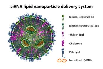
Streamlined Method Development for Efficient and Reliable Lipid Nanoparticle Analysis
3
LCGC Blog: Rewiring the Fundamentals
4
Best of the Week: The Scientific Method in the Age of AI, Rewiring the Fundamentals
5