Light Scattering for the Masses® Monoclonal Antibodies and the Preparative Use of Flow Field-Flow Fractionation (AF4)
The Application Notebook
Wyatt Technology Corporation
A.J. Freitag, K. Wittmann, G. Winter, and J. Myschik, Wyatt Technology Corporation
Proteins — especially monoclonal antibodies (MABs) — have become increasingly important in pharmaceutical work. However, there are some important differences between conventional, chemically-synthesized drugs and proteins. Because of the complex and weak structure of proteins, even a slight change in conditions, such as pH value, temperature, or mechanical stress, may lead to aggregation and a loss of activity or stability.
A separation of MABs before analysis is preferred because a detailed investigation of a fraction would be much easier and provide a better insight into the physical and chemical properties, compared to an analysis in the presence of a coexisting monomeric protein.
Asymmetrical flow field-flow fractionation (AF4) is a well-established method for sizing and quantifying different aggregate species in protein formulations. A major advantage of AF4 is the use of the formulation buffer of the protein as the mobile phase. The separation is independent of the ionic strength of the buffer.
The capability of AF4 as a preparative tool for the separation of aggregates, fragments, and monomeric species was investigated in this study. The system consisted of an Eclipse (Wyatt Technology Europe GmbH, Dernbach, Germany), equipped with DAWN multi-angle light scattering (MALS), refractive index (RI) and ultraviolet (UV) detectors, and a fraction collector. The separation was achieved by using a semi-preparative SP channel with 350 μm height.
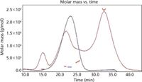
A monoclonal antibody sample was exposed to light, resulting in the formation of fragments and aggregates (Figure 1). A sample of 1 mg of protein per run was injected into the channel and collected in tubes made of glass, using a Gilson FC-203B fraction collector. The collected fractions were concentrated and the final concentration was determined by UV absorbance at 280 nm using a Micro-BCA Assay.

Figure 1: UV 280 nm fractograms and MALS detection of light exposed MAb1 (red line) and native MAb1 (blue line).
AF4 is a useful method for analytical and preparative separation of protein aggregates since the separation can be performed in each buffer or even pure water, thus facilitating subsequent activities. Furthermore, an investigation of possible immune responses triggered by protein aggregates in comparison to monomeric proteins can be performed, which is currently of major interest in administrative organizations and the pharmaceutical industry at large.

Table 1: Molecular weight of the detected species of MAb1.
The results presented in this application note have been published before in A.J. Freitag, K. Wittmann, G. Winter, and J. Myschik, LCGC Europe 24(3), 134–140 (2011).

Wyatt Technology Corporation
6300 Hollister Ave., Santa Barbara, California 93117, USA
Tel: +1 805 681 9009 fax: +1 805 681 0123
E-mail: info@wyatt.com
Website: www.wyatt.com

SEC-MALS of Antibody Therapeutics—A Robust Method for In-Depth Sample Characterization
June 1st 2022Monoclonal antibodies (mAbs) are effective therapeutics for cancers, auto-immune diseases, viral infections, and other diseases. Recent developments in antibody therapeutics aim to add more specific binding regions (bi- and multi-specificity) to increase their effectiveness and/or to downsize the molecule to the specific binding regions (for example, scFv or Fab fragment) to achieve better penetration of the tissue. As the molecule gets more complex, the possible high and low molecular weight (H/LMW) impurities become more complex, too. In order to accurately analyze the various species, more advanced detection than ultraviolet (UV) is required to characterize a mAb sample.


