Characterizing Protein-Nucleic Acid Conjugates with Light Scattering
The Column
This article presents two case studies regarding the characterization of protein-DNA complexes using two complementary multi-angle light scattering (MALS) techniques, namely size-exclusion chromatography (SEC–MALS) to determine absolute molar mass of each component, and composition-gradient MALS (CG–MALS) to quantify stoichiometry and affinity at binding sites in solution.
Photo Credit: lumikk555 /stock.adobe.com

In this article, two case studies regarding the characterization of protein-DNA complexes are presented, using two complementary multi-angle light scattering (MALS) techniques: SEC–MALS-to determine absolute molar mass of each component, with size-exclusion chromatography, and CG–MALS-to quantify stoichiometry and affinity of the interaction, label-free and in solution, with composition gradients.
Analysis of Protein-Nucleic Acid Complexes
The role of nucleic acids in biotherapeutics has been increasing. The emergence of cancer immunotherapies designed to deliver nucleic acids directly to immune cells is expanding the realm of biotherapeutics, from antibody and peptide conjugates to now include DNA and RNA. Engineered nucleic acids are often conjugated to receptor proteins, such as lectins, for the purposes of targeted delivery to cells and their therapeutic function will typically involve induction and inhibition of genes or mediated gene silencing (1).
Nucleic acids present many of the same challenges present in traditional biotherapeutics: the delivery efficiency of a biotherapeutic may depend on the amount of conjugation present, while purification or formulation conditions may cause formation of nucleic acid duplexes or aggregation. In addition, the interactions between nucleic acids and host proteins may involve cooperative binding, allosteric modulation, or other complex phenomena. Hence biophysical analysis of protein conjugate size, molar mass, composition, and conformation, as well as the interactions between components, is essential to developing stable and efficacious therapies. Strategies for characterizing these properties are widely applicable to biopharmaceutical analysis and would be particularly advantageous for protein-nucleic acid drug formats.
Electrophoretic mobility shift assay (EMSA), often referred to as gel-shift assay, is one of the traditional methods for characterizing proteinânucleic acid complexes. Because the rate of DNA migration is slowed, or shifted, when bound to protein and the gel provides a cagingâeffect for stabilizing interactions, the technique enables the study of binding affinity. Historically requiring DNA with radiolabelled 32P, the assay can now also be performed with biotinylated or fluorescently-labelled DNA probes, though major limitations include the challenges in quantitating complexes and the assumption that labelling does not influence the data.
Another strategy, using multi-angle light scattering (MALS), enables quantitative, direct analysis without the need for fluorescent or radioactive labels that can affect results. Protein-nucleic acid conjugate analysis with MALS has equal or better resolution than gel-shift assay and can be performed entirely in solution using two complementary and powerful techniques. The first is size-exclusion chromatography (SEC)–MALS, the coupling of SEC with light scattering (LS), ultraviolet (UV), and differential refractive index (dRI) detectors, that can be used to quantify both the absolute molar mass and the mass fraction of the protein and nucleic acid. The second is composition gradient multi-angle light scattering (CG–MALS) to quantify the affinity and stoichiometry of protein-nucleic acid interactions.
This article presents two case studies utilizing SEC–MALS and CG–MALS to analyze protein-nucleic acid complexes to demonstrate the compatibility of these techniques with emerging classes of biotherapeutic conjugates. These strategies have wide-ranging compatibility with biopharmaceutical drug formats.
Methods
Analysis of protein conjugates combines simultaneous collection of MALS, RI, and UV signals and uses each component’s distinct extinction coefficient (ε) and refractive index increment (dn/dc). The first case study is based on the work by Gupta et al. (2) employing a 10 mm × 300 mm, 200-µm Superdex GL column (GE Healthcare) with an inline UV detector, DAWN MALS detector (Wyatt Technology Corporation), and an Optilab differential refractive index detector (Wyatt). The data were analyzed using system methods in the ASTRA 7 software package (Wyatt). This includes the UV extinction coefficient from RI peak analysis to determine the UV extinction coefficient for each component (0.75 and 13.2 g/L cm for PFV and U5, respectively) from the RI signal using a protein dn/dc 0.185 mL/g and a nucleic acid dn/dc of 0.170 mL/g. Subsequently the protein conjugate analysis was used to determine the mass fraction contribution of each component, termed the protein and the modifier.
The CG–MALS experiments for the second case study were automated using the Calypso composition gradient system (Wyatt) to ensure accurate and particle-free mixtures as reported by Kenrick et al. (3) Aliquots of each sample and buffer were pre-mixed by the instrument and injected into a downstream DAWN MALS detector (Wyatt) and a UV detector (Shimadzu) to determine molar mass and concentration, respectively. The data were analyzed using the Calypso software package (Wyatt) to find the most appropriate equilibrium model and to determine the affinity and stoichiometry of the interaction.
SEC–MALS for Absolute Molar Mass Determination of Complexes
SEC–MALS overcomes many of the limitations of traditional analytical SEC, which estimates molar mass entirely by peak retention time. This method is vulnerable to non-ideal elution and errors in molar mass estimation from molecular conformation, density, column interactions, and variability of loading or concentration. The enhancement of SEC with MALS can conveniently determine the absolute molar mass regardless of elution behaviour. This is particularly advantageous for biopharmaceutical analysis where gel-shift assay is limited for quantifying complexes. In addition, separation of components enables absolute measurements of distributions of molar mass and radius in a single fluid pathway.
Using the concentration detectors to determine the mass fractions and the LS detector to determine the total molar mass of the complex, the molar mass of each constituent can be determined. This process is compatible with a wide range of conjugates and copolymers (4–6), but will be described in this case study with a protein-nucleic acid complex: the protein, foamy virus integrase, was bound to DNA. Characterization of the complex was performed by K. Gupta et al. (2) using SEC–MALS, with the goal of confirming that the observed crystal structure represents true solution behaviour.
When prototype foamy virus integrase (PFVâIN) was bound to U5 viral DNA, the intasome complex eluted as a single peak in the resulting chromatogram. Because each component was determined to have a different UV extinction coefficient at a 280 nm wavelength and the distinct dn/dc values described above, the mass fraction of each component can be determined in the chromatogram. When coupled with MALS data, the molar mass of each constituent (protein and DNA) can be determined for each data slice. The results from the authors after analysis is provided in Figure 1, where the molar mass of the total complex (PFV-IN and U5 DNA) as well as the molar masses of each component are provided across the peak.
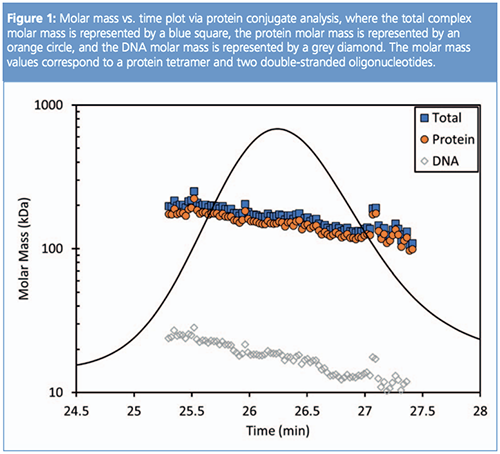
Overall, the weight-average molar mass of the complex was 194 kDa, consisting of a protein fraction of roughly 172 kDa, which corresponds to four integrase proteins, and a DNA fraction of 22 kDa, which represents two double-stranded oligonucleotides per complex. This combination of four protein molecules and two DNA molecules corresponds with the crystal structure, confirming that this is also the relevant structure in solution. This highlights the advantage of SEC–MALS to provide structural data in solution as a means of reconciling observations from crystal structures and orthogonal measurements (that is, smallâangle X-ray scattering [SAXS] and smallâangle neutron scattering [SANS]). Interestingly, the molar mass is not constant across the peak, which ultimately suggests some degree of polydispersity or onâcolumn dissociation. Investigation of binding affinity and stoichiometry via the second complementary light scattering technique will explore potential interactions in more detail.
Composition Gradient Multi-Angle Light Scattering (CG–MALS) for Quantification of the Affinity and Stoichiometry of Protein-Nucleic Acid Interactions
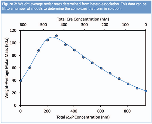
CG–MALS quantifies equilibrium association in unfractionated or batch solutions. In this technique, a series of freshly made compositions are injected into a MALS and concentration detector. The MALS detector determines the weight-average molar mass of the sample at each composition, where the relationship of the change in molar mass to the composition relates to the formation of complexes. This case study elucidated the interactions of the Cre recombinase protein and IoxP DNA recognition site as reported by Kenrick et al. (3). In this study, Cre and IoxP were first investigated using CG–MALS to determine the molar mass, native oligomeric state, and identify possible self-associations via gradients, where it was determined that neither molecule self-associates under experimental conditions. For analyzing two components, CG–MALS operates under the principles that if no complexes are formed, the molar mass will ultimately be a simple weight average of the two species; however, as complexes form, the weight-average molar mass increases. Stronger interactions result in more sustained complex formation, leading to sharper and more pronounced curves, whereas the position of the peak apex corresponds to the stoichiometry of the interaction. This relationship can be fit by a suitable equilibrium model to determine the affinity and stoichiometry of the interaction.
Hetero-association gradients using 13 freshly prepared mixtures of Cre and IoxP were used to quantify association affinity and stoichiometry of these two molecules (3). Upon infusion into the MALS flow cell, flow was stopped to enable equilibration of the mixture for determining equilibrium molar mass, ultimately coming to equilibrium in 15 min. From the raw light scattering intensity and the concentration of each species, the resulting data were converted to weight-average molar mass for each data point. In Figure 2, the molar mass data for the complex is plotted, which suggests a higher order stoichiometry than 1:1 is present and that a deeper analysis is required to fully understand the complex stoichiometry.
In order to obtain the appropriate fit for the CG–MALS data, three distinct binding events (1:1; 2:1, and 4:2 Cre:IoxP) were characterized. Under these conditions, the first binding event (1:1) has an affinity of 170 nM, the second binding event (2:1) has an affinity of 17 nM, and the third binding event (4:2), the dimerization, has an affinity of 400 nM. The various complexes and their molar composition are shown in Figure 3, with the free monomer excluded for clarity.
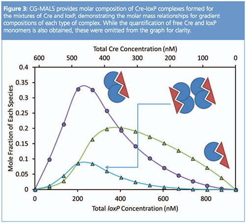
In contrast to gel shift assays, CG–MALS enables straightforward and rapid measurements with excess Cre or excess IoxP to quantify multiple interactions and was successful in quantifying all three complexes that formed in solution. Furthermore, CG–MALS enables time-dependent analysis of equilibrium through measurements of molar mass versus time to help quantify association kinetics.
Conclusion
Light scattering provides uniquely powerful and complementary techniques for effective characterization of protein-nucleic acid complexes to determine absolute molar mass, conjugation ratio, interactions, binding mechanisms, and stoichiometry, as well as elucidating the attributes of higher-order structures. These simple but robust techniques enable facile analysis of protein-nucleic acid interactions and can be widely applicable to other biomolecular complexes.
References
- J.L. Nair et al., J. Am. Chem. Soc.136(49), 16958–16961 (2014).
- K. Gupta et al., Structure20, 1918–1928 (2012).
- S. Kenrick and M. Chen, “SEC–MALS and CG–MALS: Complementary Techniques to Characterize Protein-DNA Complexes,” 2017. [Online]. Available: https://www.wyatt.com/library/application-notes/wp3001-characterizing-protein-dna-interactions.html
- Y. Peng and L. Zhang, Journal of Biochemical and Biophysical Methods30, 243–252 (2003).
- S. Kenrick and M. Chen, “A light scattering toolbox for characterizing transport proteins and their interactions,” 2014. [Online]. Available: http://www.wyatt.com/files/literature/appnotes/sec-mals-proteins/transport-protein-sec-cgmals-poster.pdf.
- D.J. Slotboom et al., Methods46(2), 73–82 (2008).
Nemal Gobalasingham is an applications scientist at Wyatt Technology in the customer support and service division, working with customers on a diverse range of applications. Nemal received his B.S. degree in chemistry from Syracuse University (Syracuse, New York, USA) and his doctorate in chemistry from the University of Southern California (Los Angeles, California, USA), specializing in polymer chemistry and materials science.
Sophia Kenrick is senior applications scientist at Wyatt Technology where she supports multiple applications for Wyatt instrumentation, especially in the field of molecular recognition and biomolecular interactions. Sophia received a bachelor’s degree in chemical engineering from Arizona State University (Arizona, USA), a doctorate in chemical engineering from the University of California, Santa Barbara (USA), and has been with Wyatt since 2010. She is also the Dean of Light Scattering University.
E-mail:info@wyatt.comWebsite:www.wyatt.com

Regulatory Deadlines and Supply Chain Challenges Take Center Stage in Nitrosamine Discussion
April 10th 2025During an LCGC International peer exchange, Aloka Srinivasan, Mayank Bhanti, and Amber Burch discussed the regulatory deadlines and supply chain challenges that come with nitrosamine analysis.
Polysorbate Quantification and Degradation Analysis via LC and Charged Aerosol Detection
April 9th 2025Scientists from ThermoFisher Scientific published a review article in the Journal of Chromatography A that provided an overview of HPLC analysis using charged aerosol detection can help with polysorbate quantification.
Removing Double-Stranded RNA Impurities Using Chromatography
April 8th 2025Researchers from Agency for Science, Technology and Research in Singapore recently published a review article exploring how chromatography can be used to remove double-stranded RNA impurities during mRNA therapeutics production.


