Characterization Studies of PEGylated Lysozyme Using TSK-GEL® HPLC Columns
Chemical modification of therapeutic proteins in order to enhance their biological activity is of increasing interest.
Jessica Christel, Werner Conze, Achim Sprauer, Volker Noedinger, and Egbert Mueller, Tosoh Bioscience GmbH
Chemical modification of therapeutic proteins in order to enhance their biological activity is of increasing interest. One of the most frequently used protein modification methods is the covalent attachment of poly (ethylene glycol), which is referred to as PEGylation. This polymeric modification changes the biochemical and physicochemical properties of the protein, which can result in several important benefits, among them more effective target delivery, slower in-vivo clearance, and reduced toxicity and immunogenicity of therapeutic proteins. After PEGylation reaction, the mixture has to be purified in order to remove nonreacted protein and undesired reaction products. Liquid chromatography is the most common purification method of PEGylation reaction products. Since the extent of PEGylation influences masking and shielding effects of the covalently linked PEG molecule, there is increasing demand for chromatographic methods to separate the modified isoforms from the native protein. This application note describes the use of size exclusion and ion exchange chromatography for the characterization of PEGylated lysozyme.
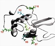
Lysozyme is a well known standard protein and is often used to determine the dynamic binding capacity of ion exchange chromatography (IEC) resins; therefore we decided to use PEG-lysozyme as a model protein in our study. PEGylated lysozyme was synthesized from methoxy PEG aldehyde (with a MW of 5 kDa, 10 kDa, and 30 kDa) and chicken egg white lysozyme in phosphate buffer in the presence of sodium cyanoborohydride (NaCNBH3) as a reducing agent. The PEGylation reaction takes place between the aldehyde group of methoxy PEG aldehyde and the free amino acid group (NH2-group) of lysine residues within the lysozyme molecule (see Figure 1).

Figure 1: Lysozyme has six lysine residues as possible PEGylation reaction sites.
The product mixture was analyzed with a TSKgel® G3000SWXL size exclusion column, SDS-PAGE (not shown), a TSKgel SP-5PW strong cation exchange column, a TSKgel SP-NPR strong cation exchange column and subsequent MALDI-TOF MS analysis (not shown).
Materials & Methods
PEGylation of egg white lysozyme:
5, 10, 30 kDa methoxy PEG aldehyde; 100 mmol/L phosphate buffer (Na2HPO4, NaH2PO4) pH 6.0; PEGylation by reductive alkylation; 20 mmol/L NaCNBH3 to reduce a Schiff base; 100 mmol/L HCl to stop PEGylation reaction.
Size Exclusion:
Column: TSKgel G3000SWXL, 5 µm, 7.8 mm X 30 cm
Mobile phase: 0.1 mol/L phosphate buffer, 0.1 mol/L Na2SO4, pH 6.7
Flow rate: 1.0 mL/min
Detection: UV@ 280 nm
Injection vol.: 20 μL
Ion Exchange (low pressure):
Column: TSKgel SP-5PW, 20 μm, 6.6 mm ID × 22 cm
Buffer A: 25 mmol/L phosphate buffer, 0.1 mol/L Na2SO4, pH 6.0
Buffer B: A + 0.5 mol/L NaCl
Flow rate: 0.85 mL/min
Detection: UV@ 280 nm
Injection vol.: 100 μL
Ion Exchange:
Column: TSKgel SP-NPR, 2.5 μm, 4.6 mm ID × 3.5 cm
Buffer A: 25 mmol/L phosphate buffer, 0.1 mol/L Na2SO4, pH 6.0
Buffer B: A + 0.5 mol/L NaCl
Flow rate: 1.0 mL/min
Detection: UV@ 280 nm
Injection vol.: 5 μL
Results
PEGylation of lysozyme
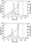
Figure 2 shows a typical chromatogram of a reaction mixture of PEGylated lysozyme separated on a TSKgel SP-5PW column. PEG chain lengths of 5 kDa and 30 kDa are shown. The profiles indicate a similar reaction characteristic. Nonreacted lysozyme remained in the reaction mixture; mono-PEGylated lysozyme as well as poly-PEGylated lysozyme was formed during the reaction.

Figure 2: Separation of PEGylated lysozymes on an analytical TSKgel SP-5PW column: mPEG5-aldehydes (A) and mPEG30-aldehydes (B). Peaks were identified by MALDI-TOF analysis. Identical sizes were numbered consecutively.
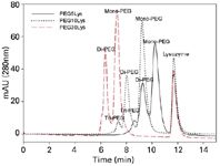
Size exclusion chromatography was performed as shown in Figure 3. The retention volumes of PEGylated lysozymes were used to assign the peaks in Figure 3 based on a standard calibration curve.

Figure 3: SEC analysis of reaction mixtures performed with a TSKgel G3000SWXL column. Lysozyme and PEGylated lysozyme derivates are shown.
Selectivity
As expected, particle size greatly influenced resolution, while differences in particle and ligand chemistry contributed to selectivity differences. The nonporous particle resin of the prepacked TSKgel SP-NPR column in particular showed very high resolution; with a number of mono-PEGylated isoforms resolved while two isoforms were visible for di-PEGylated lysozyme. TSK-GEL SP-5PW (20 μm) is a polishing resin with a particle size that is almost ten times larger than the TSK-GEL SP-NPR matrix and also a much higher binding capacity as the TSK-GEL SP-5PW particles are fully porous. As shown in Figure 4, although the resolution on the TSKgel SP-5PW column is lower, two mono-PEGylated isoforms remained visible.

Figure 4: Resolution dependency on particle size shown with 5 kDa PEGylated lysozyme reaction mixture. (A) TSKgel SP-NPR, (B) TSKgel SP-5PW; (1) poly-PEG5Lys, (2) 1-mono-PEG5Lys, (3) 2-mono-PEG5Lys, (4) 3-mono-PEG5Lys and (5) lysozyme.
Discussion
Lysozyme, as the model protein, was PEGylated to examine the behavior of PEGylated proteins in cation exchange chromatography. A random PEGylation of lysozyme using methoxy PEG aldehyde of sizes 5 kDa, 10 kDa, and 30 kDa was performed.
As a result of PEGylation, we observed a large increase in the size of lysozyme by size exclusion chromatography. The SEC elution position of lysozyme modified with a 30 kDa PEG was equivalent to that of a 450 kDa globular protein. There was a linear correlation between the theoretical MW of PEGylated protein and the MW calculated from SEC. This result illustrates the strong effect that PEG has on the hydrodynamic radius of the resulting PEGylated protein.
Selectivity comparison
Cation exchange chromatography was capable of resolving the PEGylated isomers, which are products of the random PEGylation. Best resolution was obtained with a nonporous TSKgel SP-NPR column. This is due to the faster mass transfer kinetics for large molecules on small, nonporous particles. Despite lower resolution, a porous resin with larger particle size for the first chromatographic step was useful because of higher capacity and better pressure-flow characteristics.
Conclusion
The selectivity of various cation exchange resins were evaluated with random PEGylated lysozyme (chicken egg white). It is shown that the selectivity for PEG-modified proteins depends on particle size of the resin. All PEGylated lysozyme species could be resolved on a TSKgel SP-NPR column with a particle size of 2.5 μm and on a TSKgel SP-5PW column packed with 20 μm particles.
Acknowledgement
We thank our colleagues from Institute of Bioprocess Engineering, Friedrich-Alexander University Erlangen-Nuernberg, for carrying out the MALDI-TOF analysis. This work was supported by the Federal Ministry of Education and Research (BMBF), Germany.
Reference
(1) A. Moosmann et al.; J. Chromatogr. A, 1217 (2), 209–215, (2010).

Tosoh Bioscience LLC
3604 Horizon Drive, Suite 100, King of Prussia, PA 19406
tel. (484) 805-1219, (800) 366-4875; fax (610) 272-3028
Website: www.tosohbioscience.com

SEC-MALS of Antibody Therapeutics—A Robust Method for In-Depth Sample Characterization
June 1st 2022Monoclonal antibodies (mAbs) are effective therapeutics for cancers, auto-immune diseases, viral infections, and other diseases. Recent developments in antibody therapeutics aim to add more specific binding regions (bi- and multi-specificity) to increase their effectiveness and/or to downsize the molecule to the specific binding regions (for example, scFv or Fab fragment) to achieve better penetration of the tissue. As the molecule gets more complex, the possible high and low molecular weight (H/LMW) impurities become more complex, too. In order to accurately analyze the various species, more advanced detection than ultraviolet (UV) is required to characterize a mAb sample.


