Review of Volatile Perfluorocarboxylic Acids as Ion Pair Reagents in LC: Part II
LCGC North America
In Part II, we review MS ionization suppression; column, pH, and temperature selection; and system peaks and column equilibration issues associated with the use of five volatile perfluorocarboxylic acid reagents in LC.
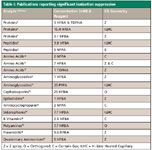
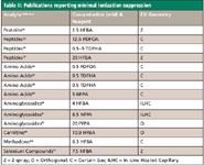
Ionization suppression is a complex phenomenon associated with atmospheric pressure-based ionization techniques such as electrospray ionization (ESI) that affects mass spectrometry (MS) detection (1–4). The differences in ionization response in ESI have been commonly attributed to deprotonation, increase in surface tension, increase in droplet size, presence of nonvolatile components, and in complex samples, large amounts of competing species that take some of the available ions. Here we summarize, in chronological order, some observations on ionization suppression with these reagents. Approximately one third of the reviewed publications mention ionization suppression, and of these, about 55% observed significant ionization suppression, while approximately 45% reported no significant ionization suppression. For ease of comparison, Tables I and II list some key parameters of the experimental and instrument setup of these investigators, but note that differences in sample matrix also could affect ionization suppression.

Roughly 70% of the publications that reported significant ionization suppression used HFBA, three used NFPA, and one each used TDFHA and PFPA. Among the publications that reported no significant ionization suppression, only 43% used HFBA; 36% used PDFOA or TDFHA; two used NFPA; and one used PFPA. Amino acids and aminoglycosides accounted for a total of 28% of the significant ionization suppression group, versus 50% of the nonsignificant ionization suppression group. Proteins and peptides accounted for 28% of both groups. Diverse analytes accounted for 44% and 21% of the significant ionization suppression and nonsignificant ionization suppression groups, respectively.

Table I: Publications reporting significant ionization suppression
Keever and colleagues (15) noted that HFBA produced some signal suppression, but postcolumn addition of propionic acid enhanced response 10-fold for a cephalosporin antibiotic. Venkateshwaran and colleagues (25) noted that peptides and proteins are very susceptible to denaturation at extremes of pH, which leads to unfolding of the biomolecule and a change in the ESI charge state intensities towards lower m/z values due to an increase in the number of exposed amino acids, which increases the number of ionizable sites in the molecule. Chaimbault and colleagues (26), in a study of underivatized amino acids, using 0.25–5 mM PDFOA, reported that an increase in the surfactant concentration increased the intensity of the positive ion MS signal; that is, no suppression, but ionization enhancement. Watt and colleagues (16) observed that buffered 1 mM HFBA (pH 7.4) gave lower sensitivity, possibly due to reduced protonation of epibatidine. Petritis and colleagues (10) observed intense background signals at m/z 102 with 2 mM NFPA.

Table II: Publications reporting minimal ionization suppression
Ishihama and colleagues (23) reported that perfluorocarboxylic acids have low surface energy, which is associated with weak intermolecular interaction and high volatility. However, PDFOA, but not TFA, is a surface active agent. In this case, surface tension had little effect on signal suppression. For myoglobin, PDFOA yielded almost the same signal as formic acid and did not change the charge distribution significantly, which reflects the conformation of a protein. Heller and colleagues (30) reported ESI is more sensitive for aminoglycosides than thermospray, and found ionization suppression to be far less severe with HFBA than with TFA. Gustavsson and colleagues (5) studied signal suppression using microflow injection analysis with 3 mM HFBA and 3 mM TDFHA on eight amines. The ESI–MS response decreased to only 30% for HFBA and to 15% for TDFHA, compared with formic acid, with no degradation of the ESI interface performance after 24 h of continuous infusion. With constant pH (3.4–3.7) and ionic strength, there was no correlation between the analytes' pKa values and their ESI responses. Kwon and colleagues (31) compared sensitivity of detection of amino acids with 6.5 mM NFPA and HFBA, and NFPA provided less background chemical noise. Shoji and colleagues (36) obtained satisfactory results with a C30 column and 20 mM HFBA versus C18 and 60 mM, which had significantly suppressed the ion intensity in tetrodotoxin analysis.
Garcia and colleagues (6) obtained very small responses with 15.4 mM HFBA in flow injection analysis experiments with intact proteins, compared with formic acid. Petritis and colleagues (24) compared HFBA, NFPA, TDFHA, and PDFOA and found that 10 mM perfluorocarboxylic acids with ESI–MS created an ionization suppression of 1.5–2 fold in underivatized amino acids, and was almost the same in small peptides. However, in the 0.5–2 mM range, it was not significant (less than 1.4-fold). Lindemann and colleagues (35) found 7 mM HFBA produced no significant signal suppression in the ESI mode. McCormack and colleagues (17) reported 7.7 mM HFBA suppressed the response of all the siderophores studied, relative to formic acid.
Piraud and colleagues (27) reported that under neutral conditions, amino acids are delivered to the source as zwitterions, which probably limits their ionization, but in 0.5 mM TDFHA, they showed an enhanced signal that was molecule-dependent. Keevil and colleagues (13) used a step gradient with a decreasing concentration of HFBA (starting with 1 mM), where tobramycin and sisomycin were eluted into the mass spectrometer at nearly zero HFBA concentration with some loss of sensitivity due to signal suppression. Teerlink and colleagues (29) analyzed carboxymethyl lysine with a gradient of 5 mM NFPA as solvent A and acetonitrile as solvent B and reported no significant ionization suppression.
Dillon and colleagues (37) analyzed recombinant antibodies and observed MS signal reduction with the combination of 0.2% TFA and 2.3 mM HFBA. Consequently, they reduced the acids to 0.05% TFA and 0.4 mM HFBA. In a study of peptides and proteins, Walcher and colleagues (8) noted the absolute MS signal intensities were more than three times higher with HFBA versus TFA, but a significant increase in the noise level was obvious with HFBA. Wang and colleagues (22) in a study of intact protein separations reported concentrations greater than 1.5 mM HFBA (versus TFA) dramatically reduced protein ion intensities.
Piraud and colleagues (28) analyzed underivatized amino acids with gradients containing 4 mM NFPA, 0.5 mM and 1.5 mM TDFHA, or 0.5 mM PDFOA, and did not observe significant variation of the MS signal. Midttun and colleagues (38) used a ternary gradient with an effective constant concentration of 2.5 mM HFBA and noted that ionization suppression was most pronounced for pyridoxine 5'-phosphate and pyridoxamine 5'-phosphate.
Gao and colleagues (34), in the determination of methadone in human plasma, added 6.3 mM HFBA, NFBA, TDFHA, and PDFOA to the samples, but adding 93 mM HFBA to the sample reconstitution solution gave a 10.7-fold higher response. Naldi and colleagues (7) found a C4 column and 3.1 mM HFBA in the analysis of histone proteins gave better selectivity than 0.4% formic acid, but the ionization suppression was higher with HFBA. Lecaroz and colleagues (32) used 20 mM PFPA and samples prepared in mobile phase containing PFPA. By using a step gradient with a decreasing concentration of PFPA, gentamicin was eluted into the mass spectrometer at nearly zero PFPA concentration, solving the problem of ionization suppression. Rinne and colleagues (39) tested acetic acid, TFA, PFPA, and HFBA for the analysis of catecholamines on a porous graphitic carbon column and observed severe signal suppression with TFA, PFPA, and HFBA, compared to acetic acid. Jin and colleagues (21), in the analysis of diallyldimethylammonium chloride, chose 5 mM HFBA because 20 mM suppressed the signal.
Hakkinen and colleagues (19) analyzed underivatized polyamines and found 0.1% HFBA gave less signal suppression than 0.1% PFPA. Ding and colleagues (20) compared PFPA and HFBA and selected 9.6 mM HFBA because it gave a better separation of synephrine, but they observed ionization suppression.
Cherlet and colleagues (14) found that 20 mM PFPA reduced the signals of dihydrostreptomycin and streptomycin to 50% and 56%, respectively, compared to direct infusion without PFPA. Gu and colleagues (12) analyzed amino acids using 1 mM TDFHA as solvent A and acetonitrile as solvent B. High variation in intra-and interday precision of peak area ratios was attributed to poor ionization of glycine, likely due to interferences from solvent clusters. Orioli and colleagues (9) analyzed histidine and peptide adducts in urine samples, comparing HFBA and formic acid to 5 mM NFPA, which significantly suppressed the extracted ion current, but less than HFBA (a 15–40% increase with NFPA versus HFBA). Shen and colleagues (40) tested five different perfluorocarboxylic acids, and as the chain length increased, the observed atmospheric pressure chemical ionization (APCI) — not ESI — ion current increased.
De Person and colleagues (11) compared the performance of two MS instruments that differed in their atmospheric pressure sources, using amino acids and several perfluorocarboxylic acids. Previous authors observed a higher signal decrease with instruments of linear geometry in comparison with mass spectrometers of orthogonal or even Z-spray geometry. Whatever the spectrometer, the large differences in the response factors for all amino acids infused under the same conditions indicated that ion desolvation was highly dependent on the nature of the amino acid residue. Differences between the interfaces suggested that ion transmission was strongly dependent upon the ion source geometry. The MS signal increased up to NFPA compared with shorter chain homologs but decreased dramatically with TDFHA and PDFOA, with the Z-spray interface. However, with the curtain gas interface, the highest relative intensity was obtained with TDFHA and PDFOA, yielding significantly increased amino acid MS response over their shorter chain homologs. Signal suppression was evaluated with the curtain gas interface using only NFPA, and the optimum concentration was between 10 and 20 mM. However, with the Z-spray interface, the greater the NFPA concentration, the higher the amino acid signal, and therefore it was compatible with very high surfactant concentrations. They concluded that the Z-spray is more sensitive than the curtain gas interface to the concentration of ion-pair reagent and consequently is a less robust instrument for this type of analysis. However, these authors have not accounted for the partitioning effects of the ESI probe, where some amino acids move to the fringe of the spray plume while some remain in the core. The momentum effect coupled to electrostatics causes dispersion in the mass-to-charges within the plume. The Z-spray requires a 90° turn while the curtain gas interface requires in excess of a 90° turn to get into the sample orifice, which can cause differences in sampling and significantly bias the intensity. This is akin to comparing lenses with different focal points. If a "common" ESI probe was pointed at the orifice, then one could isolate whether it is the Z-spray or curtain gas geometry that plays a role. Without this experiment, the results are inconclusive regarding the ESI geometry.
Column, pH, and Temperature Selection
Approximately 60% of all the reviewed publications used C18 columns of varying dimensions (from semipreparative to nanospray) packed with particle sizes of 3–5 μm, and only one used a column packed with sub-2-μm particles (40). About 20% used C8 columns, and with only two exceptions (32,41), all C8 columns were used with HFBA. Six (8,6,17,42–44) used polystyrene–divinylbenzene (PSDVB) columns, five (17,39,45–47) used columns packed with porous graphitic carbon, two (36,48) used C30, and one each used C12 (9), C4 (7), cyano (49), and chiral (16) packing in columns. Because of the very acidic pH of the unbuffered perfluorocarboxylic acids, earlier publications reported some silica stationary phase degradation over time, but later publications used newer silica stationary phases that are resistant to acid and heat. PSDVB and porous graphitic carbon are more resistant to acid hydrolysis than silica. However, Rinne and colleagues (39) reported that 0.1% PFPA in the mobile phase was involved in on-column oxidation of catecholamines, by slow oxidation of the porous graphitic carbon material (to ionic graphite salts).
One third of all the reviewed publications used isocratic elution and two thirds used gradient elution. In some instances of gradient elution, the concentration of the ion-pair reagent was kept constant, but in most cases, it decreased. Although nearly 20 reviewed publications report system peaks, column equilibration issues, and peak drift, about 80% do not report these phenomena, in spite of the disruption of the dynamic equilibrium with gradient elution. This might be due to the increasing popularity of "fast" liquid chromatography (LC)–MS-MS, where "interferences" can be "tuned out" and system peaks are not as obvious, although peak drift and equilibration issues could persist.
Only 25% of the reviewed publications reported a pH value for the mobile phase or for any portion of the mobile phase used in the analyses. The majority of the reported pH values for mobile phases are "s/w pH" (older definition was "apparent pH") values and half are buffered mobile phases (pH 3.0–4.0, with a few exceptions); 30% are unbuffered mobile phases (pH 1.5–3.0); and 20% are mixtures of perfluorocarboxylic acids and TFA, for example (pH 1.7–2.8). For a discussion of pH measurements in aqueous buffer–organic mobile phases, there are several recent reviews (50–55).
We have measured the pH of aqueous PDFOA at various concentrations in our laboratory as: 0.01%, pH 4.4; 0.02%, pH 3.1; 0.06%, pH 2.9; and 0.08%, pH 2.8. For comparison, Kotrebai and colleagues (56) report aqueous 0.1% TFA, pH 2.05; 0.1% PFPA, pH 2.21; and 0.1% HFBA, pH 2.55. Garcia and colleagues (6) report a pH of 2.5 for 0.2% formic acid and a pH of 2.5 for 0.025% TFA. Shen and colleagues (40) state that "TFA is the strongest acid with a pKa of 0.3, while PDFOA is the weakest of the series, with a pKa of 2.5." These investigators prepared two mobile phases with the same concentration of NFPA, but adjusted the s/w pH value to 6.8 and 2 and tested them in the LC separation of diastereomers. Very little shift (5%) in retention time was observed. However, Snoble and colleagues (57) did detect slight retention shifts for a nine-peptide test mixture with two-day-old, room-temperature mobile phases containing dilute amounts of formic acid and methanol.
Goss (58) reported that the literature has used a pKa of 2.8 for PDFOA, based upon a measurement of 50:50 (v/v) ethanol–water. For existing compound data, a pKa value measured in a 50:50 alcohol–water mixture is typically about 1 log unit higher than what is measured in water. An artifact that leads to overestimation of the pKa value of PDFOA, sorption to interfaces such as the water surface or the walls of a glass vessel can occur to an extent that is unknown for ordinary molecules. The titrations will be shifted significantly toward higher pH values. The calculated pKa of PDFOA is –0.5, while the calculated pKa of HFBA is approximately 0.4.
Only half of all the reviewed publications report a controlled column temperature, which is surprising because it is especially important with ion pairing in reversed-phase chromatography, as Dolan (59) notes. A total of 16 publications report column temperatures above 30 °C; 13 between 35–55 °C; and one each at 60 °C (42), 70 °C (37), and 100 °C (40). Among the potential advantages of elevated column temperatures are faster separations with reduced backpressure, sharper peaks, and potentially reduced ionization suppression in ESI–MS detection. Selectivity differences also have been reported with different column temperatures. For example, Castro and colleagues (60) reported that a change in a C8 column temperature from ambient to 50 °C using 15 mM HFBA gradient elution "completely changed the elution order of several quaternary ammonium herbicides."
Several recent publications (61–63) review elevated temperature chromatography and its potential advantages. In our review, we have not found any obvious patterns in the relatively small number of articles using the perfluorocarboxylic acids at elevated column temperatures, especially above 55 °C. However, because most analytes are eluted faster with higher column temperature, it could be useful to reexamine equilibration times (that is, mobile phase volumes), selectivity, and mobile phase ion-pair concentrations with TDFHA and PDFOA on the newer acid-and heat-resistant C18 stationary phases.
System Peaks and Column Equilibration
Five reviewed publications discuss system peaks in detail, while six report equilibration issues, and eight mention peak drift with perfluorocarboxylic acids. System peaks are not unique to ion-pairing chromatography and can have many causes. Several recent reviews (64–66) discuss this phenomenon in detail, so we will limit our discussion of "ghost peaks, pseudo peaks, eigenpeaks, vacancy peaks, induced peaks, artifact peaks, dip peaks, perturbation peaks, or spurious peaks," to reports of this phenomenon specific to these reagents. Interestingly, system peaks are not reported after 2002 in the reviewed literature, although equilibration issues and peak drift are reported as recently as 2007 and 2008, respectively.
Chaimbault and colleagues (26) used gradient elution for basic amino acids, based upon simultaneously increasing the concentration of acetonitrile and decreasing the concentration of the ion-pair reagent. At the end of each chromatogram, a system peak was observed, in addition to the solute peaks. "System peaks are observed when an injection is made in a chromatographic system previously equilibrated with a mobile phase consisting of a solution with a constant concentration of an additive that is involved in the retention mechanism. These peaks appear after perturbation of the equilibrium, when some of the adsorbed additive molecules are desorbed and migrate along the column as an excess additive band." System peaks can mask the elution of one or more analytes with evaporative light scattering detection, but MS can detect the analytes under a system peak. PDFOA induced two system peaks: one near, but before the end of the chromatogram, and the second at the end of the chromatogram, probably due to impurities in PDFOA that accumulated during column equilibration and were desorbed at the same time as the surfactant.
Petritis and colleagues (67) ensured that no irreversible surface modification occurred between each experiment by using extensive column washes (3 h to flush the column). Previous experiments using various perfluorocarboxylic acids with UV detection had problems with baseline drifts and UV spikes due to UV-absorbing contaminants. With evaporative light scattering detection (ELSD) and ESI–MS, however, if the contaminants are volatile, they will not be a problem. The adsorption of different perfluorocarboxylic acids on the stationary phase was determined by measuring breakthrough with a conductivity meter. Adsorbed quantities of surfactant increased with surfactant concentration in the mobile phase, as well as with the surfactant alkyl chain length on all three C18 columns tested. Whatever C18 column was used and the column dimensions, equilibration times were almost the same for a given surfactant at a given concentration, and the total adsorbed quantities of surfactant were similar on the three different columns tested. Moreover, for a given perfluorocarboxylic acid, the equilibration time was independent of the surfactant concentration in the mobile phase. These surfactants had the following approximate equilibration times: PDFOA, 3 h; TDFHA, 1.25 h; NFPA, 15 min; and HFBA, 5 min. Note that all of these experiments were conducted with unthermostated columns at room temperature. Furthermore, it is the volume of the mobile phase at a given concentration of ion-pair reagent (not time) that passes through the column that is important (50). However, because a range of flow rates are used in the literature, equilibration time is referenced here for convenient comparison of different ion-pair reagents.
Chaimbault and colleagues (45) conducted further experiments with this "breakthrough system" using a porous graphitic carbon column at 10 °C. HFBA and NFPA had short equilibration times whatever the surfactant concentration range (0.5–15 mM). However, TDFHA required 35 min and PDFOA required 105 min to be equilibrated. On this column with NFPA, there were no system peaks during gradient elution, perhaps because of the very low quantities of NFPA adsorbed on the porous graphitic carbon surface, in contrast with that of PDFOA.
Petritis and colleagues (24,68,69) continued work using the "breakthrough system" with underivatized amino acids on a C18 column at 25 °C and indirect conductivity detection. System peaks can appear in a chromatographic system containing more than one eluent component after injection of a solution with a composition different from the bulk eluent. For example, an injection of 10 μL of water into the equilibrated system caused a small positive peak in the void volume and an intense negative peak later (main system peak). For TDFHA, a small system peak appeared after about one hour and for PDFOA after about 3 h. There was a linear relationship between the mobile phase concentration of reagent and the peak area of the system peak. Moreover, the system peak always was eluted at the same time as the breakthrough of the adsorption isotherms. Even with ELSD, system peaks can be observed after a violent gradient, or after violent desorption from a chromatographic support of high concentrations of volatile compounds. The detector signal given by these peaks is attributed to sudden differences in mobile phase volatility. However, small differences in mobile phase composition occurring after perturbation of an equilibrated chromatographic system are not detectable with ELSD. Later analysis of underivatized small peptides using ESI–MS and TDFHA with an acetonitrile gradient elution produced a system peak that increased in size with increasing TDFHA concentration at the end of the chromatogram and masked the elution of some peptides. Reequilibration time was about 80 min with TDFHA, but only 15 min with NFPA. In the analysis of underivatized amino acids, the elution order was always the same whatever the concentration of PDFOA. However, with some reagents such as TDFHA, inversions in the elution order of some amino acids occurred with increasing TDFHA concentration.
Zhu and colleagues (70) tested HFBA, NFPA, and PDFOA in the analysis of acetylcholine, and all gave good resolution. However, because HFBA had a shorter equilibration time, it was chosen. Kwon and colleagues (31) used 6.5 mM NFPA in the analysis of underivatized amino acids with gradient elution and a 20-min equilibration time. The retention time reproducibility for the 20 amino acids tested was better than 5%. In the analysis of 76 amino acids, Piraud and colleagues (28) used a 15-min equilibration time with 0.5 mM TDFHA before the first gradient, as well as between two gradients, which gave good repeatability of the retention times, even after 20 consecutive gradients. However, the first gradient had to be discarded. Gao and colleagues (34) added HFBA, NFPA, TDFHA, and PDFOA to the sample and found the uptake of the reagents on the C18 column mainly determined the retention of analytes. With HFBA or NFPA in the reconstitution solution, gradient LC did not require extensive equilibration times and carryover was reduced. However, it was difficult for systems using TDFHA and PDFOA to equilibrate. This method also might potentially address the carryover problem for other polar compounds (for example, aminoglycosides) that form ion pairs. Spacil and colleagues (71) used 3.2 mM NFPA and found in cases of gradient elution, a 15-min interval was required between runs for column equilibration.
Peak drift can occur in LC using these reagents, even with standard solutions after several injections over time, or be exacerbated by sample matrices. With one exception (72), all the publications mentioned in the following reported controlled column temperatures ranging from 20 °C to 40 °C. Cherlet and colleagues (72) observed important matrix effects on retention time stability and peak shape for the analysis of gentamicin in tissue samples using isocratic elution with 20 mM PFPA. A shift of retention times and peak tailing were observed when comparing the sample and standard chromatograms. Gammelgaard and colleagues (41), using isocratic elution with 3 mM NFPA, noted the retention times and sensitivity changed slowly during the lifetime of the columns. Wybraniec and colleagues (73) observed with 0–5 mM PFPA and HFBA, there was some shifting of retention time for betacyanins, using gradient elution. Qu and colleagues (74) used 1 mM PDFOA and gradient elution for the analysis of glutamine and other amino acids and attributed peak drift to the accumulation of PDFOA in the column, which slowly modified the surface of the C18 phase. To avoid this, they flushed the column after every six chromatographic runs. In a later article (75), they used even more extensive flushing when 0.8 mM TDFHA was used with gradient elution. Piraud and colleagues (76), using 0.5 mM TDFHA with gradient elution, observed a slight drift of retention time after analyzing many biological samples for amino acids. However, this was limited by using acetonitrile column washing every 14–16 samples. Heller and colleagues (77) used column temperature regulation (30 °C) and isocratic elution with 55 mM TFA to eliminate occasional retention time drift, which contributed to peak-shape variations. Heavily used columns produced ghost peaks in the analysis of control samples of gentamicin. Liu and colleagues (78), using 5 mM NFPA with gradient elution, reported that long-term injection of amino acids lead to retention time drifts. A thorough column cleaning with methanol was necessary when the retention time of amino acids was seen to decrease after weeks of sample injections. The retention time shift in this study is opposite to what was reported by Qu and Piraud. Eckstein and colleagues (79) reported that the chromatographic durability with gradient elution using HFBA versus PDFOA was improved, as thousands of cerebrospinal fluid samples were analyzed for glutamine and related compounds, with no change in retention or durability. Andrieux and colleagues (33), using isocratic elution with 10.9 mM HFBA, reported that column washing after every 50 samples preserved column life for more than 1000 injections with no shift in retention time or degradation of peak shape.
Conclusions
The volatile perfluorocarboxylic acids offer a range of choices for selectivity and are valuable and versatile ion-pair reagents. They also provide good MS-compatible (and a host of other detection methods) LC gradient and isocratic separations for an increasingly diverse selection of polar analytes (without requiring derivatization), as well as proteins and peptides. In recent years, nearly half of all the reviewed publications covered analytes other than aminoglycosides, amino acids, proteins, and peptides.
While some significant ionization suppression has been reported in about 20% of the reviewed publications, mostly with HFBA, this disadvantage has been manageable in most cases. The longer alkyl chain homologs, used at lower concentrations, offer the potential for reduced ionization suppression. Furthermore, knowing whether the MS instrument ESI geometry truly has an effect on the ionization suppression of specific analyte–ion pair reagent combinations might help to minimize this suppression.
Good column temperature control (including mobile phase prewarming for elevated temperatures) and proper mobile-phase pH adjustment and measurement are essential for reproducibility in all LC methods, and ion-pairing LC is no exception. Beginning with the nature of the sample, one should ask: Where did it come from? Is it stable when dissolved in acidic mobile phase? Will the ion-pair reagent in the sample reduce carryover (for example, gentamicin), eliminate double peaks, show "matrix effects," or affect the column equilibrium? Also consider LC column stationary phase compatibility with very acidic mobile phases, potentially at elevated temperatures; column equilibration; gradients used; flushing; storage; and dedication of the column to a specific application.
Faster LC separations and acid-and heat-resistant C18 stationary phases offer new opportunities to explore the performance of PDFOA and TDFHA at column temperatures up to 90 °C. With a fast separation, the thermal stability of various polar analytes might not limit such an approach (64), and column equilibration times with these reagents might be reduced dramatically. Indeed, it can be advantageous to delay the too-rapid elution of some very polar analytes under such conditions with these reagents. Furthermore, PDFOA has shown greater selectivity than HFBA and NFPA for amino acids, for example, and is an effective reagent at lower concentrations than NFPA or HFBA (for example, 0.5 mM vs. 5 mM, or 4–8 mM, respectively). Also, PDFOA often is associated with less ionization suppression than HFBA and NFPA (11), potentially increasing sensitivity in ESI–MS detection. Daily preparation of fresh mobile phase containing methanol or the use of acetonitrile as an organic modifier could prevent the formation of esters in the mobile phase (40,57).
According to Desmet (80), the advantage of sub-2-μm particles diminishes if columns are run at elevated temperatures. Nevertheless, Shen and colleagues (40) achieved fast baseline resolution of diastereomers with a column temperature of 100 °C using sub-2-μ particles and NFPA. The purity of PDFOA and all ion-pair reagents, including the potential effects of branched isomers, should be considered carefully in future investigations. Although LC methods using hydrophilic interaction chromatography and other techniques are becoming increasingly popular, we believe the perfluorocarboxylic acids have much to offer and their use, especially in LC–ESI-MS, will continue to increase.
References
(1) R. King, R. Bonfiglio, C. Fernandez-Metzler, C. Miller-Stein, and T. Olah, J. Am. Soc. Mass Spectrom. 11, 942–950 (2000).
(2) K. Tang, J.S. Page, and R.D. Smith, J. Am. Soc. Mass Spectrom. 15, 1416–1423 (2004).
(3) F. Beaudry and P. Vachon, Biomed. Chromatogr. 20, 200–205 (2006).
(4) M.P. Balogh, LCGC 24(12), 1284–1289 (2006).
(5) S.A. Gustavsson, J. Samskog, K.E. Markides, and B. Langstrom, J. Chromatogr., A 937, 41–47 (2001).
(6) M.C. Garcia, A.C. Hogenboom, H. Zappey, and H. Irth, J. Chromatogr., A 957(2), 187–199 (2002).
(7) M. Naldi, V. Andrisano, J. Fiori, N. Calonghi, E. Pagnotta, C. Parolin, G. Pieraccini, and L. Masotti, J. Chromatogr., A 1129, 73–81 (2006).
(8) W. Walcher, H. Toll, A. Ingendoh, and C.G. Huber, J. Chromatogr., A 1053(1–2), 107-117 (2004).
(9) M. Orioli, G. Aldini, M.C. Benfatto, R.M. Facino, and M. Carini, Anal. Chem. 79, 9174–9184 (2007).
(10) K. Petritis, V. Dourtoglou, C. Elfakir, and M. Dreux, J. Chromatogr., A 896(1–2), 335–341 (2000).
(11) M. de Person, P. Chaimbault, and C. Elfakir, J. Mass Spectrom. 43, 204–215 (2008).
(12) L. Gu, A.D. Jones, and R.L. Last, Anal. Chem. 79, 8067–8075 (2007).
(13) B.G. Keevil, S.J. Lockhart, and D.P. Cooper, J. Chromatogr. B 794, 329–335 (2003).
(14) M. Cherlet, S. De Baere, and P. De Backer, J. Mass Spectrom. 42, 647–656 (2007).
(15) J. Keever, R.D. Voyksner, and K.L. Tyczkowska, J. Chromatogr., A 794(1–2), 57–62 (1998).
(16) A.P. Watt, L. Hitzel, D. Morrison, and K,L, Locker, J. Chromatogr., A 896(1–2), 229–238 (2000).
(17) P. McCormack, P.J. Worsfold, and M. Gledhill, Anal. Chem. 75, 2647–2652 (2003).
(18) O. Midttun, S. Hustad, E. Solheim, J. Schneede, and P.M. Ueland, Clin. Chem. 51(7), 1206–1216 (2005).
(19) M.R. Hakkinen, T.A. Keinanen, J. Vepsalainen, A.R. Khomutov, L. Alhonen, J. Janne, and S. Auriola, J. Pharm. Biomed. Anal. 45, 625–634 (2007).
(20) L. Ding, X. Luo, F. Tang, J. Yuan, Q. Liu, and S. Yao, J. Chromatogr. B 857, 202–209 (2007).
(21) F. Jin, J. Hu, M. Yang, X. Jin, W. He, and H. Han, J. Chromatogr. A, 1101, 222–225 (2006).
(22) Y. Wang, B.M. Balgley, P.A. Rudnick, and C.S. Lee, J. Chromatogr., A 1073(1–2), 35–41 (1–2).
(23) Y. Ishihama, H. Katayama, and N. Asakawa, Anal. Biocem. 287, 45–54 (2000).
(24) K. Petritis, S. Brussaux, S. Guenu, C. Elfakir, and M. Dreux, J. Chromatogr. A 957(2), 173–185 (2002).
(25) T.G. Venkateshwaran, J.T. Stewart, R.T. Bishop, J.A. de Haseth, M.G. Bartlett, J. Pharm. Biomed. Anal. 17, 57–67 (1998).
(26) P. Chaimbault, K. Petritis, C. Elfakir, and M. Dreux, J. Chromatogr., A 855, 191–202 (1999).
(27) M. Piraud, C. Vianey-Saban, K. Petritis, C. Elfakir, J. Steghens, A. Morla, and D. Bouchu, Rapid Commun. Mass Spectrom. 17, 1297–1311 (2003).
(28) M. Piraud, C. Vianey-Saban, K. Petritis, C. Elfakir, J. Steghens, and D. Bouchu, Rapid Commun. Mass Spectrom. 19, 1587–1602 (2005).
(29) T. Teerlink, R. Barto, H.J. ten Brink, and C.G. Schalkwijk, Clinical Chemistry 50, 1222–1228 (2004).
(30) D.N. Heller, S.B. Clark, and H.F. Righter, J. Mass Spectrom. 35, 39–49 (2000).
(31) J. Kwon, and M. Moini, J. Am. Soc. Mass Spectrom. 12, 117–122 (2001).
(32) C. Lecaroz, M.A. Campanero, C. Gamazo, and M.J. Blanco-Prieto, J. Antimicrob. Chemo. 58, 557–563 (2006).
(33) P. Andrieux, T. Kilinc, C. Perrin, and E. Campos-Gimenez, J. AOAC Int. 91(4), 777–785 (2008).
(34) S. Gao, S. Bhoopathy, Z. Zhang, D.S. Wright, R. Jenkins, and H.T. Karnes, J. Pharm. Biomed. Anal. 40(3), 679–688 (2006).
(35) T. Lindemann and H. Hintelmann, Anal. Bioanal. Chem. 372, 486-490 (2002).
(36) Y. Shoji, M. Yotsu-Yamashita, T. Miyazawa, and T. Yasumoto, Anal. Biochem. 290, 10–17 (2001).
(37) T.M. Dillon, P.V. Bondarenko, and M.S. Ricci, J. Chromatogr., A 1053(1–2), 299–305 (2004).
(38) O. Midttun, S. Hustad, E. Solheim, J. Schneede, and P.M. Ueland, Clinical Chemistry 51, 1206–1216 (2005).
(39) S. Rinne, A. Holm, E. Lundanes, and T. Greibrokk, J. Chromatogr., A 1119, 285–293 (2006).
(40) J.X. Shen, E.A. Merka, D.P. Dreyer, R.P. Clement, and R.N. Hayes, J. Sep. Sci. 31, 242–254 (2008).
(41) B. Gammelgaard, L. Bendahl, U. Sidenius, and O. Jons, J. Anal. At. Spectrom. 17, 570–575 (2002).
(42) S. Delgado, M.C. Garcia, M.L. Marina, and M. Torre, J. Sep. Sci. 26, 1363–1375 (2003).
(43) M.C. Carson. And D.N. Heller, J. Chromatogr. B 718, 95–102 (1998).
(44) A. Tholey, H. Toll, and C.G. Hubel, Anal. Chem. 77, 4618–4625 (2005).
(45) P. Chaimbault, K. Petritis, C. Elfakir, and M. Dreux, J. Chromatogr. A 870, 245–254 (2000).
(46) Y. Aoki, H. Hakamata, Y. Igarashi, K. Uchida, H. Kobayashi, N. Hirayama, A. Kotani, and F. Kusu, J. Chromatogr., B 858, 135–142 (2007).
(47) K. Petritis, P. Chaimbault, C. Elfakir, and M. Dreux, J. Chromatogr. A 896(1–2), 253–263 (2000).
(48) T. Hashimoto, S. Nishio, N. Nishibori, S. Yoshioka, and T. Noguchi, J. Food Hyg. Soc. Japan 43(3), 144–147.
(49) J. Lu, Y. Chan, S.D. Poppitt, and G.J.S. Cooper, J. Chromatogr., B 859(1), 62–68 (2007).
(50) C. Duda, Current Separations 13(1), 36–37 (1994).
(51) M. Roses, J. Chromatogr., A 1037, 283–298 (2004).
(52) D. Sykora, E. Tesarova, and D.W. Armstrong, LCGC 20(10), 974–981 (2002).
(53) G.W. Tindall, LCGC 20(11), 1028–1032 (2002).
(54) G.W. Tindall, LCGC 20(12), 1114–1118 (2002).
(55) G.W. Tindall, LCGC 21(1), 28–32 (2003).
(56) M. Kotrebai, J.F. Tyson, E. Block, and P.C. Uden, J. Chromatogr., A 866, 51–63 (2000).
(57) K.A.J. Snoble, N.M. Fox, S.M. Lorenz, A.R. Kemperman, and J.T. Przybytek, LCGC 26(9), 946–950 (2008).
(58) K. Goss, Environ. Sci. Technol. 42, 456–458 (2008).
(59) J.W. Dolan, LCGC 26(2), 170–174 (2008).
(60) R. Castro, E. Moyano, and M.T. Galceran, J. Chromatogr., A 830(1), 145–154 (1999).
(61) P. Sandra, and G. Vanhoenacker, J. Sep. Sci. 30, 241–244 (2007).
(62) G. Vanhoenacker, and P. Sandra, J. Sep. Sci. 29, 1822–1835 (2006).
(63) Y. Yang, LCGC Supplement 26(S4), 36–42 (2008).
(64) S. Williams, J. Chromatogr., A 1052, 1–11 (2004).
(65) F. Gritti, and G. Guiochon, Anal. Chem. 76, 7310–7322 (2004).
(66) J. Srbek, P. Cufal, Z. Bosakova, and E. Tesarova, J. Sep Sci. 28, 1263–1270 (2005).
(67) K.N. Petritis, P. Chaimbault, C. Elfakir, and M. Dreux, J. Chromatogr. A 833, 147–155 (1999).
(68) K. Petritis, P. Chaimbault, C. Elfakir, and M. Dreux, J. Sep. Sci. 24, 397–405 (2001).
(69) K.N. Petritis, P. Chaimbault, C. Elfakir, and M. Dreux, J. Chromatogr., A 833, 147–155 (1999).
(70) Y. Zhu, P.S.H. Wong, M. Cregor, J.F. Gitzen, L.A. Coury, and P.T. Kissinger, Rapid Commun. Mass Spectrom. 14, 1695–1700 (2000).
(71) Z. Spacil, J. Folbrova, N. Megoulas, P. Solich, and M. Koupparis, Anal. Chim. Acta 583(2), 239–245 (2007).
(72) M. Cherlet, S. De Baere, and P. De Backer, J. Mass Spectrom. 35, 1342–1350 (2000).
(73) S. Wybraniec, and Y. Mizrahi, J. Chromatogr., A 1029(1–2), 97–101 (2004).
(74) J. Qu, W. Chen, Y. Wang, S. Xiao, Z. Ling, and G. Chen, Analyst 127, 66–69 (2002).
(75) J. Qu, Y. Wang, G. Luo, Z. Wu, and C. Yang, Anal. Chem. 74, 2034–2040 (2002).
(76) M. Piraud, C. Vianey-Saban, C. Bourdin, C. Acquaviva-Bourdain, S. Boyer, C. Elfakir, and D. Bouchu, Rapid Commun. Mass Spectrom. 19, 3287–3297 (2005).
(77) D.N. Heller, J.O. Peggins, C.B. Nochetto, M.L. Smith, O.A. Chiesa, and K. Moulton, J. Chromatogr. B 821, 22–30 (2005).
(78) D. Liu, L.W. Beegle, and I. Kanik, Astrobiology 8(2), 229–241 (2008).
(79) J.A. Eckstein, G.M. Ammerman, J.M. Reveles, and B.L. Ackerman, J. Neurosci. Methods 171, 190–196 (2008).
(80) G. Desmet, LCGC 26(6), 514–530 (2008).

New Study Reviews Chromatography Methods for Flavonoid Analysis
April 21st 2025Flavonoids are widely used metabolites that carry out various functions in different industries, such as food and cosmetics. Detecting, separating, and quantifying them in fruit species can be a complicated process.


