Investigating Cheetah Metabolite Profiles Using GC–MS
Adrian Tordiffe from the Veterinary Faculty of the University of Pretoria in South Africa, is working to establish baseline metabolic profiles for captive and free-ranging cheetahs to investigate the unusual medical conditions that the animals develop in captivity. He spoke to The Column about his work and about the role that chromatography plays.
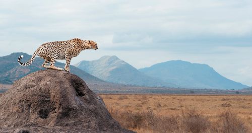
Photo Credit: Volodymyr Burdiak/Shutterstock.com

Adrian Tordiffe from the Veterinary Faculty of the University of Pretoria in South Africa, is working to establish baseline metabolic profiles for captive and free-ranging cheetahs to investigate the unusual medical conditions that the animals develop in captivity. He spoke to The Column about his work and about the role that chromatography plays. -Interview by Kate Mosford
Q. Your work focuses on the investigation of chronic medical conditions that cheetahs develop in captivity (1). What led you to begin this research?
A: Several years ago I worked at the National Zoological Gardens of South Africa as a clinical veterinarian. During that time, I treated several cheetahs with gastrointestinal or kidney problems and evaluated the scientific literature for answers on how to prevent or treat these conditions. I was amazed that, despite concerted efforts to investigate these diseases over the last 30 years, very little progress had been made in identifying causal factors. It was clear to me that we had a limited understanding of cheetah physiology and that traditional approaches and diagnostic methods would be inadequate in unravelling the mysteries surrounding these unusual diseases. In 2011, I was introduced to the field of metabolomics and at the same time became very interested in “systems biology” as a holistic way of understanding complex biological interactions and processes. Convinced that this approach might provide much needed answers for the cheetah, I made it the subject of my Ph.D. study.
Q. What were the main analytical challenges you encountered and how did you overcome them?
A: One of the first challenges was to find a way of adjusting the urine metabolite concentrations to correct for variations in hydration status and urine production in the cheetahs. In most species, including humans, urine metabolite concentrations are corrected in spot samples by adjusting them relative to urine creatinine concentrations. Creatinine is thought to be produced primarily by muscle tissue at a fairly constant rate and excreted unchanged through the kidneys. I soon realized that there was up to an eightfold difference in the urine creatinine concentrations of young adult cheetahs compared to older individuals. It became clear that creatinine was not being produced at a constant rate relative to muscle mass in cheetahs and therefore it could not be used for metabolite concentration adjustments. After a careful evaluation of the literature, I decided to use urine-specific gravity values to adjust the metabolite concentrations and this seems to provide more accurate results.
An ongoing challenge is the identification of several novel organic acids in cheetah urine. The two most abundant organic acids in the urine of captive cheetahs could initially not be matched to any compound on any metabolite database. After some time, we were able to identify the structure of one of these organic acids, but the identity of the other still remains unknown. Novel metabolite identification still seems to be a significant challenge, even for experienced researchers in this field.
Q. Can you comment on the sample preparation and gas chromatography–mass spectrometry (GC–MS) method you developed for this project?
A: We used virtually the same methods for the cheetah sample analyses as for the human samples analyzed in our laboratory (Potchefstroom Laboratory for Inborn Errors of Metabolism). The urine organic acid analysis is very similar to the method described by Piero Rinaldo in reference 2, with some minor variations. Basically, the organic acids were extracted with ethyl acetate and diethyl ether. The organic phase was removed and dried under nitrogen and then derivatized with BSTFA:TMCS:pyridine before injection into the GC–MS system. For the reasons described above, we used a fixed volume (1 mL) of urine for the analysis rather than a variable quantity based on the creatinine concentration.
For the serum fatty acids, the samples were subject to both acidic and alkaline hydrolysis to ensure quantification of the total fatty acids present in the serum as described by Wanders (3) for very long-chain fatty acids and phytanic acid analysis. The protocol was, however, modified for the additional quantification of long-chain fatty acids by adding an additional stable isotope standard (eicosanoic acid-d39) to the internal standard solution. The derivatization was achieved with MTBSTFA as described by the same author.
Q. Are there any external factors that must be taken into consideration when conducting this kind of investigation with wild animals?
A: In the initial stages of my study, I realized that several external factors influenced the results. For example, cheetahs are easily stressed during the capture and immobilization process and it soon became clear that the urine metabolite profiles were modified if the animals remained in a prolonged stressed state prior to sample collection. I therefore had to exclude the urine samples of any free-ranging cheetahs that had been caught in cage traps because I could not control for the period of time spent in the traps prior to immobilization.
Another factor I had to consider was the impact of a recent meal on the serum and urine metabolome. In the captive individuals we could ensure that they were starved for 24 to 48 h prior to sampling, but in wild cheetahs this was more difficult.
Q. Could you talk a little about your research on the serum fatty acid profiles of cheetahs (4).
A: The wild cheetahs in Namibia are largely free of the diseases suffered by their captive counterparts and therefore they provided me with a great opportunity to document the serum fatty acid profiles of healthy freeâliving individuals that consume a truly natural diet. These profiles were then compared with those of a group of captive cheetahs in Namibia. The serum fatty acid concentrations are known to largely reflect dietary fatty acid intake. I therefore expected the captive cheetahs to have higher polyunsaturated fatty acid (PUFA) concentrations, since they are fed meat from monogastric animals (horses and donkeys), which are relatively high in PUFAs. In contrast, wild cheetahs mostly eat small ruminants, which have a higher saturated fat content and lower levels of PUFAs. Although the results were consistent with this prediction, the fatty acid profile differences between the captive and wild cheetahs were more dramatic than I expected. These healthy wild cheetahs had very low serum PUFA concentrations with a surprisingly high mean omega-6:omega-3 fatty acid ratio (281:1). We suggest in our published article that cheetahs may have adapted to a diet that is high in saturated fat and low in PUFAs and that when they are presented with diets with a different fatty acid composition, certain problems may arise (4). PUFAs are prone to oxidation and cheetahs may not have the antioxidant capacity to deal with oxidized fatty acids in meat that is not entirely fresh.
Q. Why do captive cheetahs suffer with these diseases over other large captive cats?
A: This is a fascinating question. Cheetahs are the only living member of the Acinonyx genus and are considered to be somewhat intermediate between the wolf and other large felids. They are thought to be highly specialized and therefore less adaptable to changes in their diet and environment. In the wild, they mostly hunt small antelope and consume most of the carcass components rapidly before other stronger predators, such as lions, leopards, or hyenas, arrive to steal their prey. After they have gorged themselves, they will often fast for a few days before they hunt again. In captivity, however, they are often only fed the skeletal components (muscle meat with or without bones) of domestically farmed species. The internal organs, blood, and skin are usually removed and discarded as these components of the carcass spoil quite rapidly. In captivity, cheetahs are often fed daily and the meat they get is not always very fresh. Lions, leopards, and other large carnivores typically prey on a much wider range of species and consume their prey over a much longer period of time. It therefore seems likely that such species would have a greater metabolic flexibility than the cheetah.
Q. Can anything be done to limit the impact of these diseases or prevent these diseases in the future? What kind of work needs to be done?
A: The simple answer would be yes; ensure that cheetahs are kept in large camps, minimize their exposure to stressors, and feed them a natural diet of freshly killed small to medium size antelope (or a similar domestic ruminant). This is, however, not possible to achieve consistently at most captive cheetah facilities and I think it becomes really important to get a better understanding of the pathophysiology of these diseases in the cheetah so that we can identify alternative interventions to reduce the incidence of disease in these animals. My research on this species has certainly raised more questions than answers, but the platform of data that has been generated so far has provided several interesting leads to follow. We are currently busy with a long-term study on gastritis in captive cheetahs at the AfriCat Foundation in Namibia and I am hoping to publish the results from that study soon.
Q. With the status of cheetahs listed as vulnerable and the impact that living in captivity obviously has, is there a case to be made against their captivity?
A: If wild cheetah numbers continue to decline, the captive population may become increasingly important in the future. Rather than phasing out the keeping of cheetahs in captivity, I think it is more important to emphasize the specialized requirements of this species in captivity. Standards for the nutrition and husbandry of these animals need to be maintained at the highest level, and only institutions with adequate facilities and well trained staff should be allowed to house them.
References
- A.S.W. Tordiffe, M. van Reenen, F. Reyers, and L.J. Mienie, Journal of Chromatography B1049–1050, 8–15 (2017).
- P. Rinaldo, in Laboratory Guide to the Methods in Biochemical Genetics, N. Blau, M. Duran, and K.M. Gibson, Eds. (Springer-Verlag Berlin Heidelberg, 2008), pp. 137–169.
- R.J.A. Wanders and M. Duran, in Laboratory Guide to the Methods in Biochemical Genetics, N. Blau, M. Duran, and K.M. Gibson, Eds. (Springer-Verlag Berlin Heidelberg, 2008), pp. 221–231.
- A.S.W. Tordiffe, B. Wachter, S.K. Heinrich, F. Reyers, and L.J. Mienie, PLoS ONE11(12), e0167608 (2016).

Adrian Tordiffe graduated with a degree in veterinary science from the University of Pretoria in 1997 and then spent the next 8 years in small animal private practice in the United Kingdom. In 2005 he returned to South Africa and completed a master’s degree in African mammalogy. He then worked at the National Zoological Gardens of South Africa as a clinical and research veterinarian until April 2015. He currently teaches veterinary pharmacology at the Veterinary Faculty of the University of Pretoria and has recently submitted his PhD thesis in biochemistry in which he evaluated the serum and urine metabolomic profiles of captive and free-ranging cheetahs. His research interests focus on the physiology, metabolism, nutrition, stress, and anaesthesia of captive and wild carnivores.
E-mail:adrian.tordiffe@up.ac.za

Common Challenges in Nitrosamine Analysis: An LCGC International Peer Exchange
April 15th 2025A recent roundtable discussion featuring Aloka Srinivasan of Raaha, Mayank Bhanti of the United States Pharmacopeia (USP), and Amber Burch of Purisys discussed the challenges surrounding nitrosamine analysis in pharmaceuticals.
Silvia Radenkovic on Building Connections in the Scientific Community
April 11th 2025In the second part of our conversation with Silvia Radenkovic, she shares insights into her involvement in scientific organizations and offers advice for young scientists looking to engage more in scientific organizations.

.png&w=3840&q=75)

.png&w=3840&q=75)


.png&w=3840&q=75)


.png&w=3840&q=75)


