Improved Aggregate Quantitation of Ig-G Antibodies Using BioSep GFC Columns
When one is assessing the structure and function of a protein there are numerous analytical tests that are performed to fully characterize a protein of interest.
Michael McGinley and Vita Knudsen, Phenomenex, Inc.
When one is assessing the structure and function of a protein there are numerous analytical tests that are performed to fully characterize a protein of interest. Of particular interest to organizations developing protein therapeutics are analyses used to detect post-translational modifications (PTM), which are chemical and structural modifications that can occur with a protein. Aggregation is a particular class of PTMs that are of special concern in that protein aggregation is known to lead to reduced protein efficacy as well as an increased possibility of adverse immunogenic response. The standard method for analyzing and quantitating protein aggregation is gel filtration chromatography (GFC), which is an HPLC method run under native conditions where protein samples are separated by differences in their relative sizes in solution. Since the molecular weight of a protein is directly related to its size in solution, gel filtration is a good method for quantitating dimers, trimers, and larger order assemblies of a protein in solution; comparisons of the different peak areas to the monomer peak allow one to determine an "aggregation percent" for each particular species. High performance GFC columns are generally diol-based phases bound to a silica support; the diol stationary phase is used to create inertness in an aqueous solution resulting in analytes separating solely by their ability to penetrate the porous inner structure of the silica particle. Recently process improvements were introduced with Phenomenex's BioSep SEC media to improve the separation performance and reproducibility of the columns. Studies were performed on BioSep columns to investigate the inertness of the media and assess its performance for aggregation analysis of Ig-G antibodies, an application of particular interest to researchers developing Ig-G based protein therapeutics.
Material and Methods
All proteins, analytes, and mobile phase buffer additives were purchased from Sigma Chemical (St. Louis, MO). All solvents used were obtained from EMD Scientific (San Diego, CA). HPLC columns (BioSep-SEC-S 2000 of 300 × 7.8 mm or BioSep-SEC-S 3000 of 300 × 4.6 mm) were obtained from Phenomenex (Torrance, CA). An Agilent HP-1100 (Palo Alto, CA) was used for all analyses. The HPLC system consisted of a quaternary pump, solvent degasser, autosampler, column oven, and MWD detector. Data was collected using Chemstation Software (Agilent, Palo Alto, CA). All separations were performed using isocratic conditions with a mobile phase of 100 mM sodium phosphate pH 6.8 at a flow rate of 1.0 mL/min (0.35 mL/min for 4.6 mm ID). For inertness studies 0.2 M arginine was added to the mobile phase and different volumes of sample were injected. Protein elution was monitored at either 214 nm or 280 nm based on whether or not arginine was used in the mobile phase (arginine absorbs strongly so 280 nm must be used).

Figure 1
Results and Conclusion
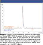
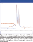
The goal of any aggregate analysis is to verify that adequate separation is being achieved between the dimer and monomer peaks of a protein therapeutic; an additional requirement is that adequate recovery of dimer and aggregate is realized. Recent publications by Arakawa et al have shown that some GFC columns underrepresented aggregate percentage, possibly due to nonspecific binding of aggregate to GFC columns with poor inertness (1). This poor recovery of aggregate can be mediated by adding arginine to the mobile phase resulting in improved recovery of dimer and other aggregate peaks. Optimized BioSep columns were evaluated using Ig-G antibody samples to assess their inertness; chromatograms were overlaid between standard mobile phase and mobile phase with added arginine. Any significant increases in aggregate recovery in arginine-containing mobile phase indicate significant secondary interactions between aggregate and the stationary phase of the column. In Figure 1, chromatograms of a murine Ig-G1 antibody run on a BioSep-SEC-S 2000 column are overlaid showing essentially identical recovery of dimer from both runs, with only a very slight increase in large molecular weight aggregates in the sample run using arginine-containing mobile phase. These results show that the BioSep 2000 column is a very inert phase giving accurate quantitation of dimer content for antibody aggregate applications. A similar result is shown for the BioSep-SEC-S 3000 in Figure 2. In this example, a mixture of Ig-G and albumin is injected on the BioSep 3000 column; mobile phase containing and omitting arginine are overlaid. Again, similar to results seen with the BioSep 2000 column, the dimer peak is nearly identical for the two analyses; the large molecular weight aggregate peak is only slightly increased when arginine-containing buffer is used for separations on the BioSep 3000 column indicating a very inert phase appropriate for Ig-G antibody applications. As this application is performed using BioSep columns of different molecular-weight ranges, one has resolving options based on the molecular weight range one wishes to focus on; unlike other offerings the BioSep 2000 can sometimes be a better solution for Ig-G applications because of its ability to focus heterogeneous high molecular weight aggregates. On the other hand the BioSep 3000 column offers the possibility of further resolving different degrees of Ig-G aggregation allowing one to better understand the biochemistry of their protein aggregation. However, in both cases good recovery (suggesting good inertness) and resolution make optimized BioSep a good solution for aggregation analysis of Ig-G protein therapeutics.

Figure 2
References
(1) T. Arakawa, J. S. Philo, D. Ejima, K. Tsumoto, and F. Arisaka, BioProcess International November 2006, 32–42.

Phenomenex, Inc.
411 Madrid Ave., Torrance, CA 90501
tel. (310)212-0555, (310)328-7768
Website: www.phenomenex.com

SEC-MALS of Antibody Therapeutics—A Robust Method for In-Depth Sample Characterization
June 1st 2022Monoclonal antibodies (mAbs) are effective therapeutics for cancers, auto-immune diseases, viral infections, and other diseases. Recent developments in antibody therapeutics aim to add more specific binding regions (bi- and multi-specificity) to increase their effectiveness and/or to downsize the molecule to the specific binding regions (for example, scFv or Fab fragment) to achieve better penetration of the tissue. As the molecule gets more complex, the possible high and low molecular weight (H/LMW) impurities become more complex, too. In order to accurately analyze the various species, more advanced detection than ultraviolet (UV) is required to characterize a mAb sample.


