Extraction and Detection of Antibiotics in the Rhizosphere Metabolome
Special Issues
Root diseases caused by soilborne plant pathogens are responsible for billions of dollars of losses annually in food, fiber, ornamental, and biofuel crops. The use of pesticides often is not an option to control plant diseases because of economic factors or potential adverse effects on the environment or human health. For this reason, many Americans are now buying pesticide-free organic foods. Organic agriculture has few options for controlling pests and thus must make full use of natural microbial biological control agents in soils that suppress diseases.
An important mechanism of natural disease suppression is the production of antibiotics by soil microbes. Antibiotics are small organic molecules of microbial origin that at low concentrations are deleterious to the growth or metabolism of other microorganisms. Soilborne pathogens are sensitive to many antibiotics produced by soil microbes on the roots of plants. Phenazines, 2,4-diacetylphloroglucinol (DAPG), pyrrolnitrin, and pyoluteorin are some of the most common of these antibiotics, but they are just a small fraction of the thousands that comprise the rhizosphere metabolome. Rhizosphere metabolomics is the collective chemical fingerprint of the rhizosphere, consisting of root exudates and the compounds produced by rhizosphere organisms. The rhizosphere metabolome is a reservoir of novel antibiotics owing to the millions of unique microorganisms found in soil and enriched in the rhizosphere. Knowledge of the rhizosphere metabolome, its wealth of novel antibiotics, and its role in natural disease suppression has been limited by the fact that soil is the most difficult matrix from which to isolate antibiotics due to its complexity and variation in chemical and physical properties. Methods of sample preparation have been time consuming, and antibiotic extraction efficiency and reproducibility vary from soil to soil. This review focuses on the extraction and quantification of soil antibiotics, the evolution in technology to detect and quantify them, and the use of ultrahigh performance liquid chromatography–time-of-flight mass spectrometry (UHPLC–TOF-MS).
Antibiotics Produced in the Rhizosphere
The role of antibiotics in microbial fitness and plant disease suppression was debated for most of the last century because of the lack of definitive evidence of antibiotic production in natural habitats. Antibiotic production was considered to be an in vitro phenomenon because nutrients in soil were thought to be insufficient to support production. Classic studies in the 1980s with Pseudomonas fluorescens 2-79, which produces the antibiotic phenazine-1-carboxylic acid (PCA), utilized both molecular and bioanalytical approaches to demonstrate unequivocally the production of PCA in the rhizosphere (1). These findings precipitated a flood of studies of antibiotics in situ, resulting in the detection of a broad spectrum of compounds (2). Especially notable is the finding that rhizosphere populations of DAPG-producing P. fluorescens are responsible for the natural suppression of take-all disease of wheat and other diseases (3). Antibiotics are produced in microsites on the root surface and in the rhizosphere, where nutrients are sufficient to support growth of the bacteria.
Isolation From the Soil Matrix
Collection of Samples
The detection of antibiotics in soil is influenced by the physical properties of the sample as well as the procedures used to process it before and during extraction. For rhizosphere samples, it is important to note whether specific portions of the root system have been harvested, the amounts of plant tissue and soil, and how the tissue and soil have been separated. Rhizosphere samples that cannot be extracted immediately are frozen to prevent losses resulting from microbial degradation and sensitivity to heat or light. Bulk soil samples are collected to a known depth, sieved, and stored frozen or dried before extraction, depending upon the stability of the antibiotic. The extraction efficiency and the sensitivity of detection are major factors in determining the sample size, with 25–30 g of roots with adhering soil commonly used (4).
Sorption
The distribution of antibiotics between soil solids and water is a topic of considerable interest both for antibiotics produced in the rhizosphere and those introduced to soil via waste from animals treated with antibiotics. Generally, antibiotics adsorb rapidly into the surfaces of soil particles and dissolved organic matter. Recovery declines over time, but many antibiotics remain active even when tightly adsorbed to clay particles (5). Sorption is influenced by the physicochemical properties of an antibiotic and the composition and structure of the soil. Sorption coefficients (ratio of the concentrations of a compound in the sorbent and aqueous phases at equilibrium) can differ by several orders of magnitude from one soil to another (6).
Extraction
Principles
In an efficient extraction process, the distribution of an antibiotic in the soil is shifted from the sorbed form to a solvent. This process is facilitated by stirring, shaking, sonication, or pressurized liquid extraction (7). The composition of the liquid phase is determined by the solubility and charge properties of the target antibiotics, and to a lesser extent by the need to minimize the coextraction of soil organic compounds that can interfere with subsequent purification and analysis. The liquid phase for soil and rhizosphere antibiotics typically is a mixture consisting of a polar organic solvent in water, adjusted to a pH below the pKa of the antibiotics to facilitate partitioning into the solvent. The extractant solution also might contain agents (for example disodium EDTA, citric acid, sodium chloride, McIlvine buffer) to improve the recovery of antibiotics that form strong complexes with di-and trivalent metal ions in soil (4,8).
Field Samples
Procedures for the extraction of the most common antibiotics produced in the rhizosphere by Pseudomonas spp. (9,10) can be adapted for other antibiotics by adjusting the amount of sample required and selecting appropriate solvents. One widely used method recovers PCA, its hydroxyphenazine derivatives, pyrrolnitrin, pyoluteorin, and DAPG from field samples with recoveries of > 60%. Briefly, samples are shaken in 80% acetone acidified to pH 2.0 with 10% trifluoroacetic acid, followed by filtration to remove plant material and centrifugation at 4 °C to remove residual soil particles. The solvent is then evaporated and the remaining aqueous fraction is again acidified to pH 2.0, extracted twice with ethyl acetate, and evaporated to dryness.
Extraction efficiency typically is tracked by adding an internal standard to samples before extraction. Standards should be chemically similar to the target antibiotic but should not occur naturally in the sample matrix and must not interfere with subsequent analyses. Before analysis, dried samples are suspended in 1 mL of 35% acetonitrile–0.1% trifluoroacetic acid, centrifuged, and subjected to further cleanup as necessary to remove soil residues that can foul chromatography equipment and interfere with UV detection.
Solid phase extraction (SPE) cartridges that exploit pH-dependent ionic speciation and polarity differences among antibiotics are now replacing liquid–liquid extraction procedures. SPE offers several advantages: less solvent waste, reduced operator exposure to solvents, and more rapid and efficient isolation and concentration of analytes. SPE sorbents frequently employed for antibiotic recovery include polymeric Oasis HLB (lipophilic divinylbenzene + hydrophilic N-vinyl pyrrolidone) (tolerates a broad pH range, greater capacity than alkyl-bonded silicas, and good recovery of both polar and nonpolar antibiotics), and Oasis MCX cartridges containing a mixed-mode sorbent with cation-exchange and reversed-phase characteristics effective for polar to medium-polar compounds (both from Waters Corp., Milford, Massachusetts) (4,8).
Technology Developments
Early studies of antibiotics in the rhizosphere metabolome often utilized chromatographic techniques such as thin-layer chromatography (TLC) that did not require sophisticated instrumentation. Subsequently, the application of HPLC, coupled with photodiode-array detection, provided the first definitive evidence of antibiotics in the rhizosphere metabolome and insight into the wide variety of antibiotics present. This technique was adaptable to a wide variety of analytes and offered a high degree of reproducibility, resolving capability, sensitivity, and quantitative accuracy. Considerations in developing and optimizing a chromatographic system include selection of the column, the mobile phase, the elution profile, and the mode of detection. Reversed-phase columns with octadecyl (C18) or octyl (C8)-bonded silica packing and gradient elution with acidified acetonitrile–water or methanol–water are commonly used. For high-throughput applications, isocratic elution avoids time- and solvent-consuming column re-equilibration between samples if satisfactory resolution can be achieved. Shorter columns also can speed up analysis, albeit with the risk of reduced separation. Retention time and peak shape typically are optimized via the solvent composition and elution profile (4).
More in-depth probing into the rhizosphere metabolome requires simultaneous analysis and quantification of different classes of antibiotics, and enhanced throughput by reducing sample preparation. Because soil extracts typically include organic material that reduces sensitivity and interferes with quantification, they are now analyzed by MS-MS, TOF, or triple-quadrupole (Q-TOF) MS. The technical differences and relative merits among these mass analyzers, which increase detection sensitivity by providing an additional degree of chemical separation of analytes from interfering compounds in the matrix, have been considered in several reviews (8,11). Q-TOF-MS has become an especially important analytical tool because it can provide high mass-accuracy data and full MS-MS spectra, enabling both screening and confirmation of analytes in a single run (4).
Example of DAPG and PCA Detection in the Rhizosphere of Field-Grown Wheat
Extractions of DAPG and PCA were performed as described above (9,10) with 2 μg of phenazine as an internal standard. Each sample was brought up to 1 mL with 98% acetonitrile–2% acetic acid. The analysis used 30-μL injections on a C18 reversed-phase Symmetry column (5 μm, 150 mm × 3.5 mm, Waters). HPLC-QTOF-2 mass spectrometer conditions consisted of a corona current of 2.7 μA, cone voltage of 25 V, collision energy of 12, source temperature of 120 °C, and probe temperature of 650 °C for exact (TOF) mass measurements. For MS-MS profiles, the cone voltage was 35 V and the collision energy was 25.

Table I: Isolation of DAPG and PCA from the rhizosphere of field-grown wheat colonized by recombinant Pseudomonas fluorescens strains Z30-97 or Z15-02 using TOF exact mass and MSâMS library matching*
HPLC–photodiode array gradient profiles (2-min initiation at 10% water–2% acetic acid, solvent A, followed by a 20-min linear gradient to 100% acetonitrile–2% acetic acid, solvent B) were monitored simultaneously using a QTOF-2 at a photodiode array detection range of 180–470 nm with screening for DAPG at 270 nm and PCA at 248 nm. QTOF-2 profiles were monitored at 211.0606 (DAPG) and 225.0664 (PCA) exact mass (TOF) positive ion mode, atmospheric chemical ionization (APCI) with an Ion Sabra probe (nitrogen flow at 1.2, Waters). Using six derivatives of phenazine or DAPG, resolution was at 8000 or greater with an average sensitivity of 0.16 mDa (0.70 ppm) exact mass (data not shown). For MS-MS profiles, library matching was used with a forward matching range of 600–991 and a reverse matching range of 810–991, where a perfect library match of 100% equals 1000 for both forward and reverse library matching (Table I, Figures 1 and 2).
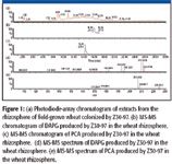
Figure 1
Strains Z30-97 and Z15-02 produce PCA and DAPG; Z15-02 also produces 2-hydroxy PCA. Bacteria, introduced onto the seeds (105 cells/seed), colonized the rhizosphere. For MS-MS library matching, the forward match value, which applies to pure chromatographic peaks, is reduced when mass peaks (background noise, contaminant peaks) are present in the spectrum of the unknown but not in that of the library, or present in the library spectrum but not in that of the unknown. In reverse library matching, which applies to peaks containing mixtures, the match value is reduced by mass peaks present in the library but not in the unknown spectrum. Contaminant peaks in the unknown spectrum but not present in the library spectrum will have no effect on the reverse hit (perfect fit = 1000). As a general rule, confirmation of high mass accuracy (TOF), together with forward–reverse match values of 500, 600–700, and 800–1000, correspond to compound identification at confidence levels of 70%, 85%, and 100%, respectively.
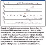
Figure 2
Quantification
Standard curves consisting of five data points (in triplicate) for both DAPG and phenazine were generated by spiking 5 g of roots plus rhizosphere soil with 200 ng to 5 μg of both standards. The relationship between peak areas (y) and spiked standards (x) was linear, as described by the equation y = 73.9378x – 3.09562, with r = 0.991842 and r2 = 0.983750 (DAPG), and y = 119.387x + 0.566344, with r = 0.929664 and r2 = 0.894274 (PCA) (Figures 3 and 4). In our hands, the detection limits by QTOF-2 mass spectrometry of DAPG and PCA produced in the rhizosphere of wheat are 15 ng and 800 pg, respectively, using the standard APCI probe (Waters); eightfold greater sensitivity occurs with the use of the Ion Sabra probe (Waters).
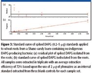
Figure 3
Conclusions
The rhizosphere metabolome is one of the most complex in nature and is a treasure trove of novel antibiotics and other bioactive compounds that potentially can be exploited for use in human and veterinary medicine. It also plays a key role in natural plant disease suppression, allowing crops to be grown more sustainably and free of certain diseases without inputs of chemical pesticides. Our understanding of antibiotics in the rhizosphere metabolome has evolved over several decades as bioanalytical techniques and separation science have progressed from classical TLC to HPLC with photodiode-array detection to HPLC-TOF-MS. Techniques such as SPE and QTOF-MS are facilitating reductions in sample preparation time and increases in sample throughput. These techniques, coupled with molecular approaches such as transcriptional analyses of antibiotic gene expression, will allow in-depth analyses of the kinetics of antibiotic production and degradation in soil and the rhizosphere, and continue to peel back the layers of the rhizosphere metabolome.
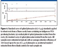
Figure 4
References
(1) D.V. Mavrodi, W. Blankenfeldt, and L.S. Thomashow, Annu. Rev. Phytopathol. 44, 417–445 (2006).
(2) L.S. Thomashow, R.F. Bonsall, and D.M. Weller, in Manual of Environmental Microbiology, 2nd ed., C.J. Hurst, R.L. Crawford, G.R. Knudsen, M.J. McInerney, and L.D. Stetzenbach, Eds. (ASM Press, Washington, D.C., 2002), pp. 636–647.
(3) D.M. Weller, B.B. Landa, O.V. Mavrodi, K.L. Schroeder, L. De La Fuente, S. Blouin Bankhead, R. Allende Molar, R.F. Bonsall, D.V. Mavrodi, and L.S. Thomashow, Plant Biol. 9, 4–20 (2007).
(4) L.S. Thomashow, R.F. Bonsall, and D.M. Weller, in Secondary Metabolites in Soil Ecology, P. Karlovsky, Ed. (Springer, Germany, in press).
(5) Y. Chander, K. Kumar, S.M. Goyal, and S.C. Gupta, J. Environ. Qual. 34, 1952–1957 (2005).
(6) T.L. ter Laak, W.A. Gebbink, and J. Tolls, Environ. Toxicol. Chem. 25, 933–941 (2006).
(7) L. Ramos, E.M. Kristenson, and U.A.T. Brinkman, J. Chromatogr., A 975, 3–29 (2002).
(8) M. Petrovic, M.D. Hernando, M.S. M.S. Díaz-Cruz, and D. Barceló, J. Chromatogr., A 1067, 1–14 (2005).
(9) R.F. Bonsall, D.M. Weller, and L.S. Thomashow, Appl. Environ. Microbiol. 63, 951–955 (1997).
(10) J.M. Raaijmakers, R.F. Bonsall, and D.M. Weller, Phytopathology 89, 470–475 (1999).
(11) S.C. Kim and K. Carlson, Water Res. 40, 2549–2560 (2006).
Robert F. Bonsall and Dmitri V. Mavrodi are with the Department of Plant Pathology, Washington State University, Pullman, Washington. Linda S. Thomashow and David M. Weller are with the USDA, Agricultural Research Service, Root Disease and Biological Control Research Unit, Pullman, Washington. Please direct correspondence to: wellerd@wsu.edu
Separation of Ultra-Short and Long Chain PFAS Compounds Using a Positive Charge Surface Column
December 11th 2024A separation of ultra-short and long chain PFAS (C1-C18) is performed on a HALO®PCS Phenyl-Hexyl column along with a HALO®PFAS Delay column which demonstrates excellent retention for both hydrophilic and hydrophobic analytes.


