Drug Development: Overcoming Glycan Screening Bottlenecks for Clone Selection and Cell Culture Optimization
The Column
This article focuses on ways to accelerate glycan screening data analysis, while keeping high reproducibility.A major focus of the pharmaceutical industry is the production and development of biologics-therapeutics produced via biological means-to provide novel treatments for diseases with unmet clinical needs. A large percentage of biologics under development are proteins, such as monoclonal antibodies (mAbs), fusion proteins, antibody–drug conjugates (ADCs), and enzymes. The structures of these protein drugs are made more complex by post‑translational modifications (PTMs).
Elena Pankova/stock.adobe.com
Glycosylation is one of the most common protein post-translational modifications (PTMs), so detecting changes in glycosylation is important for researchers to determine glycan profiles that may affect the function, efficacy, and clearance of novel biologic drugs. As glycans are so important for biological function, screening glycan profiles early in the clone selection and cell culture optimization process of drug development can provide better information for selecting the right clones. Doing this, however, has traditionally meant setting aside an entire day for sample preparation before performing the analysis the next day. In order to reduce the challenges of tedious sample preparation, long separation times, and data analysis, researchers need to accelerate the workflow to allow screening of more glycan samples per day, and obtain visual, actionable data without the need for time-consuming interpretation. This article focuses on ways to accelerate glycan screening data analysis, while keeping high reproducibility.A major focus of the pharmaceutical industry is the production and development of biologics-therapeutics produced via biological means-to provide novel treatments for diseases with unmet clinical needs. A large percentage of biologics under development are proteins, such as monoclonal antibodies (mAbs), fusion proteins, antibody–drug conjugates (ADCs), and enzymes. The structures of these protein drugs are made more complex by postâtranslational modifications (PTMs).
Glycosylation is a particularly prevalent and structurally complex PTM that results in the addition of carbohydrate-based moieties called glycans. Glycan chains markedly affect the protein stability, activity, antigenicity, and pharmacodynamics, and are therefore of major importance to the development of a successful biologic that exhibits optimal therapeutic efficacy (1).
To ensure the safety and potency of commercial biologics before regulatory approval, appropriate glycosylation must be demonstrated (2). It is also necessary to demonstrate that the manufacturing process is robust and can be reproduced for batch release. Glycosylation is likely among the most crucial quality attributes and the most difficult parameter to control (3).
There are a variety of analytical methods available for characterization at the intact protein level, including chromatography, electrophoresis, and mass spectrometry (MS). Recent advances in capillary electrophoresis (CE) techniques have been used for detailed characterization of glycans attached to biotherapeutic proteins. As CE is a versatile analytical platform, it offers a rapid, yet simple method for exhaustive carbohydrate screening (4,5).
The Challenges
Although there have been important advances, glycan screening remains a challenge in the biopharmaceutical industry. The sample preparation can be tedious, labour-intensive, and technique-dependent. Separation of carbohydrates is demanding because of the complex, diversified structure of glycans; they also lack chromophore or fluorophore groups and most glycans have a neutral charge. Furthermore, the complexity of glycans that can be added to a protein contributes to its heterogeneity, leading to questions as to the biological significance of the multitude of glycan peaks that can appear during profiling. Beyond the experimental difficulties associated with glycan analysis, data interpretation and structural identification are perhaps the greatest bottlenecks for streamlined glycan analysis (6,7).
Here we present an automated, highâthroughput glycan screening method using qualitative, multiplexed CE. By using a fully automated magnetic bead-based sample preparation, the glycosylation pattern of a therapeutic antibody under investigation was separated and identified by CE.
Acceleration and Reproducibility of High-Throughput Glycan Screening
Glycan acceptance criteria need to be quick and intuitive and allow real-time decisions on clone selection and cell-line optimization. Furthermore, automated glycan identification would eliminate the need for tedious glycan database searches, thus minimizing the potential for human error and delivering a more robust workflow.
To test a high-throughput screening method for speed and reproducibility, a multiplexed capillary electrophoresis platform providing the screening of large numbers of candidates quickly enough to allow for realâtime adjustments was evaluated (8).
Method
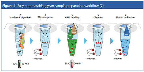
A purified antibody-MAK33-obtained from Roche Diagnostics was diluted to 10 mg/mL before sample preparation. Automated sample preparation including denaturation, digestion, and glycan labelling was performed with a setup for 96-well plates (Beckman Coulter Biomek i5). The antibodies were denatured for 8 min at 60 °C using the denaturing solution master mix (Figure 1, Step A) and digested for 20 min at 60 °C using PNGase F (Figure 1, Step B). The released glycans were captured by magnetic beads, and then labelled with 1-aminopyrene-3,6,8- trisulfonate (APTS) for 20 min at 60 °C (Figure 1, Step C). Excess APTS was removed from the reaction mixture using a magnetic-bead-mediated series of washes (Figure 1, step D). The labelled N-linked glycans were eluted from the beads by 50 μL of water (Figure 1, Step E).
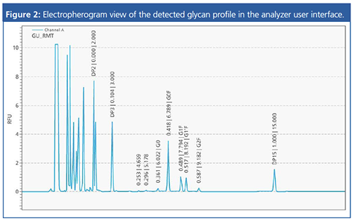
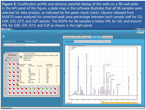
At this point, the samples were ready to be analyzed by electrophoretic separation. The C100HT Biologics Analyzer (Sciex) was set up with a pre-filled buffer tray, sample plate, and gel-preloaded multicapillary separation cartridge. Following that, the appropriate preâconfigured process profile was selected and sample information entered. Analysis of a full 96-well plate took approximately 170 min and the separation and detection of the glycan profile was visualized by the anchor peaks in the electropherogram (Figure 2). A qualification profile for each glycan of interest was pre-set in order to define the glycan composition of each sample and presented as pass or fail (Figure 3). DataReviewer Software in the C100HT from Sciex was used to analyze the data (8.9).
Results and Conclusions
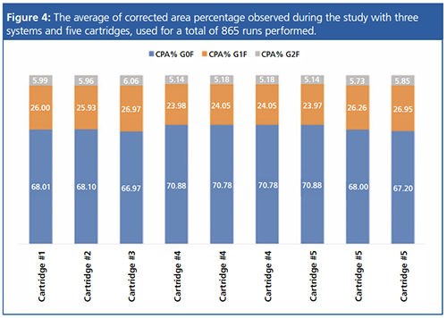
An intermediate precision study (Figure 4) was carried out using three systems, five cartridges with total 865 runs to illustrate the robustness and reproducibility of this method. The chart shows the consistency in corrected area percentage of three major glycan species found in MAK33. The method can screen glycan expression profiles for up to five 96-well plates of glycans samples in one day. Automated APTS labelling and cleanup reduces the need for human intervention while providing better precision and minimizing variation between samples.
Producing optimal and consistent glycosylation requires an integrated approach for successful biopharmaceutical drug development with systematic glycosylation analysis throughout the manufacturing processes. A dramatically shortened screening time enables clones to be selected from large sample populations quickly and cell culture optimization to be performed in parallel, thereby increasing the likelihood of selecting the best clones and manufacturing conditions. From a productivity perspective, good quality high-throughput data allow confident decisions on clone selection and cell optimization in real time, early in the process, providing more cost-efficient workflows.
For Research Use Only. Not for use in diagnostic procedures. RUO-MKT-19-9184-A
References
- P. Zhang, S. Woen, T. Wang, B. Liau, S. Zhao, C. Chen, Y. Yang, Z. Song, M.R. Wormald, C. Yu, and P.M. Rudd, Drug Discovery Today21(5), 740–765 (2016).
- WHO Technical Report Series No. 977, 2013, http://apps.who.int/medicinedocs/documents/s19941en/s19941en.pdf
- L. Hajba, A. Szekrenyes, B. Borza, and A. Guttman, Drug Discovery Today23, 616–625 (2018).
- G. Lu, C.L. Crihfield, S. Gattu, L.M. Veltri, and L.A. Holland, Chem. Rev.118, 7867−7885 (2018).
- Z. Szabo, A. Guttman, T. Rejtar, and B.L. Karger, Electrophoresis 31(8), 1389–1395 (2010).
- F. Li, N. Vijayasankaran, A. Shen, R. Kiss, and A. Amanullah, Mabs 2(5), 466–477 (2010).
- M. Szigeti, C. Lew, K. Roby, and A. Guttman, JALA21, 281–286 (2016).
- M. Lies, M. Santos, and T. Li, Sciex Separations, Brea, California, USA. High-Throughput Glycan Screening to Remove Biopharmaceutical Development Bottlenecks
- M. Santos, T. Li, M. Gutierrez, A. Lou, and C. Lew, Sciex Separations, Brea, California, USA. Repeatability of C100ht Biologics Analyzer, for High Throughput Glycan Screening
Sahana Mollah is a Senior Manager, Biologics Global Collaboration and CE Technical Marketing Coordinator.
Mark Lies is a Senior Product Manager.
Marcia Santos is a Staff Applications Scientist.
Tingting Li is a Senior Biopharma Application Development Scientist.
Kelly May is a Senior Global Commercial Marketing Manager.
E-mail: customercare@sciex.comWebsite:https://sciex.com/request-support
Separating Impurities from Oligonucleotides Using Supercritical Fluid Chromatography
February 21st 2025Supercritical fluid chromatography (SFC) has been optimized for the analysis of 5-, 10-, 15-, and 18-mer oligonucleotides (ONs) and evaluated for its effectiveness in separating impurities from ONs.

.png&w=3840&q=75)

.png&w=3840&q=75)


.png&w=3840&q=75)


.png&w=3840&q=75)


