Comparison of LC–MS and GC–MS for the Analysis of Pharmaceuticals and Personal Care Products in Surface Water and Treated Wastewaters
Special Issues
This study of a selected group of PPCP contaminants in eastern North Carolina demonstrates the advantages and disadvantages of LC–MS and GC–MS as well as SPE and liquid–liquid extraction.
Water samples were obtained from the Tar River and a local water treatment plant in eastern North Carolina in spring 2013 and fall 2015 to monitor the presence of a panel of pharmaceutical and personal care products (PPCPs). Samples were extracted by solid-phase extraction (SPE) or liquid–liquid extraction and analyzed for parent PPCPs and their metabolites by high performance liquid chromatography-time-of-flight mass spectrometry (HPLC–TOF-MS) and gas chromatography–mass spectrometry (GC–MS). Both extraction and detection methods were compared by their recoveries and detection limits for each compound. Many parent PPCPs and their metabolites were detected including: carbamazepine, iminostillbene, oxcarbazepine, epiandrosterone, loratadine, β-estradiol, triclosan, and others. Liquid–liquid extraction was found to provide overall superior recoveries. Furthermore, HPLC–TOF-MS yielded lower detection limits than GC–MS. Library searching of additional peaks identified further compounds with biological activity. Additionally, the effectiveness of the treatment plant on the removal of the compounds of interest is discussed.
Pharmaceuticals and personal care products (PPCPs) have been found as contaminants in drinking and wastewater worldwide (1–4) and can pose a toxicological risk to humans as well as wildlife (5–8). Although water treatment plants incorporate a wide variety of methods to remove these compounds, many PPCPs have been shown to persist posttreatment, allowing for their accumulation in the environment (9–12). Because of the ubiquitous nature of these contaminants and their wide variety of effects on biological organisms, detection and tracking of PPCPs has become an area of increasing research interest.
Statistics from the Center of Disease Control and Prevention show that Eastern North Carolina has the highest occurrence of stroke and heart disease compared to other regions in the United States (8). Although this phenomenon may be attributable to factors such as socioeconomic status, ethnic distribution, or dietary trends, exposure to contaminants like PPCPs may be a large contributing factor based on the wide range of health effects that they can impart. Data on the prevalence of PPCPs in this region is currently limited to only one study detecting a total of four PPCPs using gas chromatography (GC) (13).
Chromatography and mass spectrometry (MS) are commonly used analytical techniques to identify and quantify water contaminants such as PPCPs. Previous studies have used liquid chromatography–MS (LC–MS) (3,4,10,14–17), GC–MS (1,18,19), or both (2,9,20,21) to detect compounds within this class. In terms of sample preparation, many studies use solid-phase extraction (SPE) (1–4,10,14,15,17–19,21); however, some studies use liquid–liquid extraction (20), or both (16). The PPCPs analyzed in these studies vary greatly not only with respect to their biological mechanisms of action, but also to their chemical–physical properties such as acid-base properties, volatilities, thermal stabilities, and polarities. These differences, combined with the matrices of surface and wastewaters, influence the efficiencies of these extraction methods. PPCP contaminants are commonly present in the nanogram-per-liter range, so low extraction recoveries can often lead to many undetected compounds. Furthermore, liquid and gas chromatography can possess drastically different detection limits based on the types of compounds being analyzed, which can exacerbate the issue of undetected contaminants. Data comparing these two instrumental and extraction techniques on PPCPs is lacking, making it unclear which combination of methods would be best suited for this type of analysis.
The goal of this study is to uncover the advantages and disadvantages of LC–MS and GC–MS as well as SPE and liquid–liquid extraction on a selected group of PPCP contaminants in Eastern North Carolina. These data would uncover the advantages and disadvantages to these commonly used techniques, revealing some insight about which instrumental and extraction techniques are most suitable for these PPCPs. Also, this study aims to use this information to uncover some of the additional emerging PPCPs in Eastern North Carolina and determine their relationship to any underlying diseases in the area as well as any seasonal variations in detected compounds. It is expected that these findings will be translatable to similar studies detecting PPCPs in other geographical regions, improving the detection of these compounds.
Methods
Chemicals
Solvents for standard preparation and instrumental analysis (acetonitrile, methylene chloride, methanol, and water) were LC–MS-grade and were purchased from Sigma-Aldrich. ENVI-Disk C18 SPE disks (47 mm in diameter) were purchased from Supelco. β-estradiol (≥98%), caffeine (≥98.5%), ketoprofen (≥98%), naproxen (≥98%), ibuprofen (≥98%), triclocarban (≥99%), diphenylhydramine (≥98%), and napropamide (≥98%) were purchased from Sigma Aldrich. Carbamzepine (≥99%), oxcarbazepine (≥97.5), and loratadine (≥98%) were purchased from Fluka. Iminostilbene (97%) was purchased from Chem Service. Epiandrosterone (≥99%) was purchased from MP Biomedicals.
Instrumental Conditions
The GC–MS system consisted of an Agilent 7890A gas chromatograph set in splitless injection mode and an Agilent 5975B mass spectrometer with an electron ionization source. A 30 m x 0.25 mm, 0.5-µm df DB-5MS column (J&W Scientific) was used in this study. The carrier gas (helium) was kept at a flow of 0.8 mL/min. The injection volume for all samples was 1 µL using a 7683B series Agilent liquid injector. Temperature programming was as follows: Hold initially at 150 °C for 5 min, ramp at 10 °C/min to 300 °C, and hold at 300 °C for 10 min. The source temperature was kept at 230 °C, and the electron multiplier detector was set at a voltage of 2012 V, absolute. For quantitative analysis, selected ion monitoring (SIM) was used for the measurement of each compound. For each compound, the most abundant m/z fragment was used for quantification and two additional characteristic ions were used as qualifiers. Data were collected and analyzed using MSD ChemStation E.02.00.493 software.
The high performance liquid chromatography-time-of-flight mass spectrometry (HPLC–TOF-MS) system consisted of an Agilent 1200 series HPLC system coupled to an Agilent 6220 TOF-MS system with a dual electrospray ionization (ESI) interface set to positive ion mode. The HPLC column used was a 150 mm x 2.1 mm, 3.5-µm df Agilent Zorbax Eclipse Plus C18 column. Mobile-phase A was HPLC-grade water with 1% formic acid, and mobile-phase B was acetonitrile with 1% formic acid. The injection volume used for all samples was 3 µL. Solvent programming was as follows: 20–80% B over 20 min, hold at 80% B for 5 min. The column temperature was set to 35 °C. The drying gas (nitrogen) temperature was set to 335 °C at a rate of 10 L/min with the nebulizer pressure kept at 35 psig. The capillary voltage was set to 3300 V with the fragmentor voltage at 185 V and the skimmer voltage at 30 V. Identification and quantification of each compound was based off of [M+H]+ values. Agilent MassHunter Workstation software version B.02.01 was used to collect and analyze data.
Comparison of Detection Limits
A mixture containing 10 µg/mL each of β-estradiol, caffeine, carbamazepine, diphenylhydramine, epiandrosterone, ibuprofen, iminostilbene, ketoprofen, loratadine, naproxen, oxcarbazepine, and triclocarban was prepared in acetonitrile. Compounds were selected based on preliminary screening of local water sites (data not shown). Calibration curves were constructed for each compound by making dilutions between 5 µg/mL to 25 ng/mL. Each dilution was analyzed by GC–MS and HPLC–TOF-MS. For compounds that were detectable at 25 ng/mL, 3(S0) was used as the limit of detection, where S0 = standard deviation of zero concentration. For compounds that were not detected at 25 ng/mL, the lowest dilution in which they were detected was set as the detection limit.
Comparison of Extraction Methods
Standard Mixture Preparation
A mixture of 1 µg/mL each of β-estradiol, caffeine, carbamazepine, diphenylhydramine, epiandrosterone, ibuprofen, iminostilbene, ketoprofen, loratadine, naproxen, oxcarbazepine, and triclocarban was prepared in acetonitrile. Then 1 mL of this standard mixture was spiked into 500 mL of deionized water and mixed by shaking.
Solid-Phase Extraction
The SPE disks were conditioned using 10 mL of acetonitrile, 10 mL of methanol, and 10 mL of deionized water with 2 min of equilibration time in between each solvent. The 500 mL of spiked water (described above) was loaded onto the conditioned SPE disk at 0.15–0.2 mL/min and then dried under vacuum for 20 min. For elution, 10 mL of acetonitrile was added to the disk, allowed to sit for 5 min, and then eluted at 0.1 mL/min. The eluate was collected in a graduated 15-mL conical test tube and then dried under nitrogen to a volume less than 1 mL. This process was repeated a total of three times and the extracts were analyzed using both GC–MS and HPLC–TOF-MS.
Liquid–Liquid Extraction
The 2000 mL of spiked water (described above) was loaded into a separatory funnel along with 100 mL of methylene chloride. The solution was shaken by hand for 1 min and then the methylene chloride layer was collected into a round-bottom flask. This process was repeated a total of three times, giving a final volume of 300 mL of methylene chloride. The methylene chloride extract was combined in a 500-mL round-bottom flask, which was then loaded onto a rotary evaporator where the extract was concentrated to a few milliliters. The concentrated extract was quantitatively transferred to a 15-mL graduated test tube and the final volume was adjusted to 1 mL, by repeated washing, transfer, and evaporation. This process was repeated a total of three times and the extracts were analyzed using both GC–MS and HPLC–TOF-MS. Analyte recoveries for both liquid–liquid extraction and SPE were calculated using the following formula:
(Analyte mass in extract/Analyte mass in spiked water) x 100 = Analyte recovery [1]
Analysis of PPCPs in Surface and Wastewater
Sample Collection
Two sampling sites within the Greenville, North Carolina, wastewater treatment plant were chosen for analysis of PPCPs: influent wastewater and effluent water (cleaned end-product). At each of those sites, 4-L grab samples were collected during the spring of 2013 and the fall of 2015. Samples were transported back to the laboratory on ice within 30 min of collection. Amber, 4-L HPLC solvent bottles that were previously cleaned using methanol and acetonitrile were used for sample collection and storage. For samples that were not immediately analyzed, the pH was adjusted to 2 using hydrochloric acid to prevent bacterial growth and subsequent breakdown of PPCPs. Samples were then stored at 4 °C until analysis.
Extraction and Measurement of PPCPs
Samples from 2013
First, 2 L of sample material was filtered using a 0.45-µm glass fiber filter and extracted using the liquid–liquid extraction method detailed earlier. Extracts were analyzed using HPLC–TOF-MS and original sample concentrations were calculated based off of determined recovery values. Both influent and effluent sites were analyzed in duplicate. GC–MS was also used to analyze extracts to qualitatively identify additional PPCPs using the National Software Reference Library (NIST) Mass Spectral Search Program software version 2.0. Peaks were identified based on the compounds with the highest matching spectral score given by the software.
Samples from 2015
First, 500 mL of sample material was filtered through a 0.45-µm glass fiber filter and spiked with 1 mL of napropamide as an internal standard. PPCPs were extracted from 500-mL aliquots using SPE disks according to the method outlined earlier (n = 5 and n = 6 for influent and effluent samples, respectively). Extracts were analyzed using HPLC–TOF-MS and quantified using the internal standard peak.
Results and Discussion
Table I displays a panel of 12 PPCPs that were selected based on preliminary screening of water samples (data not shown). For GC–MS analysis, compounds were quantified using a target ion that was the most abundant m/z value, and the next two most abundant fragments were used as qualifier ions (Q1 and Q2). For HPLC–TOF-MS, compounds were quantified using their [M+H]+ adduct masses, with the exception of diphenylhydramine, which has a much more abundant characteristic mass of 167.0876 that was produced by the fragmentor. Standard mixtures were prepared and analyzed using both GC–MS and HPLC–TOF-MS with the methods detailed above. Representative total ion current (TIC) chromatograms showing chromatographic separation of these 12 compounds are shown in Figures 1a (GC–MS) and 1b (HPLC–TOF-MS). Of the selected compounds, triclocarban and ketoprofen were unable to be detected using the GC–MS method; however, the HPLC–TOF-MS method could detect all 12.
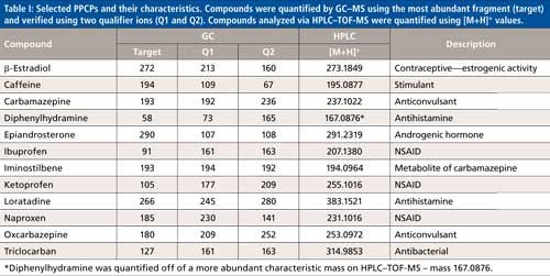
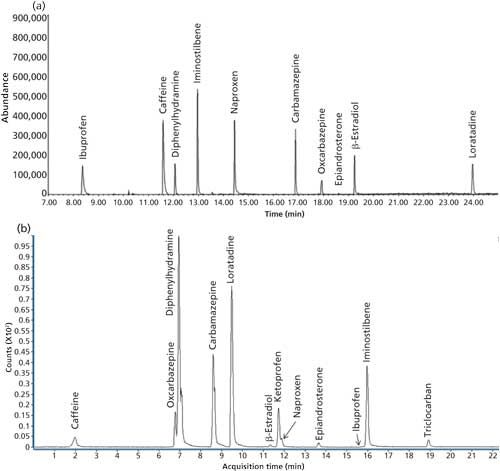
Figure 1: Representative TIC chromatograms of 12 selected PPCPs using (a) GC–MS and (b) HPLC–TOF-MS.
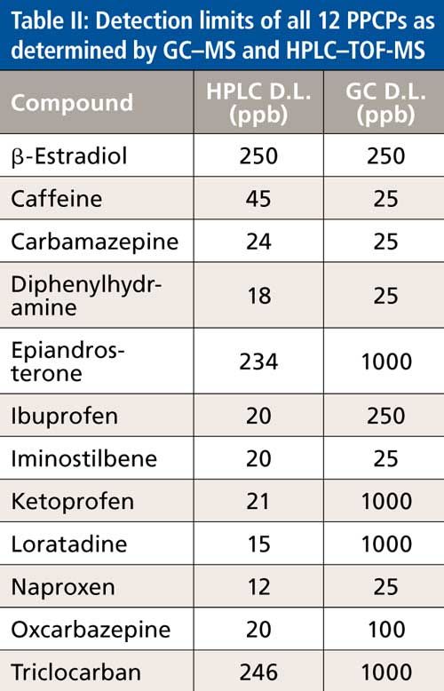
Detection limits were calculated to further compare the suitability of each instrument for the selected compounds. Because PPCPs are typically present in nanogram-per-liter concentrations in surface water and wastewater, detection limits are often one of the greatest challenges to overcome when analyzing these samples. As shown in Table II, HPLC–TOF-MS achieved lower detection limits for all compounds with the exception of caffeine and β-estradiol. Caffeine’s detection limit was 1.8 times lower on the GC–MS and β-estradiol had the same detection limit between both instruments. In general, more polar compounds are more amenable for liquid chromatography, so superior detection limits were expected with HPLC. Less-polar compounds like caffeine and β-estradiol are typically well-suited for GC analysis, which was reflected in our results. Overall, HPLC–TOF-MS was able to detect lower amounts of the selected compounds and was therefore chosen to quantify all samples for the remainder of the study.
Both liquid–liquid extraction (using methylene chloride) and C18 SPE were compared on their ability to recover each of the 12 analytes. As shown in Figure 2, liquid–liquid extraction produced higher recoveries for all 12 compounds as compared to SPE. In this scenario, the use of SPE for surface and wastewater samples may lead to a reduced ability to detect PPCPs because of lower recovery rates, particularly with iminostilbene and diphenylhydramine. Although more sample loss was observed, SPE produced less variation than liquid–liquid extraction. This increased precision combined with higher throughput may allow SPE to be more advantageous than liquid–liquid extraction in circumstances where these properties are favored. Another key difference between the two extraction methods is that liquid–liquid extraction had a higher capacity for sample volume. Amounts greater than 500 mL led to sample breakthrough on the SPE disks, whereas liquid–liquid extraction could potentially handle volumes greater than the 2000 mL used in this study. Higher sample capacity could assist in detecting trace analytes by allowing for a larger degree of concentration.
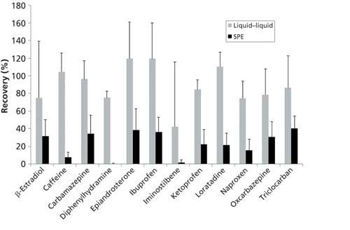
Figure 2: Comparison of recoveries obtained from extracting PPCPs using liquid–liquid extraction and SPE. Recovery values were quantified using HPLC–TOF-MS.
The two extraction methods, liquid–liquid extraction and SPE, were next compared on their ability to extract PPCPs from surface and wastewater field samples. Samples were taken from the wastewater treatment plant in the city of Greenville, North Carolina in the spring of 2013 (extracted using liquid–liquid extraction) and the fall of 2015 (extracted using SPE). This also allowed us to compare seasonal differences in the amounts of PPCPs detected. The treatment plant involved three main methods of treatment to produce clean drinking water. The primary treatment involves the use of screens to remove physical objects (for example, plastic, wood, sand, and so forth). The secondary treatment uses a microorganism chamber to break down organic matter and remove nitrogen as well as phosphorus. Finally, a tertiary treatment uses a deep-bed sand filter and UV light to further disinfect and purify. Samples were collected from the influent as well as effluent water to not only determine PPCPs present in untreated water, but also to see the effectiveness of these treatment methods on removal of these compounds.
Using HPLC–TOF-MS as the preferred instrumental technique for quantification, extracts from influent and effluent waters were analyzed for all 12 analytes. Figure 3a shows that in the spring of 2013, all compounds were detected in the influent water with carbamazepine as the most prominent contaminant at 3.47 µg/L using liquid–liquid extraction. Carbamazepine has been found at the microgram-per-liter concentration level in a number of other studies, particularly in European countries (22–26). Carbamazepine has been found to be particularly resilient toward removal by water treatment methods (27,28) as well as having reduced biodegradability (29). The high rates of seizures in the population of this region may be an additional explanation as to why this drug is found at such high levels along with its metabolite, iminostilbene, as well as oxcarbazepine. Additionally, these data show that the treatment plant was effective at reducing the levels of all of these compounds, with β-estradiol and epiandrosterone being undetectable in effluent samples. Overall, samples collected in the fall of 2015 also showed a decrease in PPCP levels in influent samples as compared to effluent samples after extraction by SPE with carbamazepine as the most prominent contaminant at 27.0 µg/L (Figure 3b). Two exceptions to this decrease were carbamazepine and β-estradiol, which both had increased levels in effluent samples. It is well known that many PPCPs heavily partition into suspended solids in wastewater samples (30–38), which can heavily influence their detectable concentration in the aqueous phase. Differences in solid composition and amount may suppress the detectable concentration in the aqueous phase using this method. Additionally, the liquid–liquid extraction method may have been better able to extract adsorbed compounds in any residual solid material left in the water samples. When comparing the two sets of samples, water from the fall of 2015 had higher amounts of all PPCPs except for ibuprofen and iminostilbene (ibuprofen was unable to be detected in either influent or effluent samples from 2015). Differences in the two sample sets may be due to variations in prescribed drugs at both times, city population, as well as any differences in water treatment processes. These results highlight the degree of variation of PPCPs within surface and wastewaters over time.
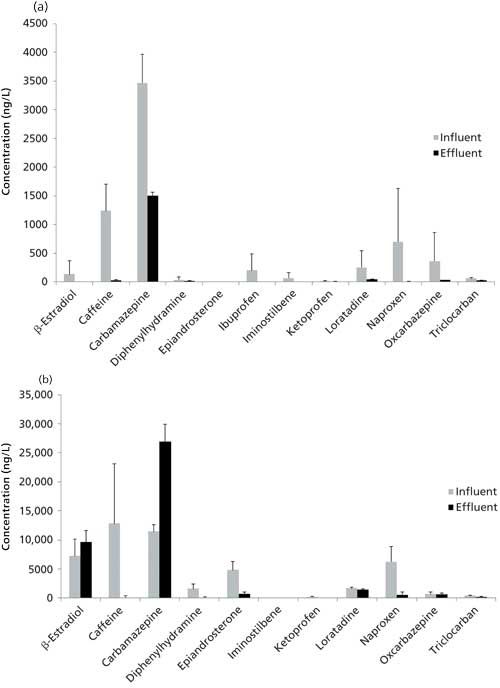
Figure 3: PPCPs detected using HPLC–TOF-MS in influent and effluent waters in a local wastewater treatment plant in Greenville, North Carolina. (a) Samples were collected in the spring of 2013 and extracted using liquid–liquid extraction. (b) Samples were collected in the fall of 2015 and extracted using SPE.
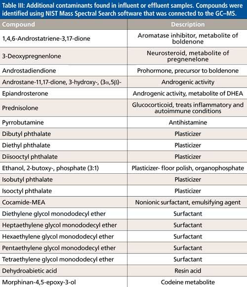
In addition to the 12 compounds selected for this study, other contaminants were identified in the spring 2013 samples using NIST library searching that was coupled to the GC–MS. Using this software, unidentified peaks could be qualitatively identified, further characterizing the compounds that were detected in these samples using these methods. Table III lists the major additional compounds detected across all samples along with a brief description of their class and any notable biological activity. Many of these contaminants had endocrine activities or were plasticizers or surfactants. Future studies may apply the methods used here on these additional compounds or other PPCPs.
Conclusions
The data provided in this study provide evidence for the advantages of different combinations of instrumental and extraction techniques for the analysis of PPCPs in surface and wastewaters. For the compounds chosen in this study, HPLC–TOF-MS was overall more sensitive and allowed for the detection of more compounds than GC–MS. Additionally, liquid–liquid extraction achieved higher recoveries for the measured PPCPs than SPE, although SPE showed less variation. Also, the method of water treatment used in this study was overall shown to be effective at reducing or eliminating this panel of PPCPs. The results provided in this study highlight the importance of selecting the appropriate extraction and analysis techniques to best analyze PPCPs water samples.
Acknowledgments
The authors would like acknowledge the help of the Mr. Jeff Camp and other administrative personnel from Greenville Utilities, for facilitating sample collection for this project. Funding support for this work was provided by East Carolina University, Office of the Vice President for Research, East-West Project. We also would like to thank Dr. Siddhartha Mitra for providing some of the analytical standards used in this work.
References
- V. Koutsouba, T. Heberer, B. Fuhrmann, K. Schmidt-Baumler, D. Tsipi, and A. Hiskia, Chemosphere51, 69–75 (2003).
- M.J. Benotti, R.A. Trenholm, B.J. Vanderford, J.C. Holady, B.D. Stanford, and S.A. Snyder, Environ. Sci. Technol.43, 597–603 (2009).
- S.D. Kim, J. Cho, I.S. Kim, B.J. Vanderford, and S.A. Snyder, Water Res.41, 1013–1021 (2007).
- M. Kuster, M.J. de Alda, M.D. Hernando, M. Petrovic, J. Martin-Alonso, and D. Barcelo, J. Hydrol.358, 112–123 (2008).
- S.A. Snyder, P. Westerhoff, Y. Yoon, and D.L. Sedlak, Environ. Eng. Sci. 20, 449–469 (2003).
- J.W. Kim, H. Ishibashi, R. Yamauchi, N. Ichikawa, Y. Takao, M. Hirano, M. Koga, and K. Arizono, J. Toxicol. Sci.34, 227–232 (2009).
- F. Gagné, C. Blaise, and C. André, Ecotoxicol. Environ. Saf. 64, 329–336 (2006).
- A. Nikolau, S. Meric, and D. Fatta, Anal. Bioanal. Chem. 387, 1225–1234 (2007).
- P.E. Stackelberg, J. Gibs, E.T. Furlong, M.T. Meyer, S.D. Zaugg, and R.L. Lippincott, Sci. Total Environ. 377, 255–272 (2007).
- T. Herberer, Toxicol. Lett.131, 5–17 (2002).
- T.A. Ternes, M. Meisenheimer, D. McDowell, F. Sacher, H.J. Bruach, B. Haist-Gulde, G. Preuss, U. Wilme, and N. Zulei-Seibert, Environ. Sci. Technol. 36, 3855–3863 (2002).
- K.L. Del Rosario, S. Mitra, CP. Humphrey Jr., and M.A. O’Driscoll, Sci. Total Environ.487, 216–223 (2014).
- N.M. Vieno, H. Härkki, T. Tuhkanen, and L. Kronberg, Environ. Sci. Technol. 41, 5077–5084 (2007).
- B.J. Vanderford, R.A. Pearson, D.J. Rexing, and S.A. Snyder, Anal. Chem. 75, 6265–6274 (2003).
- P.E. Stackelberg, E.T. Furlong, M.T. Meyer, S.D. Zaugg, A.K. Henderson, and D.B. Reissman, Sci. Total Environ.329, 99–113 (2004)
- M.J. Hilton, and K.V. Thomas, J. Chromatogr. A.1015, 129–141 (2003).
- S. Ollers, H.P. Singer, P. Fässler, and S.R. Müller, J. Chromatogr. A.911, 225–234 (2001).
- H.B. Lee, T.E. Peart, and M.L. Svoboda, J. Chromatogr. A.1094, 122–129 (2005).
- P. Westerhoff, Y. Yoon, S. Snyder, and E. Wert, Environ. Sci. Technol.39, 6649–6663 (2005).
- C. Hao, X. Zhao, and P. Yang, Trends Anal. Chem.26, 569–580 (2007).
- D. Bendz, N.A. Paxéus, T.R. Ginn, and F.J. Loge, J. Hazard Mater.122, 195–204 (2005).
- M. Clara, B. Strenn, O. Gans, E. Martinez, N. Kreuzinger, and H. Kroiss, Water Res. 39, 4797–4807 (2005).
- U. Hass, U. Dünnbier, G. Massmann, and A. Pekdeger, Anal. Methods3, 902–910 (2011).
- T. Herberer, J. Hydrol.266, 175–189 (2002).
- M. Leclercg, O. Mathieu, E. Gomez, C. Casellas, H. Fenet, and D. Hillaire-Buys, Arch. Environ. Contam. Toxicol. 56, 408–415 (2009).
- S. Castiglioni, R. Bagnati, R. Fanelli, F. Pomati, D. Calamari, and E. Zuccato, Environ. Sci. Technol. 40, 357–363 (2006).
- M.J. Gómez, M. Petrovic, A.R. Fernández-Alba, and D. Barceló, J. Chromatogr. A.1114, 224–233 (2006).
- J.L. Santos, I. Aparicio, and E. Alonso, Environ. Int.33, 596–601 (2007).
- Y. Chen, G. Yu, Q. Cao, H. Zhang, Q. Lin, and Y. Hong, Chemosphere93, 1765–1772 (2013).
- W. Chenxi, A.L. Spongberg, and J.D. Witter, Chemosphere73, 511–518 (2008).
- P. Falås, A, Ballion-Dhumez, H.R. Andersen, A. Ledin, and J. la Cour Jansen, Water Res.46, 1167–1175 (2012).
- B.F. da Silva, A. Jelic, R. López-Serna, A.A. Mozeto, M. Petrovic, and D. Barceló, Chemosphere85, 1331–1339 (2011).
- A. Jelic, M. Gros, A. Ginebreda, R. Cespedes-Sánchez, F. Ventura, M. Petrovic, and D. Barcelo, Water Res.45, 1165–1176 (2011).
- T.L. Jones-Lepp, and R. Stevens, Anal. Bioanal. Chem. 387, 1173–83 (2007).
- A. Nieto, F. Borrull, R.M. Marcé, and E. Pocurull, J. Chromatogr. A. 1216, 5619–5625 (2009).
- A. Nieto, F. Borrul, E. Pocurull, and R.M. Marcé, Trends Anal. Chem. 29, 752–764 (2010).
- K. Xia, A. Bhandari, K. Das, and G. Pillar, J. Environ. Qual. 34, 91–104 (2005).
Blake Rushing, Ashley Wooten, Marcus Shawky, and Mustafa I. Selim are with the Department of Pharmacology and Toxicology in the Brody School of Medicine at East Carolina University in Greenville, North Carolina. Direct correspondence to: rushingb13@students.ecu.edu
Erratum: The print issue version of this article was published with an error in the following sentence in the Results and Discussion section: "Overall, samples collected in the fall of 2015 also showed a decrease in PPCP levels in influent samples as compared to effluent samples after extraction by SPE with carbamazepine as the most prominent contaminant at 27.0 µg/L (Figure 3b)." The concentration was expressed incorrectly as 27.0 µg/mL. The error has been corrected in the on-line version of the article.
New Study Reviews Chromatography Methods for Flavonoid Analysis
April 21st 2025Flavonoids are widely used metabolites that carry out various functions in different industries, such as food and cosmetics. Detecting, separating, and quantifying them in fruit species can be a complicated process.

.png&w=3840&q=75)

.png&w=3840&q=75)


.png&w=3840&q=75)


.png&w=3840&q=75)


