Choice of Buffer for the Analysis of Basic Peptides in Reversed-Phase HPLC
LCGC North America
Formic acid often is used for the analysis of peptides in proteomic studies by HPLC-MS, due to its volatility and reduced signal suppression. However, poorer chromatographic performance can be obtained in comparison with trifluoroacetic acid or nonvolatile phosphate buffers due to increased overloading, which can occur even for extremely small sample masses. Comparison of a highly inert silica-ODS and a wholly polymeric phase indicated that overloading effects on both are very similar and caused by the mutual repulsion of solute ions on the hydrophobic column surface.
There has been a wealth of recent interest in proteomics, which involves the global analysis of protein expression and function. Identification of complex protein mixtures is a difficult process that can first involve, for example, two-dimensional gel electrophoresis to resolve individual proteins. These separated proteins then can be hydrolyzed with enzymes such as trypsin to their constituent peptides, which can be identified by high performance liquid chromatography (HPLC) with tandem mass spectrometry (MS) (1). A number of factors need to be taken into account to achieve high-resolution chromatographic separation and identification of peptides:
- Low pH is preferred because the ionization of silanol groups on silica-based reversed-phase columns is suppressed along with their detrimental interactions with the charged peptides. Also, sensitivity is enhanced in positive electrospray ionization MS (ESI-MS)
- Volatile buffers-additives such as formic acid, trifluoroacetic acid, or heptafluorobutyric acid are used because they do not lead to contamination of the source, which occurs with involatile inorganic buffers such as phosphate.
- A low concentration of additives is preferred, which leads to less signal suppression in the mass spectrometer even when volatile substances are used.
- Formic acid generally is considered to give less signal suppression in ESI-MS than trifluoroacetic acid. One of the reasons for this finding is the possibility of ion-pairing between charged peptides and trifluoroacetic acid, which contributes to this loss of sensitivity.
Relatively little work has been published on the chromatographic effects of changing between the buffers conventionally used in HPLC-UV analysis (phosphate, for example) and these volatile buffers (particularly formic acid and trifluoroacetic acid) necessary for use with MS detection. For proteins, Huber and co-workers reported peak widths at half height 69-104% larger when using 0.5% formic acid compared with 0.1% trifluoroacetic acid. Peak shape was worse still when using 0.1% formic acid. However, formic acid was still recommended due to its reduced signal suppression effects (2). In a later paper (3), the same group recommended the use of trifluoroacetic acid due to the relatively poor resolution of peptides obtained with formic acid. They suggested that silanol activity might be the cause of poor peak shape and that these interactions were reduced when using trifluoroacetic acid.
In this article, we describe a study of the origins of the differences in peak shape obtained when using different buffers-additives. We chose the Alberta test, a commercially available mixture of four basic peptides synthesized specifically for column evaluation (4), and also a mixture of bradykinin and analogues. The Alberta mixture contains peptides with 1-4 basic lysine residues, whereas the bradykinins contained 1-3 basic arginine residues; the amino acid sequence of these peptides is shown in Table I. These mixtures represent a rather severe test of silanol activity, because they contain solutes with multiple positive charges at low pH that could interact with dissociated negatively charged silanols on the column surface. We decided to compare the performance of a silica-based reversed-phase column with that of a totally polymeric column. The latter, being a polystyrene-divinylbenzene-based material, has no silanol groups and allows the possible contribution of silanols to peak shape on the silica column to be deduced by comparison of results. However, the possibility of finding ionic groups on such polymeric surfaces cannot be entirely discounted and is considered later.
Despite the frequent attribution of poor peak shapes of peptides and other protonated analytes to silanol activity, we have found some intriguing results in the study of peak shapes of basic drugs in reversed-phase chromatography. For example, buffer cation concentration was shown to have little effect on the retention of such drugs when using some Type B reversed-phase columns, based upon very pure silicas (5). This result suggests that ionic interactions with silanols might not be involved in the retention process, otherwise increased buffer cation concentration should decrease retention. If silanols are not retention sites, then it is also difficult to see such sites as being the cause of overloading on silica reversed-phase columns. Furthermore, wholly polymeric columns gave very similar profiles of loss in column efficiency with increasing sample load for basic drugs as silica-based columns (6). These studies led us to propose that overload of the hydrophobic surface of the stationary phase caused by mutual repulsion of similarly charged species was the major contributory factor to poor peak shape of basic drugs at low pH when very inert examples of Type B columns are used (7). Overloading was shown to occur with very small quantities of basic drugs (for example, 0.1 μg on a standard-sized column) when some volatile buffers compatible with MS were utilized. These previous investigations were performed solely with isocratic systems.
It is conceivable that basic peptides might behave in a similar fashion to basic drugs and that overloading might also contribute to the poor peak shapes that result when volatile buffers such as formic acid are used. The aim of the present study is to investigate this possibility. For peptide hydrolysates, gradients usually are necessary due to the widely different degree of column sorption of the individual constituents and the powerful effect on retention caused by small changes in the concentration of organic modifier in the mobile phase (8).
Experimental
A model 1100 binary high-pressure mixing gradient HPLC system (Agilent, Waldbronn, Germany) with Chemstation software, a UV detector (1-μL flow cell), and a Rheodyne (Rohnert Park, California) model 7725 valve (5-μL injections) were used in all experiments with the standard bore columns. Connections were made with minimum lengths of 0.01-cm i.d. tubing to minimize extracolumn volume. Temperature was maintained at 30 °C by immersing the column and injector in a thermostated water bath. A 3 m à 0.5 mm i.d. length of stainless steel tubing connected between the pump and injector and also immersed in the bath was used to preheat the mobile phase; flow was 1.0 cm
3
/min. Gradient retention times were not corrected for the small delay volume this procedure produces. The columns used were a 5 cm à 0.46 cm, 5-μm
d
p
Discovery C18 column with a 19-nm pore diameter (Supelco, Bellefonte, Pennsylvania) and a 15 cm à 0.46 cm, 3-μm
d
p
PLRP-S column with a 10-nm pore diameter (Polymer Laboratories, Church Stretton, UK).
Phosphate buffers were prepared by weighing out the appropriate quantity of monobasic potassium phosphate and adjusting the pH with concentrated phosphoric acid, before the addition of organic solvent. The Alberta peptide mixture (RPS-10020) and individual peptide standards were obtained from the Alberta Peptide Institute (Edmonton, Ontario, Canada). Bradykinins were obtained from Sigma-Aldrich (Poole, UK). Buffers-additives were incorporated in both "A" and "B" solvents to maintain a constant concentration throughout the gradient. Ionic strength calculations were performed using the PHoEbuS program (Analis, Orleans, France) using correction of activity coefficients according to the Debye-Hüequation (9).
Results and Discussion
The Alberta mixture (Table I) consists of related synthetic peptides with 1-4 basic lysine residues. Because the N terminal in each peptide is acetylated and the C terminal amidated, the charge on the peptide arises solely from the charge on the lysine side chains (that is, charge +1 to +4 for peptides P1-P4). The mixture is available as a qualitative test, but we determined the approximate quantitative composition of the particular batch of the supplied mix by use of pure standards of the individual peptides. Peptide hydrophobicity is stated to increase from P1 to P4 (4). Bradykinin is a naturally occurring peptide with important biological functions. Its charge and that on the related peptides studied arise at low pH from the arginine residues and the N-terminal amino acid. At pH 2.7 or less, C-terminal carboxyl groups are little ionized, having p
K
a
values in polypeptides typically 3.6 or above (10) and thus contribute little to overall charge. The peptide content of the bradykinins, determined by amino acid analysis, is available for individual batches of the material from the manufacturer, allowing accurate preparation of quantitative standards.

Table I: Amino acid structure of test peptides
As gradient elution was to be used for separation of the peptides, measurement of the column efficiency is not appropriate. Instead, the peak capacity P can be monitored in a gradient separation from the equation

where tg is the gradient time and wb the width of the peak at base (11). For Gaussian peaks, this relationship becomes

where w0.5 is the peak width at half height. For tailing or overloaded peaks, this equation can give an optimistic value of the peak capacity, just as measurement of the peak width at half height exaggerates the true number of theoretical plates in isocratic separations. However, we have used this form of the equation due to the difficulty of reproducible measurement of the peak width at baseline, as is also the case in isocratic separations.
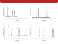
Figure 1 shows the analysis of the Alberta peptide mixture at normal working strength, as recommended by the supplier ("normal") and at 10Ã dilution using an acetonitrile gradient with 0.02 M formic acid (pH 2.7), 0.02 M formic acid-ammonium formate (pH 3.3), 0.02 M phosphate (pH 2.7), and 0.0079 M trifluoroacetic acid (pH 2.3) as the mobile phase additive, using the Discovery C18 column. The Discovery phase has been shown previously to have low silanol activity (12). Trifluoroacetic acid was used at the same weight percentage as formic acid (0.09%), because this is typical of the concentration used in MS applications. Higher concentrations of trifluoroacetic acid could lead to more serious problems of signal suppression. The pH of the trifluoroacetic acid was slightly lower than for the other buffers (pH 2.3), although it was not adjusted upwards to avoid the introduction of extraneous components into the mobile phase. When using formic acid-based buffers or phosphate, the order of elution was P1, P2, P4, P3 rather than P1, P2, P3, P4 as indicated in the original development of this test (4). It is possible that with the older silica reversed-phase used (4), the contribution to retention of ionic interactions increases in line with the increased charge of the peptide. In this case, the ionic retention of P4 would be expected to be greatest, contributing to its greater overall retention. The contribution of ionic retention might be very small or even negligible for the very inert Discovery phase at low pH, causing a somewhat different order of elution, with P4 eluted before P3. However, when trifluoroacetic acid is used, it is possible that ion-pairing effects also increase with increase in the charge of the peptides. Thus, the retention of P4 is increased more than that of the other peptides, and the order of elution is again P1, P2, P3, P4. With 0.02 M phosphate, there was some overlap between P3 and P4, necessitating separate injection of the single peptides to obtain peak capacity data.

Figure 1: (a) Analysis of Alberta peptides on Discovery C18 at normal working strength (approximately 1.3âÂÂ2.5 mg each peptide) and diluted 103 in mobile phase (dotted line); flow rate: 1 mL/min; solvent A: 0.9 g/L formic acid (0.02 M) in water (pH 2.7); solvent B: 0.9 g/L formic acid (0.02 M) in acetonitrile; gradient: 5%B to 42.5% B in 30 min (1.25% acetonitrile/min). (b) As (a), but solvent A: 0.9 g/L (0.02 M) formic acid adjusted to pH 3.3 with concentrated ammonia solution; solvent B: 50:50 (v/v) acetonitrileâÂÂ0.04 M formic acid adjusted to pH 3.3; gradient 10%B to 85% B in 30 min (1.25% acetonitrile/min, overall formic acid concentration 0.02 M). (c) As (a), but solvent A: 0.02 M phosphate buffer in water (pH 2.7); solvent B: 50:50 (v/v) acetonitrileâÂÂ0.04 M phosphate buffer in water (pH 2.7); gradient 10%B to 85% B in 30 min (1.25% acetonitrile/min.). (d) As (a), but solvent A: 0.9 g/L trifluoroacetic acid (0.0079 M) in water (pH 2.3); solvent B 0.9 g/L trifluoroacetic acid in acetonitrile.
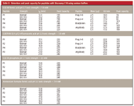
The effect of "normal" load (1.3-2.5 μg each Alberta peptide) on peak shapes for the various buffers can be observed in Figure 1. Whereas the chromatograms using phosphate (Figure 1c) or trifluoroacetic acid (Figure 1d) show little visual evidence of overloading, those with formic acid (Figure 1a) at high mass show considerably broader peaks at somewhat reduced retention time compared with the dilute solution (8). These peaks tend to a right-angle shape characteristic of overload, but there is little evidence shown of exponential tailing, which would instead be indicative of kinetic effects caused by ionic interactions with ionized silanols. However, adjustment of the pH of the formic acid mobile phase to pH 3.3 with ammonia (Figure 1b) appears to reduce the effects of overloading, with much more symmetrical peaks obtained for the normal strength peptide mix. Table II shows the peak capacities for the Alberta peptides on Discovery, giving quantitative evaluations that confirm the results visualized in the figures. Highly symmetrical peaks for each of the diluted Alberta peptides were obtained using 0.02 M phosphate buffer with asymmetry factors of only 1.1-1.2 and narrow peak widths. The asymmetry factor measured from gradient elution probably has even less theoretical significance than in isocratic separations but is still a simple and useful indicator of peak shape problems. Table II confirms that using phosphate buffer, only small differences were obtained in peak capacity for the "strong" and diluted Alberta peptides. For example, peak capacity is 248 for dilute P3 and 232 for strong (normal) P3. Use of trifluoroacetic acid gave only slightly inferior results, with peak capacities 242 and 216 for dilute and strong solutions of P3, respectively. However, major differences are shown for the formic acid mobile phase, with peak capacity for dilute and strong samples 223 and 131, respectively; the latter figure is barely half that obtained with phosphate. Thus, although peak capacities are rather similar for small peptide load (248, 242, and 223 for phosphate, trifluoroacetic acid, and formic acid, respectively), they are considerably lower for increased peptide load when formic acid is used (peak capacities 232, 216, and 131, respectively). Nevertheless, as indicated by Figure 1, there is a marked improvement in peak shape for the strong peptide solution when the pH of the formic acid is adjusted to 3.3 with ammonia. Peak capacity for P3 improves from 131 to 193, which approaches the high values shown for trifluoroacetic acid and phosphate.
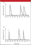
Chromatograms for the bradykinins on the Discovery column are shown for formic acid and trifluoroacetic acid in Figure 2, and the peak capacities are tabulated in Table II. The bradykinins have somewhat lower peak capacities (maximum 190) than the Alberta peptides (maximum 261) using the same mobile phase conditions. However, the trend of the results is very similar for the bradykinins. Using trifluoroacetic acid, Arg-Brady has peak capacity 190 and 163 for dilute and strong solutions, respectively, but 159 and 105 in formic acid, representing a more serious deterioration in peak shape with high sample mass when using formic acid.

Figure 2: (a) Overlaid chromatograms of 2.5 mg and 0.1 mg each bradykinins on Discovery C18; flow rate: 1 mL/min; mobile phase additive: formic acid (0.9 g/L). (b) As (a), but mobile phase additive: trifluoroacetic acid (0.9 g/L). Acetonitrile gradient as Figure 1. Peaks:1 5 bradykinin, 2 5 bradykinin fragment 1âÂÂ8, 3 5 3-Arg bradykinin.
The concentrations of each of the Alberta peptides in the mixture are of the same order. In other studies, we measured their individual gradient retention factors (k*), where

and F is the flow rate, δ%B is the gradient range expressed as the change in volume fraction of B, Vm is the column void volume, and tm is the gradient time (13). S was obtained from the variation of isocratic retention factor with acetonitrile concentration, using 0.02 M formic acid, from

where kw is the (hypothetical) retention of the solute in pure water, and Ï is the volume fraction of organic modifier in the mobile phase. Values of S were found to be much higher than those for small molecules (13). The average value for P1-P4 was 23, compared with about 4 for small molecules (8), confirming the large effect of modifier concentration on peptide retention. S and thus k* were found to be reasonably similar for each of the peptides (k* = 0.9-1.5). There is no reason, therefore, to expect very large differences in overloading behavior for the individual peptides in gradient elution with a given mobile phase, based upon their retention characteristics. However, Table II suggests that overloading effects are more serious for peptides with higher charge. For example, looking at results with formic acid, the Alberta peptides P3 and P4 (which have charge +3 and +4, respectively) show the greatest degree of overloading and the biggest falls in peak capacity for the strong compared with the dilute solutions. Alternatively, P1 and P2 (charges +1 and +2) show the smallest falls. These results are easier to see for the model Alberta peptides, because P1-P4 had similar peak widths under nonoverloaded conditions in the gradient run, which would be expected for a related group of substances (see Table II). The different bradykinins had different peak widths for small sample masses, which complicates the issue. Nevertheless, the peptide with the greatest charge (Arg-Bradykinin) showed the largest drop in peak capacity as sample mass was increased. These observations could be explained by greater mutual repulsion between more highly charged species held on the hydrophobic surface of the phase.

Table II: Retention and peak capacity for peptides with Discovery C18 using various buffers
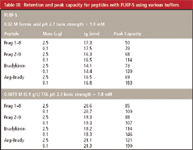
Table III shows comparative data for the bradykinins using formic acid or trifluoroacetic acid with the PLRP-S phase. If overloading on the Discovery column was taking place due to overloading of silanol groups, it would be reasonable to suppose that overloading effects might be completely different on this wholly polymeric column. We have shown that negatively charged sites might be present on such columns, possibly due to the presence of carboxyl functions on the phase (6). At a pH of 7, increasing the buffer cation concentration caused reduction in the retention of basic analytes, presumably through competition with solute cations. However, for the polymer column used in the present study at low pH, increase in the buffer cation concentration, through addition of potassium chloride, caused increases in peptide retention, probably through an ion-pair mechanism with chloride (14). This result suggests the absence of negatively charged sites on the PLRP-S phase at low pH. Care should be taken in making a detailed quantitative comparison between the PLRP-S and Discovery phases because the columns have different dimensions, different particle sizes, and are based upon different materials. Furthermore, we did not scale the gradient steepness to take into account different values of the gradient retention factor k* on these columns of different physical dimensions, which could influence overloading (8). In general, peak capacities on PLRP-S (Table III) are of the same order if slightly lower than those obtained with the silica-based phases, and confirm the suitability of the polymeric column for this analysis. Polymeric columns generally yield rather lower efficiency than silica-based columns. However, they should (in theory at least) give less stationary phase bleed in acidic mobile phases than silica-ODS phases, because they have no bonded ligands to hydrolyze - a potential advantage in HPLC-MS. They also have the advantage of greater pH stability over silica-based phases. Exactly as found previously for Discovery, the effects of overloading on PLRP-S are much more apparent with formic acid than for trifluoroacetic acid. For example, the peak capacity for high load of Arg-bradykinin is barely one third its value for low load when using 0.02 M formic acid (peak capacities 69 and 183, respectively). In comparison, this fraction increases to almost two-thirds of its low mass value using 0.0079 M trifluoroacetic acid (peak capacities 121 and 190, respectively). Figure 3 illustrates the increased overloading of bradykinins on PLRP-S when formic acid is used compared with trifluoroacetic acid, where a gradient slope of half that used in Table III was employed (0.625% acetonitrile/min instead of 1.25% /min). However, we have shown that gradient slope has a relatively minor influence on overloading for these peptides, with overloading effects increasing slightly as the gradient slope is reduced (14). Increasing the molar concentration of trifluoroacetic acid to 0.02 M (results not shown here; see [14]) to match that of the formic acid caused further decrease in the influence of overload. Peak capacity for high mass load of bradykinins was greater than three-quarters of that for low mass when 0.02 M trifluoroacetic acid was used. However, as stated earlier, such large concentrations of trifluoroacetic acid rarely are employed in HPLC-MS due to signal suppression effects.

Table III: Retention and peak capacity for peptides with PLRP-S using various buffers
The reason overloading is more problematic in formic acid than trifluoroacetic acid appears to be related to ion pairing and ionic strength effects. Mobile phases of higher ionic strength can physically screen protonated peptide cations from mutual electrostatic repulsion. A more important consideration, however, might be the formation of ion pairs between mobile phase anions and peptide cations. Those analyte ions that form "neutral" ion pairs should not undergo these mutual repulsion effects. The ion-pair properties of trifluoroacetic acid are well known and can be seen in increased peptide retention in this mobile phase (Figure 1, Table II). Note that an exactly analogous argument would hold if "dynamic ion exchange," occurred, with initial adsorption of the mobile phase anion on the stationary phase followed by interaction with the peptide cation, rather than a classic ion-pair mechanism (8). It appears also that ion pairs might form between analyte cations and inorganic anions such as chloride and perhaps even phosphate (7,13-16). Addition of potassium chloride to formic acid mobile phases was indeed shown to give improvements in column loading properties (7,13-14) for basic drugs and for peptides. Nevertheless, the addition of involatile salts to the mobile phase is hardly of practical use in applications involving MS. The adjustment of the pH of the formic acid mobile phase to 3.3 with ammonia produced a marked improvement in the column loading properties. Because formic acid is a weak acid, raising the pH in this way increases the ionic strength of the mobile phase (see Table II). Increased ionic strength gives increased opportunity for ion pairing; this effect is in addition to the physical screening of ions of the same charge. Both effects can reduce the effects of mutual repulsion and thus increase the loadability of the column. An indicator of this increased ion pairing is the increase in retention of the Alberta peptides in ammonium formate-formic acid compared with that in formic acid alone (Figure 1, Table II). Note that no exponential tailing was observed at this higher pH, which might instead have been indicative of the onset of silanol ionization. It is possible however, that competitive interactions of ammonium ions with ionized silanols could contribute to this result. The peak capacity even for dilute P3 improved from 223 to 238 and the gradient asymmetry factor from 1.74 to 1.25 using ammonium formate at higher pH indicating that silanol interactions are not a problematic factor in this particular case. Thus, use of ammonium formate buffers rather than formic acid alone can be useful in improving column loadability if scientists wish to avoid the use of trifluoroacetic acid. However, this is not likely to be true for older, more active "Type A" phases, or even all varieties of inert "Type B" phases, where silanol ionization could occur at higher pH. Clearly, some phases of even the latter group are more active than others (12).
Conclusion
Highly symmetrical peaks can be obtained when very small masses of peptides are analyzed on highly inert silica-ODS phases of standard bore using any of the buffers-additives studied here (phosphate, formic acid, or trifluoroacetic acid). However, serious deterioration in peak shape can occur as sample mass is increased. The progress of peak shape deterioration with sample mass is very similar for inert silica-ODS phases and purely polymeric phases, which contain no silanols. Thus, peak asymmetry does not seem to involve kinetic interactions between ionized silanols and peptide cations, at least on the highly inert ODS phase used in this investigation. Instead, poor peak shapes appear to involve overloading of the hydrophobic surface of the column (rather than overloading of silanols) caused by the mutual repulsion of analyte species of the same charge. A more complex situation might exist with older Type A silica phases, or even some more modern phases, where silanols can be ionized even at low pH.

Figure 3: (a) Overlaid chromatograms of 2.5 mg and 0.1 mg each bradykinins on PLRP-S; flow rate: 1 mL/min; mobile phase additive: formic acid (0.9 g/L). (b) As (a), but mobile phase additive: trifluoroacetic acid (0.9 g/L); acetonitrile gradient: 0.625%/min.
Overloading appears to be greatest for highly charged peptides, which should experience greater repulsion effects. Overloading effects are much more serious when formic acid is used as mobile phase additive rather than trifluoroacetic acid or phosphate (as favored in HPLC-UV analysis). However, loadability and peak shape is improved when using ammonium formate-formic acid buffers at somewhat higher pH, which is contra-indicative to the hypothesis that silanol ionization has begun to occur on the Discovery phase. In such a case, a reduction in performance would be expected as pH was increased. Mutual repulsion effects might be lessened due to greater ionic strength and ion-pair effects present in the higher pH buffer. Trifluoracetic acid can reduce the effect of overloading for the same reasons, particularly due to its greater ion-pair effect, even if used at lower molar concentration than formic acid (to limit MS suppression effects). At these lower concentrations, trifluoroacetic acid can give results approaching the quality of those with involatile phosphate buffers. Similar results were obtained both for a model set of related basic peptides (the Alberta peptide mixture) containing variable numbers of basic lysine residues and for a series of bradykinins containing variable numbers of basic arginine residues.
Finally, it can be noted that exactly the same arguments apply to capillary LC that apply to the standard bore columns used in this study. Injection volumes and sample masses must be decreased in proportion to the decreased surface area of these columns. We have shown that very similar overloading effects with formic acid occur for capillary columns with these peptides (13).
References
(1) R. Aebersold and M. Mann,
Nature
422
, 198-207 (2003).
(2) C.G. Huber and A . Premstaller, J. Chromatogr., A 849, 161-173 (1999).
(3) D. Corradini, C.G. Huber, A.M Timperio, and L. Zolla, J. Chromatogr., A 886, 111-121 (2000).
(4) C.R. Mant and R.S. Hodges, Chromatographia 24, 805-814 (1987).
(5) D.V. McCalley, J. Chromatogr., A 902, 311-321 (2000).
(6) S.M.C. Buckenmaier, D.V. McCalley, and M.R. Euerby, Anal. Chem.74, 4672-4681 (2002).
(7) D.V. McCalley, Anal. Chem.75, 3404-3410 (2003).
(8) L.R. Snyder, J.J. Kirkland, and J.L. Glajch, Practical HPLC Method Development (John Wiley & Sons, Hoboken, New Jersey, 1997).
(9) R.J. Beynon and J.S. Easterby, Buffer Solutions, The Basics (Oxford University Press, Oxford, UK 1996).
(10) J.D. Rawn, Biochemistry (Harper and Row, New York, New York, 1983).
(11) U.D. Neue and J.R. Mazzeo, J. Sep. Sci. 24, 921-929 (2001).
(12) D.V. McCalley, J. Chromatogr., A 844, 23-38 (1999).
(13) D.V. McCalley, J. Chromatogr., A 1038, 77-84 (2004).
(14) D.V. McCalley, J. Chromatogr., A (in press).
(15) F.Gritti and G. Guiochon, J. Chromatogr., A 1033, 57-69 (2004).
(16) F. Gritti and G. Guiochon, J. Chromatogr., A 1038, 53-66 (2004).
David V. McCalley Centre for Research in Biomedicine, University of the West of England, Frenchay, Bristol BS16 1QY, U.K., e-mail David.Mccalley@uwe.ac.uk.

Common Challenges in Nitrosamine Analysis: An LCGC International Peer Exchange
April 15th 2025A recent roundtable discussion featuring Aloka Srinivasan of Raaha, Mayank Bhanti of the United States Pharmacopeia (USP), and Amber Burch of Purisys discussed the challenges surrounding nitrosamine analysis in pharmaceuticals.
Extracting Estrogenic Hormones Using Rotating Disk and Modified Clays
April 14th 2025University of Caldas and University of Chile researchers extracted estrogenic hormones from wastewater samples using rotating disk sorption extraction. After extraction, the concentrated analytes were measured using liquid chromatography coupled with photodiode array detection (HPLC-PDA).
Silvia Radenkovic on Building Connections in the Scientific Community
April 11th 2025In the second part of our conversation with Silvia Radenkovic, she shares insights into her involvement in scientific organizations and offers advice for young scientists looking to engage more in scientific organizations.


