The Changing Face of LC–MS: From Experts to Users
Special Issues
Two decades ago, MS was the preserve of experts and skilled technicians as the instrumentation required constant attention and adjustment. At that time, liquid chromatography (LC)–MS was in its infancy and atmospheric pressure ionization (API) source interfacing was just beginning. Samples requiring analysis were passed from the requesting scientist to these "experts for analysis." The samples would be analyzed, processed, and interpreted, and the results returned via a written report. Two decades later, the users and capabilities of LC–MS have changed significantly. Now mass spectrometers and LC–MS systems are ubiquitous in the analytical laboratory, especially in the pharmaceutical industry. These instruments are used by a wide variety of scientists for a diverse range of tasks, from purity screening in medicinal chemistry, to the quantification of drugs in blood and the identification of proteins for biomarker discovery. The usability of the current MS platforms has improved..
Two decades ago, MS was the preserve of experts and skilled technicians as the instrumentation required constant attention and adjustment. At that time, liquid chromatography (LC)–MS was in its infancy and atmospheric pressure ionization (API) source interfacing was just beginning. Samples requiring analysis were passed from the requesting scientist to these "experts for analysis." The samples would be analyzed, processed, and interpreted, and the results returned via a written report. Two decades later, the users and capabilities of LC–MS have changed significantly. Now mass spectrometers and LC–MS systems are ubiquitous in the analytical laboratory, especially in the pharmaceutical industry. These instruments are used by a wide variety of scientists for a diverse range of tasks, from purity screening in medicinal chemistry, to the quantification of drugs in blood and the identification of proteins for biomarker discovery. The usability of the current MS platforms has improved dramatically, with scientists able to operate the systems remotely via the internet, carry out complex data-dependent tasks, such as purification and peptide fragmentation to open-access systems where nonanalytical chemists can queue their samples for analysis and have the results e-mailed to them without ever having to know or concern themselves about the LC–MS process.

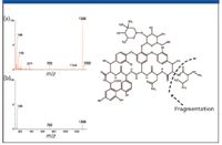
Figure 1: Daughter ion scans obtained with (top) and without (bottom) the ScanWave function.
Recent reports put the number of LC–MS systems sold per year in excess of 2500 units. This large number of units sold each year is also reflected in the increased number of users. In 1980, the number of scientists attending ASMS was around 1250. By 2002, this number had risen to greater than 4000, with a growth rate of 10% per year. This growth in LC–MS users has occurred because of the increase in the number of samples analyzed each year per user, creating larger and larger amounts of high-quality data. More and more, this data is being turned directly into information or knowledge so that decisions are made in real time. Many of these new users have little interest in becoming expert mass spectroscopists and are instead looking for the instrumentation itself to decide the appropriate experiments to be performed as well as to interpret the data automatically and recommend a course of action (for example, pass/fail, pure/impure).

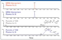
Figure 2: Mass spectra of alprazolam and a plasma sample.
The interfacing of LC with MS allowed analytical chemists access to about 80% of the chemical universe unreachable by gas chromatography (GC). It is also responsible for the phenomenal growth and interest in mass spectrometry in recent decades. A few individuals are singled out for coupling LC with MS. Beginning arguably in the 1970s, LC–MS as we know it today reached maturation in the early 1990s. Many of the devices and techniques we use today in practice are drawn directly from that time.
In its simplest form, LC relies upon the ability to predict and reproduce with great precision competing interactions between analytes in solution (the mobile or condensed phase) being passed over a bed of packed particles (the stationary phase). Development of columns packed with a variety of functional moieties in recent years and the solvent delivery systems able to precisely deliver the mobile phase has enabled LC to become the analytical backbone for many industries. Continued advances in performance since then, including development of smaller particles and greater selectivity, also saw the meaning of the acronym change to high-performance liquid chromatography. In 2004, further advances in instrumentation and column technology achieved significant increases in resolution, speed, and sensitivity in liquid chromatography. Columns packed with smaller particles (1.7 μm) and instrumentation with specialized capabilities designed to deliver the mobile phase at pressures up to 15,000 psi (1000 bar) came to be known as ultrahigh-pressure liquid chromatography (UHPLC). Much of what is embodied in this current technology was predicted by investigators such as Prof. John Knox in the 1970s.
Mass spectrometers can be smaller than a coin, or they can fill very large rooms. Although the various instrument types serve in vastly different applications, they nevertheless share certain operating fundamentals. The unit of measure has become the dalton (Da), displacing other terms such as amu. 1 Da = 1/12 of the mass of a single atom of the isotope of carbon-12 (12 C). Once employed strictly as qualitative devices — adjuncts in determining compound identity — mass spectrometers were once considered incapable of rigorous quantitation. But in more recent times, they have proved themselves as both qualitative and quantitative instruments. A mass spectrometer can measure the mass of a molecule only after it converts the molecule to a gas-phase ion. To do so, it imparts an electrical charge to molecules and converts the resultant flux of electrically charged ions into a proportional electrical current that a data system then reads. The data system converts the current to digital information, displaying it as a mass spectrum.
The ions required in MS can be created in a number of ways suited to the target analyte in question. These methods include laser ablation of a compound dissolved in a matrix on a planar surface, such as by matrix-assisted laser desorption ionization (MALDI); by interaction with an energized particle or electron, such as in electron ionization (EI); or as part of the transport process itself, as we have come to know electrospray ionization (ESI), where the eluent from a liquid chromatograph receives a high voltage, resulting in ions from an aerosol. The ions are separated, detected, and measured according to their mass-to-charge ratios (m/z). Relative ion current (signal) is plotted versus m/z, producing a mass spectrum. Small molecules typically exhibit only a single charge. The m/z is therefore some mass (m) over 1, with the "1" being a proton added in the ionization process (represented by M+H+ or M-H+ if formed by the loss of a proton), or if the ion is formed by loss of an electron, it is represented as the radical cation [M+.]. Larger molecules can capture charges in more than one location within their structure. Small peptides typically might have two charges (M+2H+), while very large molecules have numerous sites, allowing simple algorithms to deduce the mass of the ion represented in the spectrum.
The general term atmospheric pressure ionization includes the most notable technique, ESI, which itself provides the basis for various related techniques capable of creating ions at atmospheric pressure rather than in a vacuum. The sample is dissolved in a polar solvent (typically less volatile than that used with GC) and pumped through a stainless steel capillary, which carries between 500 and 4000 V. The liquid forms an aerosol as it exits the capillary at atmospheric pressure, and the desolvating droplets shed ions that flow into the mass spectrometer, induced by the combined effects of electrostatic attraction and vacuum. The mechanism by which potential transfers from the liquid to the analyte, creating ions, remains a topic of controversy. In 1968, Malcolm Dole first proposed the charge residue mechanism, in which he hypothesized that as a droplet evaporates, its charge remains unchanged. The droplet's surface tension, ultimately unable to oppose the repulsive forces from the imposed charge, explodes into many smaller droplets. These coulombic fissions occur until droplets containing a single analyte ion remain. As the solvent evaporates from the last droplet in the reduction series, a gas-phase ion forms. In 1976, Iribarne and Thomson proposed a different model, the ion evaporation mechanism, in which small droplets form by coulombic fission, similar to the way they form in Dole's model. It is possible that the two mechanisms might actually work in concert, with the charge residue mechanism dominant for masses higher than 3000 Da, and ion evaporation dominant for lower masses.
The mass analyzer is the heart of the instrument and is a means of separating or differentiating introduced ions. Both positive and negative ions (as well as uncharged, neutral species) form in the ion source. However, only one polarity is recorded at a given moment. Modern instruments can switch polarities in milliseconds, yielding high-fidelity records. As well as separating the ions, modern mass spectrometers can trap and fragment ions (MS-MS or MSn ) to produce a wealth of information about the molecule's structure. Other instruments, such as magnetic sector instruments, hybrid quadrupole–time of flight (Q-ToF), and ion cyclotron (ICR) mass spectrometers, can record the mass of a compound to 1 ppm, allowing for the elemental composition of a molecular ion or fragment ion to be deduced. The increased sensitivity afforded by modern MS over other forms of detection, such as UV and fluorescence, comes from the selectivity and specificity of the MS and MS-MS process. During these experiments, specific ions are allowed to pass through the analyzer and reach the detector. During a multiple reaction monitoring (MRM) MS-MS experiment, only ions that undergo a specific fragmentation are allowed to reach the detector. While this reduces the number of ions reaching the detector, it all but eliminates the noise, resulting in a superior signal-to-noise ratio (S/N). This improves assay sensitivity and specificity dramatically. The vast majority of quantitative experiments are performed on quadrupole-based instruments, whereas ion traps and accurate mass instruments are preferred for structural elucidation experiments.
Single-quadrupole mass spectrometers require a clean matrix to avoid the interference of unwanted ions, and they exhibit very good sensitivity. Triple quadrupoles, or tandem mass spectrometers (MS-MS), add to a single-quadrupole instrument an additional quadrupole, which can act in various ways. One way is simply to separate and detect the ions of interest in a complex mixture by the ions' unique m/z. Another way that an additional quadrupole proves useful is when used in conjunction with controlled fragmentation experiments. Such experiments involve colliding ions of interest with another molecule (typically a gas such as argon). In such an application, a precursor ion fragments into product ions, and the MS-MS instrument identifies the compound of interest by its unique constituent parts.
An ion-trap instrument operates on principles similar to those of a quadrupole instrument. Unlike the quadrupole instrument, however, which filters streaming ions, both the ion-trap and more capable ICR instrument store ions in a three-dimensional space. Before saturation occurs, the trap or cyclotron allows selected ions to be ejected, according to their masses, for detection. A series of experiments can be performed within the confines of the trap, fragmenting an ion of interest to better define the precursor by its fragments. Dynamic range is sometimes limited in ion-trap instruments, and the finite volume or capacity for ions limits the instrument's range, especially for samples in complex matrices.
Tandem quadrupole and ion-trap instruments have become the workhorses of modern analytical LC–MS(MS). The two capabilities have been incorporated into one instrument platform to produce a linear ion-trap instrument that had all the structural characterization benefits of ion-trap mass spectrometers with the quantitative capabilities of tandem MS instrumentation. These instruments have become popular with scientists who are required to perform more than one type of experiment (quantitative and qualitative) during the course of their work and require the flexibility to perform this on the same analytical platform. These tasks include impurity identification and quantification, and discovery DMPK, where both dosed parent concentration and metabolite characterization are required. Although they seem ideal, these instruments have some drawbacks, especially when using modern high-resolution chromatography. Here the chromatographic peak widths are so narrow (1–2 s) that there is not sufficient time for these ion trap mass spectrometers to select the ions for trapping, fragment the ions, and measure them to produce enough data points to define the peak accurately. Although the collection of MS-MS spectra can be performed with a standard tandem quadrupole MS instrument while still correctly defining the LC peak, the sensitivity is compromised due to the low duty cycle of the instrument.
Along with this need to improve the usage and flexibility of the instrumentation is a requirement to improve the data processing and monitoring capability. The recent introduction of a new iteration of the tandem quadrupole mass spectrometer, using a traveling wave technology (as described by Kircher in U.S. patent 5,206,506), holds the potential to resolve many of these issues. The Waters Xevo TQ mass spectrometer employs a traveling wave technology to improve MS capabilities, allowing simultaneous multifunctional data acquisition such as MRM and product ion acquisition, all within a timescale compatible with sub-2 micron LC. The instrument is equipped with a modern tool-free source, which simplifies the process of routine maintenance and cleaning; workflow is also simplified with tuning, method generation wizards, as well as real time data checking functionality to prevent sample wastage if the analytical run fails for any reason. The instrument's software also features an interactive LC-MS method database that ensures the correct method parameters are selected and used.
The mass spectrometer's T-Wave collision cell allows functionality such as rapid MRM switching, 20-ms positive ion–negative ion switching, and minimized cross-talk. This functionality makes the instrument ideal for rapid method development or use in a drug discovery environment for development of multicomponent assays.
In these drug discovery environments, mass spectrometers often serve a dual purpose, as both a quantitative and a qualitative instrument in DMPK departments. The mass spectrometer provides the flexibility to perform both of these tasks in the same analytical run, or even the same eluted peak. This functionality is called ScanWave and is achieved by maximizing the duty cycle of the instrument. In conventional mass spectrometers, the selected ions enter from the first quadrupole (Q1) into the collision cell, where they are fragmented. The resulting fragmented ions exit the collision cell and are transferred through the third quadrupole (Q3) to the detector. This quadrupole (Q3) can act either as a selective filter, as in MRM mode, only allowing ions with a specific m/z value to pass through to the detector, or it can scan across the entire m/z value range, providing a full spectrum. This full scan mode is particularly useful when performing structural analysis; unfortunately, in this mode, conventional instrumentation suffers from poor duty cycle. This is because the ions exit the collision cell simultaneously regardless of their m/z value. However, as the third quadrupole scans, it can only measure or detect one m/z value at a time. Therefore, for a scan speed of 1000 m/z per sec over a mass range of 1000 Da and a 2-s wide peak, the instrument will only spend 2 × 1/1000 s measuring each m/z value. This mass spectrometer's collision cell is designed to improve full scan sensitivity. In the last third of the collision cell, the fragmented ions are accumulated behind a DC barrier to effect ion enrichment. These ions are then released and contained between the DC barrier and an RF barrier at the end of the collision cell. The RF barrier is gradually reduced, ejecting the ions from the collision cell to the third resolving quadrupole. These ions are ejected according to their m/z ratio, with the heavier ions being ejected first. To improve the duty cycle of the instrumentation, the final quadrupole (Q3) is scanned in synchronization with the ejection of the ions from the collision cell, thus increasing the number of ions reaching the detector and hence the sensitivity.
This increased scan sensitivity can be used to address several business and scientific needs in pharmaceutical analysis. The acquisition of this high duty cycle scan can be triggered during a standard MS experiment to provide structural information on the identity of an LC peak. In the field of bioanalysis, the functionality can be employed to confirm identity of a peak (product ion confirmation, or PIC). This is carried out within an MRM analysis. PIC works by taking one high-quality, high-sensitivity spectra after the apex of a peak and before the "touch-down" of a chromatographic peak. This does not affect the fidelity or accuracy of the peak quantification but allows for the acquisition of a product ion spectra to confirm the identity of the peak.
In the disciplines of metabolite identification and impurity analysis, MS experiments such as constant neutral loss or common fragment ion analysis are often employed to detect ions that are related to the parent API molecule, or to look for particular metabolites that might be toxic, such as glutathione. Once the peaks of interested have been detected, a second analytical run is often required to obtain MS-MS structural information. The mass spectrometer can utilize its ability to perform either constant neutral loss or common fragment ion analysis and then rapidly switch to high-sensitivity MS-MS to obtain structural information. This capability removes the need for a second analytical run and hence improves productivity.
Recent regulatory guidelines on bioanalysis have placed greater emphasis upon the measurement of ion suppression and control of the matrix and measurement of drug-related metabolites. The mass spectrometer's functionality facilitates the rapid switching between matrix molecules and analytes of interest. Phospholipids are a class of endogenous molecules that have been associated with ion suppression. These molecules can be monitored by measuring the parents of the common fragment ion, m/z = 184, which is associated with the choline polar head group. The MS system allows the simultaneous MRM monitoring of the compounds of interest and precursors of m/z = 184, allowing rapid, reliable method development. This capability also can be employed to monitor the background ions during the course of a clinical trial to evaluate any differences between patients due to phenotype, gender, age, or diet. This provides extra confidence in the results and allows anomalies to be explained.
Conclusion
The usability and functionality of mass spectrometers has improved greatly over the last 15 years. Not only have they become more sensitive and capable of performing multiple experiments simultaneously, they have also become easier to use and have improved "up-time" and productivity. The advent of fast electronics and novel collision cell designs has allowed high-sensitivity full-scan MS-MS experiments to be performed at the same time as high-sensitivity MRM quantitative experiments. Concurrent with the increase in MS capability has been the move away from the expert MS scientist to the user tasked with answering a specific question, determining whether a product can be shipped, or monitoring food safety. The improved ease of use and intelligent software, which helps the general user achieve success in their task, will help push LC–MS even further into the general analytical community.
Acknowledgments
The authors would like to thank Paul Rainville and Marian Twohig for their scientific contributions.
Robert S. Plumb is Senior Applications Manager, Pharmaceutical Business Operations, Waters Corporation, Milford, Massachusetts.
Michael P. Balogh is Principal Scientist, MS Technology Department, Waters Corporation, Milford, Massachusetts.

New Study Reviews Chromatography Methods for Flavonoid Analysis
April 21st 2025Flavonoids are widely used metabolites that carry out various functions in different industries, such as food and cosmetics. Detecting, separating, and quantifying them in fruit species can be a complicated process.
Quantifying Terpenes in Hydrodistilled Cannabis sativa Essential Oil with GC-MS
April 21st 2025A recent study conducted at the University of Georgia, (Athens, Georgia) presented a validated method for quantifying 18 terpenes in Cannabis sativa essential oil, extracted via hydrodistillation. The method, utilizing gas chromatography–mass spectrometry (GC–MS) with selected ion monitoring (SIM), includes using internal standards (n-tridecane and octadecane) for accurate analysis, with key validation parameters—such as specificity, accuracy, precision, and detection limits—thoroughly assessed. LCGC International spoke to Noelle Joy of the University of Georgia, corresponding author of this paper discussing the method, about its creation and benefits it offers the analytical community.
Understanding FDA Recommendations for N-Nitrosamine Impurity Levels
April 17th 2025We spoke with Josh Hoerner, general manager of Purisys, which specializes in a small volume custom synthesis and specialized controlled substance manufacturing, to gain his perspective on FDA’s recommendations for acceptable intake limits for N-nitrosamine impurities.


