Strategies for the Detection and Elimination of Matrix Effects in Quantitative LC–MS Analysis
LCGC North America
We assessed a simple method based on recovery for the detection of matrix effects and two alternative methods for the rectification of matrix effects in LC–MS: standard addition and the coeluting internal standard method.
Currently available methods for the detection of matrix effects in liquid chromatography–mass spectrometry (LC–MS) are tedious and complex; therefore, a simpler method is required. Although there are no methods to completely eliminate matrix effects, the most well-recognized technique available to correct for matrix effects is that of internal standardization using stable isotope–labeled versions of the analytes. As this method can prove expensive, an alternative method of correction is likely to be useful. In this study, a simple method based on recovery is assessed for the detection of matrix effects. Two alternative methods for the rectification of matrix effects in LC–MS are also assessed: standard addition and the coeluting internal standard method.
High performance liquid chromatography (HPLC) coupled to mass spectrometry (MS) has become the predominant analytical method for the quantitative determination of analytes in biological matrices because of its high specificity, sensitivity, and throughput (1–3). However, matrix effects have become a major concern in quantitative liquid chromatography–mass spectrometry (LC–MS) because they detrimentally affect the accuracy, reproducibility, and sensitivity (3). Matrix effects occur when compounds that are coeluted with the analyte interfere with the ionization process in the MS detector, thereby causing ionization suppression or enhancement (2–7). Compounds with high mass, polarity, and basicity are possible candidates to cause matrix effects (4–8). However, the mechanisms involved in matrix effects have not been fully explored. One of the proposed theories to explain matrix effects is that the coelution of interfering compounds, especially basic compounds, may deprotonate and neutralize the analyte ions and, thus, reduce the formation of protonated analyte ions (2,4). Another theory postulates that less-volatile compounds may affect the efficiency of droplet formation and reduce the ability of charged droplets to convert into gas-phase ions (2–4,8). In addition, matrix effects may also be caused by high viscosity interfering compounds that could possibly increase the surface tension of the charged droplets and reduce the efficiency of droplet evaporation (2,4,6).
Several methods have been proposed for the detection and assessment of matrix effects, including postextraction spike and postcolumn infusion methods. The postextraction spike method evaluates matrix effects by comparing the signal response of an analyte in neat mobile phase with the signal response of an equivalent amount of the analyte in the blank matrix sample spiked post-extraction. The difference in response determines the extent of matrix effect (2,3,9). The major drawback of this method is that for endogenous analytes such as metabolites (for example, creatinine) blank matrix (urine or plasma) is not available. The postcolumn infusion method assesses matrix effects qualitatively. A constant flow of analyte is infused into the HPLC eluent, followed by injection of the blank sample extract. A variation in signal response of the infused analyte caused by coeluted interfering compounds indicates ionization suppression or enhancement (3,10). By identifying the ionization suppression or enhancement regions of the chromatogram, analytical methods can be developed to eliminate matrix effects by preventing the elution of the analyte peak in regions where matrix effects occur. However, the process of postcolumn infusion is time consuming and requires additional hardware, and it is not appropriate for multianalyte samples. Considering these drawbacks of existing methods, we propose a simple, fast, and reliable method to detect matrix effects that can be applied to any analyte including endogenous compounds such as creatinine and to any matrix without requiring any additional hardware.
To obtain accurate and reliable LC–MS data, several methods have been suggested to reduce or eliminate matrix effects. Matrix effects can be reduced simply by injecting small amounts of samples or by diluting samples (11,12). However, this approach can only be feasible when the sensitivity of the assay is very high (12). Methods to reduce or eliminate matrix effects include optimizing sample preparation to remove interfering compounds from the samples (1,9,10,13), changing chromatographic parameters to avoid coelution of analytes and interfering compounds (4,14–17), and changing MS conditions to reduce the occurrence of matrix effects in the ion source. However, these methods are not without their limitations. Most of the sample cleanup methods fail to remove impurities that are similar to the analyte and, hence, likely to be coeluted with the analyte (11,18). Modifying chromatographic conditions can be time-consuming, and some of the additives used in the mobile phase to improve separation have been found to suppress the electrospray signal of the analytes (3,4,9,15). Furthermore, even when the sample is devoid of coeluted substances, trace impurities present in the mobile phase can significantly suppress the analyte peak (19).
It is clear from the above that matrix effects in LC–MS cannot be completely eliminated. Therefore, the only option available is the rectification of data to eliminate the matrix effects. Calibration techniques such as the external-matched standards method, the echo-peak technique, and the most commonly used approach, the internal standard method, have been developed to correct the data (15,18–21). However, these calibration techniques also have their drawbacks. For example, the matrix-matching technique requires many blank matrices and appropriate blank matrices are not always available for the preparation of external standards (9,11,18,23). It is also impossible to match the matrix of the calibration standards with each of the samples exactly, as each sample has coeluting, interfering compounds that are thereby exposed to a different extent of ionization suppression (18). Echo-peak does not compensate for matrix effects completely because both standard and analyte peaks are not eluted at the exact same retention time (11). The stable isotope–labeled internal standards (SIL-IS) approach is the best available option but it is expensive and standards are not always commercially available for the analyte of interest (4,9,23).
The standard addition method for correcting matrix effects is widely used in spectrophotometric analysis, especially in atomic spectroscopy (24–27). However, this method is less well documented with other analytical techniques and currently there is no record of its practical use in compensating matrix effects in LC–MS. Standard addition does not require a blank matrix and is therefore appropriate for compensating matrix effects for any analyte including endogenous metabolites in biological fluids (20,28,29). In this study, we investigated the possibility of using the method of standard addition in routine LC–MS analysis to compensate for matrix effects and to thereby obtain improved data. We also investigated the use of a coeluting structural analogue of the analyte as the internal standard as an alternative to the expensive and often unavailable stable isotope–labeled internal standard for correcting matrix effects. Although coeluting structural analogue compounds are used to extend the linear range of calibration curves (30), there have been no reports of their use in compensating matrix effects in routine LC–MS analysis. In addition, we report a simple and effective method for the detection as well as the correction of matrix effects in routine LC–MS. All studies were carried out using a creatinine assay applied to human urine samples.
Experimental
Instrumentation
Separation of compounds was achieved with an Agilent 1100LC binary pump and Agilent 1100 autosampler (Agilent Technologies). The HPLC column used was a 150 mm × 2.1 mm, 4-μm dp Cogent Diamond-Hydride 100A column (MicroSolv Technology). For detection and quantification, an API 3000 tandem mass spectrometer equipped with a turbo ion spray interface and the software program Analyst 1.5 (Applied Biosystems) was used.
Materials
Creatinine, creatinine-d3 (2-amino-1-[trideuteriomethyl]imidazolidin-4-one), and cimetidine (N"-cyano-N-methyl-N'-[2[(5-methyl-1H-imidazol-4-yl)methylthio]-ethyl]guanidine) were purchased from Sigma. HPLC-grade acetonitrile and deionized water from a Milli-Q water system (Millipore) were used for mobile-phase preparation. Human urine samples were obtained from volunteers.
Chromatographic Conditions
Separations were carried out at an ambient temperature of approximately 25 °C. The flow rate was 200 μL/min. The injection volume was 10 μL. Mobile-phase A consisted of deionized water containing 0.1% (v/v) formic acid, and mobile-phase B comprised 0.1% (w/v) formic acid in acetonitrile.
Detection of Matrix Effects and Standard Addition Method
Gradient elution was used for analyte separation. The mobile-phase composition was varied from 90% B to 50% B over the first 20 min, and then maintained at 50% B for 1 min, before returning to 90% B from 21 to 24 min. The percentage of B was maintained at 90% B for 10 min for re-equilibration before the next sample was injected. The total run time was 24 min.
Coeluting Internal Standard Method
An isocratic elution method was used for elution. The mobile-phase composition was 55% B over 10 min. The total run time was 10 min.
MS Conditions
Creatinine, creatinine-d3, and cimetidine were detected in a positive multiple reaction monitoring (MRM) mode in which the transitions of m/z 113.9 to 44.0, m/z 117.0 to 47.0, and m/z 252.8 to 95.1 were monitored, respectively. The MS and electrospray ionization parameters were optimized and the analyzer settings were as follows: ion spray voltage (IS) 5000 V; entrance potential (EP) 10 V; orifice/declustering potentials (DP) 26 V (creatinine), 36 V (creatinine-d3), and 36 V (cimetidine); ring/focusing potentials (FP) 140 V (creatinine), 250 V (creatinine-d3), and 140 V (cimetidine); collision energy (CE) 29 V (creatinine, creatinine-d3 and cimetidine); collision exit potential (CXP) 8 V (creatinine, creatinine-d3, and cimetidine). Curtain gas (CUR), nebulizer gas (NEB), and the collision gas (CAD) flows were kept at 12, 8, and 4, respectively (in arbitrary units used in the instrument). The temperature of the ion spray was 300 °C. A dwell time of 150 ms was used for all transitions. Resolution of both Q1 and Q3 were 1 amu.
Preparation of Urine Samples
Human urine samples were prepared by filtering a small amount of the urine through a 0.22-μm polytetrafluoroethylene (PTFE) filter (Millipore).
1000-Fold Dilution of Urine Samples
The filtered urine sample was diluted 10-fold by mixing 30 μL of filtered urine and 270 μL of deionized water. This was followed by 100-fold dilution by mixing 900 μL of acetonitrile, 10 μL of internal standard (0.05 mM creatinine-d3 or 0.05 mM cimetidine), 10 μL of the diluted filtered urine sample, and 80 μL of deionized water to a total volume of 1000 μL.
Preparation of Standard Solutions and Quantification
Stock solutions (1.00 mg/mL) of creatinine, deuterated creatinine, and cimetidine were prepared in deionized water and stored at -20 °C.
Detection of Matrix Effects Using Recovery
Standard solutions were prepared by mixing appropriate volumes of 1.0, 0.1, and 0.01 mM creatinine, 900 μL of acetonitrile and deionized water to a final volume of 1000 μL. The concentrations of standards were 0.0001, 0.0003, 0.0005, 0.001, 0.003, 0.006, 0.01, 0.015, and 0.02 mM of creatinine in 90% (v/v) acetonitrile. A calibration curve of peak area of creatinine versus creatinine concentration was obtained.
Creatinine was added to five filtered urine samples and then diluted 1000-fold to a final added creatinine concentration of 0.006 mM. The creatinine concentrations of spiked and unspiked urine were determined using the above calibration plot to calculate the assay recovery.
Comparison of the Stable Isotope–Labeled Internal Standard and Standard Addition Methods
Standard solutions for the SIL-IS method were prepared by mixing 10 μL of 0.05 mM creatinine-d3, 900 μL of acetonitrile, appropriate volumes of 1.0, 0.1, and 0.01 mM creatinine, and deionized water to make the final volume up to 1000 μL. The final concentration of creatinine-d3 in all standards was 0.0005 mM and the concentrations of creatinine were 0.0001, 0.0003, 0.0005, 0.001, 0.003, 0.006, 0.01, 0.015, and 0.02 mM in 90% (v/v) acetonitrile. A calibration curve of the ratio of the peak area of creatinine and the peak area of creatinine-d3 versus creatinine concentration was then plotted to determine the concentration of the above five aliquots of 1000-fold diluted urine.
For the standard addition method, two levels of additions were prepared. A 0.003 mM addition was prepared by mixing 10 μL of 10-fold diluted filtered urine, 30 μL of 0.1 mM creatinine, 900 μL of acetonitrile, and 60 μL of water to make the final volume up to 1000 μL. The 0.006 mM addition was prepared by mixing 10 μL of 10-fold diluted filtered urine, 60 μL of 0.1 mM creatinine, 900 μL of acetonitrile, and 30 μL of water to make the final volume up to 1000 μL. The zero addition was prepared in the same manner but water was added in place of the 0.1 mM creatinine solution. A calibration plot of peak area of creatinine versus the added creatinine concentration was plotted for each sample and the original concentration of creatinine in five samples were estimated by extrapolating the line of best fit to zero peak area.
A negative control experiment was also carried out by running five samples containing no internal standard and determining their concentration using the calibration curve prepared in the section titled "Detection of matrix effects using recovery" above.
Comparison of the Stable Isotope–Labeled Internal Standard and Coeluting Internal Standard Methods
Standard solutions for the SIL-IS method were prepared by mixing 10 μL of 0.05 mM creatinine-d3, 900 μL of acetonitrile, appropriate volumes of 1.0, 0.1, and 0.01 mM creatinine, and deionized water to make the final volume up to 1000 μL. The final concentration of creatinine-d3 in all standards was 0.0005 mM and the concentrations of creatinine were 0.0001, 0.0003, 0.0005, 0.001, 0.003, and 0.006 mM in 90% (v/v) acetonitrile.
Standard solutions for the coeluting internal standard method were prepared by using 0.05 mM cimetidine in place of creatinine-d3 and by following the procedure mentioned in the previous section to obtain similar final concentrations of creatinine and the internal standard. A calibration curve of the ratio of the peak area of creatinine and peak area of the internal standard versus creatinine concentration were then plotted for each internal standard to calculate the concentrations of creatinine in seven aliquots of 1000-fold diluted urine. A negative control experiment was run in the same manner as described in the previous section.
Results and Discussion
Detection of Matrix Effects
To eliminate matrix effects, they should be assessed during early analytical method development. In this paper, we propose a simple and reliable method, recovery value, to estimate the extent of matrix effects in biological matrices analysis. In the recovery value test, the original concentration of creatinine in each of the five urine samples was determined using a calibration plot of the peak area of creatinine versus creatinine concentration. Each of the urine samples was then spiked with a known concentration of standard, 0.006 mM creatinine, and the total creatinine concentrations of the spiked samples were calculated using the same calibration curve. The percent recovery was determined by using the following formula:

where C0 is the added known concentration of standard; C2 is the concentration of analyte in final solution after spiking with known concentration of standard; and C1 is the original concentration of analyte in initial solution.
When percent recovery is closer to 100 (within normally expected error of ±3 standard deviations [SD]), it can be deduced that there is minimal or no matrix effect. A value of percent recovery less than 100 indicates that ionization suppression is present and a value greater than 100 indicates ionization enhancement.
As presented in Table I, the mean percentage of recoveries for the five urine samples was 65.98%. This result shows that the matrix effects profoundly affected the urine samples and thus degraded the accuracy of the quantitative LC–MS analysis. A high percentage of matrix effects was anticipated because the sample cleanup for the urine samples involved only filtration and dilution in this experiment and there was no mechanism in place to compensate for the resulting matrix effects. Furthermore, the results of subsequent experiments described later in this paper confirm the presence of matrix effects. The use of the recovery value to detect matrix effects is simple and cost-effective since it requires only two LC–MS runs. Also, it is ideal for the determination of the concentration of analytes in biological fluids that have complex matrices since it does not require a blank matrix and only involves the addition of analytes into the same sample matrix. Thus, it is certainly an easier and simpler method when compared to other methods available such as post-column infusion and post-extraction spike method, as discussed above. Unlike post-column infusion, that assesses the matrix effects in a qualitative manner, the recovery value method provides quantitative data.
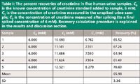
Table I: The percent recoveries of creatinine in five human urine samples. C0 is the known concentration of creatinine standard added to sample, 6 mM; C1 is the concentration of creatinine measured in the unspiked urine sample; C2 is the concentration of creatinine measured after spiking (to a final spiked concentration of 6 mM). Recovery calculation procedure is explained in the results and discussion section.
Comparison of Stable Isotope–Labeled Internal Standard and Standard Addition Methods
Because of its proven accuracy and reproducibility, the SIL-IS method was chosen as the reference (or positive control) method and was thus compared with other methods. In the comparison of SIL-IS and standard addition methods, the SIL-IS method was run and analyzed using the same chromatographic and MS conditions as that of the standard addition method. The calibration curve of the peak area ratio of creatinine to that of creatinine-d3 (SIL-IS) versus creatinine concentration was linear with an R2 value of 0.989 up to a concentration of 0.015 mM creatinine.
Although standard addition is a method that is widely used in atomic spectrophotometric analysis, it has not been so used in quantitative LC–MS analysis. In this study, we investigated the possibility of using standard addition to rectify matrix effects in LC–MS. Three runs were performed for each sample (as described in the experimental section), but there was no need for calibration standards; that is, each sample has its own calibration curve with three points. A calibration plot obtained for a typical sample in this study is shown in Figure 1. The calibration line was extrapolated to zero response to estimate the original concentration of creatinine in the urine sample.
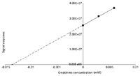
Figure 1: Standard addition method. A three-point calibration line is extrapolated to zero response to estimate the original concentration of creatinine present in the urine sample.
If the linear range of the calibration of analyte is already established, and the concentration of analyte in both the added and original samples fall within that range, an easier alternative approach for standard addition is to do only one addition and calculate the only unknown in the following equation, the original analyte concentration in the sample:

where [X]i is the original concentration of analyte in initial solution, [S] is the known concentration added, IX is the signal response from initial solution, and I(S+X) is the signal response from the final solution after the addition of a known concentration of standard.
Ellison and Thompson (31) highlighted that standard addition can only be precise and feasible when the analytical calibration curve is linear throughout the targeted concentration range. From our earlier experiments it has been demonstrated that the calibration curve for creatinine was linear to a concentration of 0.015 mM, and we were therefore able to estimate the original concentration of analyte in the samples by using equation 2.
Figure 2 illustrates the results of the comparison of methods: the mean concentrations of creatinine were 7.534 mM (negative control), 12.356 mM (positive control, SIL-IS), 13.424 mM (standard addition using extrapolated calibration curve), and 13.266 mM (standard addition using equation). As described in the experimental section, in the negative control experiment no method was used to compensate for matrix effects. The accuracies of the methods, shown as the percent error (deviation from the mean value of reference method, SIL-IS) were 39.0% (negative control), 8.64% (standard addition using extrapolative calibration curve), and 7.36% (standard addition using equation). The precision values of the methods, presented as relative standard deviations (using five replicates in each case) were 0.612 (negative control), 0.450 (positive control), 1.42 (standard addition using extrapolative calibration curve), and 1.083 (standard addition using equation).
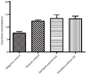
Figure 2: Comparison of approaches using negative control (external standard calibration), positive control (SIL-IS), standard addition (a) with extrapolated calibration line and standard addition (b) with equation. Detailed explanation in text.
The mean concentration values of creatinine obtained from using the standard addition method are very close to those from the SIL-IS method and therefore it may be considered as a potential alternative to the expensive and less versatile SIL-IS method. Standard addition involves the addition (spiking) of an analyte or a mixture of analytes into the sample and measuring the analyte concentrations before and after spiking. The spiking and subsequent analysis can be done once or twice depending on whether the equation or calibration curve option is used. There is no need for running many standards for the construction of calibration curves for each analyte. Thus, standard addition is especially suited for multicomponent analysis and when there are few samples to analyze. Furthermore, when the matrix is complex and the sample cleanup procedure is lengthy and matching matrix in calibration standards is impossible, standard addition provides a simple and effective alternative.
Although the SIL-IS method is generally regarded as the best option available for correcting matrix effects in LC–MS, a coeluted internal standard also suppresses the analyte signal and is therefore not the ideal option (9). Because the same concentration of SIL-IS is added to all standards and samples, in some samples the SIL-IS concentration may be significantly higher than that of the analyte, and therefore the extent of suppression by the IS may be significant. When considering this, the standard addition method can be considered as being closest to ideal since there is no additional suppression and the matrix has not been changed in the process of analysis. Other than the additional workload required (which may not be the case in multicomponent analysis) the method of standard addition can be considered as the optimal route to providing the most accurate data. Furthermore, it is a very practical and inexpensive option, because SIL-IS are not available for many analytes, and those that are available are very expensive.

Figure 3: Chemical structures of (a) creatinine and (b) cimetidine.
Comparison of Stable Isotope–Labeled Internal Standard and Coeluting Internal Standard Methods
A SIL-IS has physicochemical properties and will undergo ionization processes that are almost identical to those of the analyte of interest. Therefore, a SIL-IS is eluted at the same retention time as the analyte and experiences the same extent of matrix effects (30,32–34). However, SIL-IS is very expensive and not always commercially available (4). Structural analogue internal standards that have chemical structures, physicochemical properties, and retention times that are sufficiently close to SIL-IS may provide a cheaper and more readily available alternative to SIL-IS in compensating for matrix effects. Although structural analogues have been used in LC–MS (34) and were demonstrated to be capable of increasing the linearity of the method, both accuracy and precision were not improved as compared to the SIL-IS method. This was because the structural analogue used was not coeluted with the analyte of interest, which resulted in variable exposures to matrix effects in MS. A number of compounds such as hypoxanthine, xanthine, and quinine dihydrochloride monohydrate have been used as internal standards for creatinine analysis (35,36). The majority of such compounds are not eluted sufficiently close to the retention time of creatinine and, in addition, some of these compounds may be present in human body fluids and are therefore unsuitable as internal standards for urine samples.
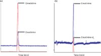
Figure 4: Chromatograms of (a) creatinine with coeluting internal standard, cimetidine (0.005 mM) and (b) creatinine with coeluted SIL-IS, creatinine-d3 (0.005 mM).
In this study, we used cimetidine as an internal standard (Figure 3) because it is a cheaper and more versatile alternative to SIL-IS (37), and it is not endogenously present in human urine. The chromatographic conditions were altered to a final composition of 55:45 acetonitrile–water in isocratic mode to produce coelution of creatinine and cimetidine and, hence, a similar matrix effect. As shown in Figure 4, the two peaks of creatinine and cimetidine overlapped to a very significant degree using these conditions. The coeluting internal standards method was then compared to the SIL-IS method, and the results are shown in Figure 5. The mean concentrations of creatinine for the negative control, SIL-IS, and coeluting internal standard methods were 9.68 mM, 19.18 mM, and 14.7 mM, respectively. The percent differences from the SIL-IS method (positive control) were 49.4% (negative control) and 23.3% (coeluting internal standard). The precision values of the methods, expressed in terms of relative standard deviation (using five replicates in each case) were 0.258 (negative control), 0.561 (reference), and 0.852 (coeluting internal standard). Although the coeluting internal standard method using cimetidine lacks accuracy when compared with the SIL-IS method, the results were still much improved when compared to the method without internal standard. It is clear that the use of compounds that merely are coeluted with the analyte of interest is insufficient for optimal compensation of matrix effects.
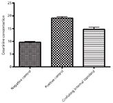
Figure 5: Comparison of approaches using negative control (external standard calibration), positive control (SIL-IS), and coeluting internal standard. The coeluting internal standard is cimetidine. Detailed explanation in text.
Conclusions
Matrix effects represent a major problem affecting the accuracy of LC–MS analysis. In this study, a simple method has been introduced for the qualitative and quantitative detection of matrix effects. This method is based on recovery calculations; if the responses are within a previously established linear range the same data can be used to quantify the analyte using the standard addition method. This means that the detection and correction of the matrix effects may be both achieved in a simple single-step process.
Two alternatives to the SIL-IS method, the standard addition method and the coeluting internal standard method, were evaluated for their capacity to correct for matrix effects. The standard addition method proved to be a viable alternative and is likely to be a better alternative in terms of accuracy and versatility. It could also be a superior method in terms of time and labor for multicomponent analysis using LC–MS such as pesticide analysis. However, the coeluting internal standard method as applied using cimetidine for the creatinine assay was found to be less effective in correcting for the matrix effect. A better structural analogue with similar chemical structure, physicochemical properties, and ionization properties may have produced better results. Because this approach was significantly better than the negative control, it could well be a good alternative for alleviating matrix effects if the appropriate compound is used as the coeluting internal standard.
References
(1) E. Chambers, D.M. Wagrowski-Diehl, Z. Lu, and J.R. Mazzeo, J. Chromatogr. B 852, 22 (2007).
(2) A. Van Eeckhaut, K. Lanckmans, S. Sarre, I. Smolders, and Y. Michotte, J. Chromatogr. B 877, 2198 (2009).
(3) P.J. Taylor, Clin. Biochem. 38, 328 (2005).
(4) D.A. Volmer, LCGC North Am. 24, 498 (2006).
(5) B.K. Matuszewski, M.L. Constanzer, and C.M. Chavez-Eng, Anal. Chem. 75, 3019 (2003).
(6) J.-P. Antignac, K. de Wasch, F. Monteau, H. De Brabander, F. Andre, and B. Le Bizec, Anal. Chim. Acta 529, 129 (2005).
(7) W.M.A. Niessen, P. Manini, and R. Andreoli, Mass Spectrom. Rev. 25, 881 (2006).
(8) T.M. Annesley, Clin. Chem. 49, 1041 (2003).
(9) H. Trufelli, P. Palma, G. Famiglini, and A. Cappiello, Mass Spectrom. Rev. 30, 491 (2011).
(10) R. Bonfiglio, R.C. King, T.V. Olah, and K. Merkle, Rapid Commun. Mass Spectrom. 13, 1175 (1999).
(11) H. Stahnke, S. Kittlaus, G. Kempe, and L. Alder, Anal. Chem. 84, 1474 (2011).
(12) C. Ferrer, A. Lozano, A. Agüera, A.J. Girón, and A.R. Fernández-Alba, J. Chromatogr. A 1218, 7634 (2011).
(13) C.R. Mallet, Z. Lu, and J.R. Mazzeo, Rapid Commun. Mass Spectrom. 18, 49 (2004).
(14) C. Cote, A. Bergeron, J.N. Mess, M. Furtado, and F. Garofolo, Bioanalysis 1, 1243 (2009).
(15) F. Gosetti, E. Mazzucco, D. Zampieri, and M.C. Gennaro, J. Chromatogr. A 1217, 3929 (2010).
(16) S. Bogialli, R. Curini, A. Di Corcia, A. Laganà, M. Nazzari, and M. Tonci, J. Chromatogr. A 1054, 351 (2004).
(17) R. Weaver and R.J. Riley, Rapid Commun. Mass Spectrom. 20, 2559 (2006).
(18) A.K. Hewavitharana, J. Chromatogr. A 1218, 359 (2011).
(19) H.M.D.R. Herath, P.N. Shaw, P. Cabot, and A.K. Hewavitharana, Rapid Commun. Mass Spectrom. 24, 1502 (2010).
(20) G. Ouyang and J. Pawliszyn, Anal. Chim. Acta 627, 184 (2008).
(21) L. Alder, S. Luderitz, K. Lindtner, and H.-J. Stan, J. Chromatogr. A 1058, 67 (2004).
(22) A.K. Hewavitharana, Crit. Rev. Anal. Chem. 39, 272 (2009).
(23) M. Stüber and T. Reemtsma, Anal. Bioanal. Chem. 378, 910 (2004).
(24) M. Bader, J. Chem. Educ. 57, 703 (1980).
(25) S. Altinöz and D. Tekeli, J. Pharm. Biomed. Anal. 24, 507 (2001).
(26) P. Koscielniak, J. Kozak, and M. Wieczorek, J. Anal. At. Spectrom. 26, 1387 (2011).
(27) P. Koscielniak, J. Kozak, M. Wieczorek, and M. Herman, Anal. Lett. 44, 411 (2011).
(28) P. Kosõcielniak and J. Kozak, Crit. Rev. Anal. Chem. 36, 27 (2006).
(29) G. Schumann, R. Klauke, and J. Büttner, Fresenius' J. Anal. Chem. 343, 89 (1992).
(30) O.A. Ismaiel, M.S. Halquist, M.Y. Elmamly, A. Shalaby, and H. Thomas Karnes, J. Chromatogr. B 875, 333 (2008).
(31) S.L.R. Ellison and M. Thompson, Analyst 133, 992 (2008).
(32) L.G. Freitas, C.W. Götz, M. Ruff, H.P. Singer, and S.R. Müller, J. Chromatogr. A 1028, 277 (2004).
(33) X. Zhao and C.D. Metcalfe, Anal. Chem. 80, 2010 (2008).
(34) K. Lanckmans, S. Sarre, I. Smolders, and Y. Michotte, Rapid Commun. Mass Spectrom. 21, 1187 (2007).
(35) Y. Zuo, C. Wang, J. Zhou, A. Sachdeva, and V.C. Ruelos, Anal. Sci. 24, 1589 (2008).
(36) S.-M. Huang and Y.-C. Huang, J. Chromatogr. B 429, 235 (1988).
(37) A.K. Hewavitharana and H.L. Bruce, J. Chromatogr. B 784, 275 (2003).
Amitha K. Hewavitharana, Swee K. Tan, and P. Nicholas Shaw are with the School of Pharmacy at The University of Queensland in Brisbane, Australia. Direct correspondence to: ahewavitharana@pharmacy.uq.edu.au

Common Challenges in Nitrosamine Analysis: An LCGC International Peer Exchange
April 15th 2025A recent roundtable discussion featuring Aloka Srinivasan of Raaha, Mayank Bhanti of the United States Pharmacopeia (USP), and Amber Burch of Purisys discussed the challenges surrounding nitrosamine analysis in pharmaceuticals.
Advances in Non-Targeted Analysis for PFAS in Environmental Matrices
March 27th 2025David Megson from Manchester Metropolitan University in Manchester, UK, spoke to LCGC International about the latest developments in non-targeted analysis (NTA) of per- and polyfluoroalkyl substances (PFAS) in environmental matrices based on a recent systematic review paper he has collaboratively published (1).


