Low-Resolution or High-Resolution MS for Clinical and Forensic Toxicology: Some Considerations from Two Real Cases
How can low-resolution mass spectrometry (LRMS) and high-resolution (HR) MS work together to provide unambiguous results in clinical and forensic toxicology?
The search for xenobiotics in analytical toxicology is based on different screening approaches depending on the context. Targeted analysis is preferred when some toxicants are suspected, but in some cases an untargeted analysis can be performed to detect compounds without preconceptions. Whatever the case, there is a strong need for certainty in the identification of compounds. In this context, the “gold standard” in analytical toxicology is liquid chromatography combined with mass spectrometry (LC–MS). Most of the time, toxicological laboratories are equipped with low-resolution mass spectrometers (LRMS). However, the principle of LRMS is that it can only discriminate compounds by their nominal mass, which can lead to false positives in the case of isobaric components. To solve this relative lack of specificity, high-resolution mass spectrometry (HRMS) is necessary to confirm (or deny) LRMS results.
Main Differences Between HR vs. LR
FIGURE 1: Resolution as the “full width at half maximum.”
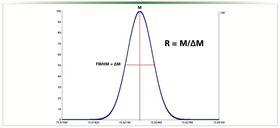
In mass spectrometry, resolution (R) can be defined by the full width at half maximum (FWHM, Figure 1). Mass spectrometers that achieve a resolution of at least 20,000 are called high-resolution mass spectrometers (HRMS). The main difference between HRMS and LRMS is the precision in the determination of the mass; the so-called “mass accuracy”. To confirm a compound identification in HRMS, error in the mass determination (Δm) has to be < 5 ppm (equation 1).

According to this, HRMS systems are known to be able to discriminate compounds by their accurate masses, contrary to LRMS systems, which can only reach a so-called “nominal mass” (± 1 Da) (1).
Roughly, the high selectivity of HRMS ensures certainty in the identification of precursors in MS1 and fragments in MS2, while LRMS cannot differentiate two different compounds when they are isobaric (same nominal masses) and/or share similar fragments and retention times (RT). However, a major drawback of HRMS is that it usually exhibits lower sensitivity and dynamic ranges (2). Consequently, LRMS and HRMS approaches can be seen as complementary tools for laboratories that have to perform screen analysis in multiple contexts. This article presents two real cases that were faced by our forensic unit and for which both LRMS and HRMS approaches were needed to obtain unambiguous results.
The Two Cases
The first analysis concerned a whole blood in a case of driving under the influence of drugs (DUID).
The second case concerned a whole blood analyzed in the context of drug‑facilitated sexual assault (DFSA). In the two cases, the objective of the HRMS analysis was to confirm the presence of compounds detected by LRMS.
Sample Preparation and Chromatographic Separation
In both cases, the following extraction procedure was applied: to 100 μL of whole blood, 200 μL of glacial acetonitrile (-20 °C) was added for sample deproteinization. Then, 40 mg of QuEChERS salts (4 g MgSO4/1 g NaCl/1 g sodium citrate dihydrate/0.5 sodium citrate sesquihydrate) supplied by UCT were added. The mixture was centrifugated and 30 μL of the upper phase was added to 90 μL of aqueous mobile phase and 5 μL was then injected.
For LRMS and HRMS approaches, the chromatographic system consisted of two Shimadzu LC-30 AD pumps (Nexera X2), a CTO 20 AC oven, and a SIL-30 AC-MP autosampler (Shimadzu Corporation). Chromatographic separation was performed on a 100 × 2.1 mm, 2.7‑μm Shim‑Pack Velox biphenyl column (Shimadzu Corporation), using a gradient of (A) ammonium formate 2 mM/0.002% formic acid buffer, and (B) methanol ammonium formate 2 mM/0.002% formic acid buffer as mobile phase at a flow-rate of 0.3 mL/min (0.6 mL/min 11–16.2 min to accelerate column conditioning and equilibration), as follows: 0.00–1.00 min, 5% (B); 1.00–2.00 min, 5 to 40% (B); 2.00–10.50 min, 40 to 100% (B); 10.50–11.00 min, 100% (B); 11.01–13.00 min, 100% (B); 13.00–13.01, 100 to 5% (B); 13.51–18 min, 5% (B). Oven temperature was set at 40 °C.
Mass Spectral Conditions for LRMS and HRMS
A fully validated and routinely employed method allowing the analysis of 48 illicit drugs was used. It was based on a multiple reaction monitoring (MRM) mode with two transitions per molecule. The LRMS system was a triple quadrupole LCMS-8050 (Shimadzu Corporation) (3).
The HRMS system was an LCMS‑9030 (quadrupole time-of-flight [QTOF]) (Shimadzu Corporation). A targeted acquisition mode was used (“MSMS mode”). Briefly, using this acquisition mode, the candidate precursor was searched using full-scan MS1 analysis, while its fragments were secondarily searched using MS2 acquisition after obtaining the exact mass of the precursor.
Case 1: Consumption of Illicit Drugs?
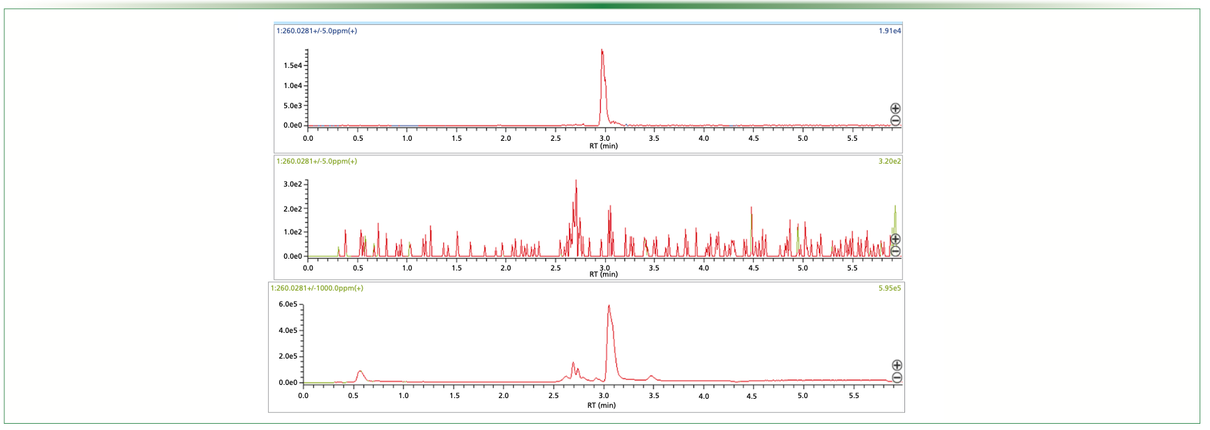
The LRMS targeted acquisition highlighted a signal corresponding to 2C-B, an amphetamine (Figure 2). The RT was consistent with that of our library, and the two transitions (260.10 > 243.05 and 260.10 > 228.10) were detected with similar ratios to those of the standard sample (± 20%). However, 2C-B consumption is not very common in France, and the expert in forensic toxicology asked for a confirmation of its presence in the whole blood of the DUID case by using HRMS (Figure 3). In a whole blood sample spiked at 100 ng/mL of 2C-B, a signal was obtained with a measured mass of 260.02759 m/z (exact mass of 2C-B = 260.0281 m/z; Δm < 2 ppm). When analyzing the whole blood of the driver, a precursor mass of 260.16391 m/z was obtained. Similarly, the fragments obtained in the patients were not those expected (243.13686 m/z vs. 243.0015 m/z; and 228.11419 m/z vs. 227.9780 m/z). The observed signals corresponded to a mass error > 500 ppm for the precursor and the fragments. As mentioned above, Δm < 5 ppm is mandatory to unambiguously identify a molecule. Consequently, the molecule detected by LRMS was not 2C-B.
FIGURE 2: LRMS screening results for 2C-B on the standard sample (upper chromatogram) and for the patient sample (bottom chromatogram).
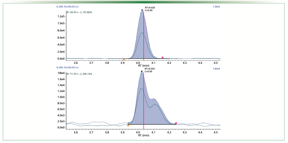
FIGURE 3: Search of the accurate mass of 2C-B on a full-scan MS1 with a tolerated mass error of 5 ppm. Signal obtained on the standard sample (upper chromatogram), and the patient sample (middle chromatogram). At the bottom, the signal obtained on the patient sample considering a mass error of 1000 ppm to the theoretical mass of 2C-B.
Case 2: Drug-Facilitated Sexual Assault?
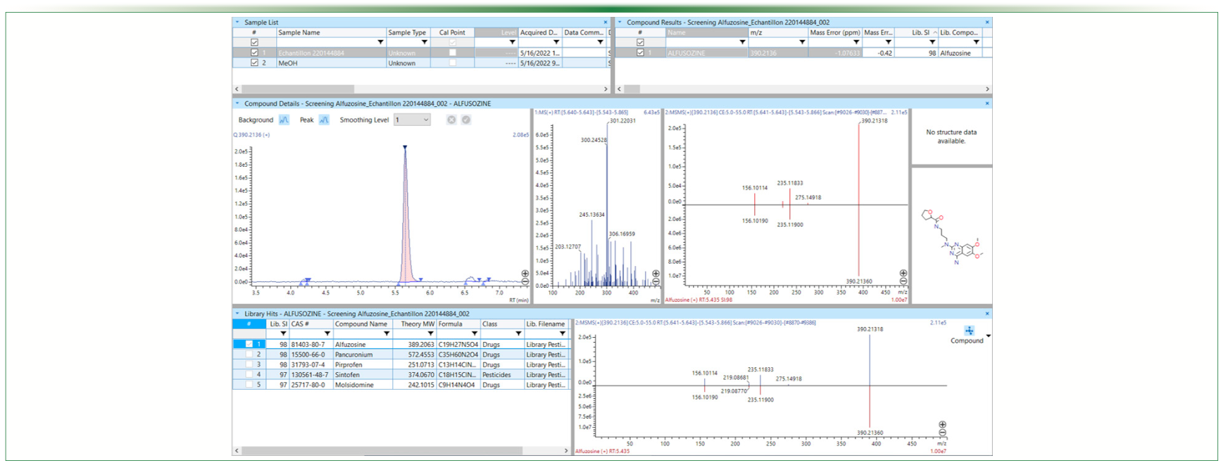
The LRMS screening procedure identified alfuzosin, an alpha-blocker used for functional symptoms of benign prostatic hyperplasia. As the presence of alfuzosin was hardly compatible in the case of a woman and was also not known as a compound employed in DFSA cases, the expert asked for a confirmation using HRMS. The analysis of the whole blood revealed a compound with a mass of 390.21291 m/z, which was favorably compared with the expected mass of alfuzosin (390.2136 m/z Δm < 2 ppm). In addition, fragments were also very close to those expected (235.11833 m/z vs. 235.11900 m/z and 156.10114 m/z vs. 156.10190 m/z). For the detected precursor ion and the fragment, Δm was < 5 ppm, which confirmed the presence of alfuzosin (Figure 4).
FIGURE 4: HRMS screening results for alfusozin in the “case-2” sample: chromatographic peak highlighting in full-scan MS1 and library comparison
Is There a Place for HRMS in Clinical or Forensic Toxicology?
These two real cases, observed in the routine activity of a forensic toxicology unit, illustrate the obvious added-value of using both LRMS and HRMS approaches in a laboratory. LRMS targeted or untargeted screening procedures ensure sensitivity, but these two cases highlighted situations where their specificity could be insufficient. In the first case, isobaric compounds with very similar precursor ions and fragments—which should have led to a false positive result without a secondary analysis by HRMS—were analyzed. In the second case, the detected compound was not consistent with the context and the expert refused to consider the molecule before confirmation by HRMS.
Depending on the laboratory, we believe that HRMS can be used either in combination with LRMS, or as a kind of all-in-one solution, taking into account the recent improvement in the sensitivity and dynamic ranges (4,5).
References
(1) Ojanperä, I.; Kolmonen, M.; Pelander, A. Current use of high-resolution mass spectrometry in drug screening relevant to clinical and forensic toxicology and doping control. Anal. Bioanal. Chem. 2012, 403 (5), 1203–1220. DOI: 10.1007/s00216-012-5726-z
(2) Kaufmann, A. The current role of high-resolution mass spectrometry in food analysis. Anal. Bioanal. Chem. 2012, 403 (5), 1233– 1249. DOI: 10.1007/s00216-011-5629-4
(3) Dulaurent, S.; El Balkhi, S.; Poncelet, L. et al. QuEChERS sample preparation prior to LC-MS/MS determination of opiates, amphetamines, and cocaine metabolites in whole blood. Anal. Bioanal. Chem. 2016, 408 (5), 1467–1474. DOI: 10.1007/s00216-015-9248-3
(4) Grund, B.; Marvin, L.; Rochat, B. Quantitative performance of a quadrupole-Orbitrap-MS in targeted LC–MS determinations of small molecules. J. Pharm. Biomed. Anal. 2016, 124, 48–56. DOI: 10.1016/j.jpba.2016.02.025
(5) Fedorova, G.; Randak, T.; Lindberg, R.H.; Grabic, R. Comparison of the quantitative performance of a Q-Exactive high-resolution mass spectrometer with that of a triple quadrupole tandem mass spectrometer for the analysis of illicit drugs in wastewater. Rapid Comm. Mass Sp. 2013, 27 (15), 1751–1762. DOI: 10.1002/rcm.6628
About the Authors
Elies Zarrouk, Souleiman El Balkhi, and Franck Saint-Marcoux are with the Unit of Biological and Forensic Toxicology, Department of Pharmacology and Toxicology, at Limoges University Hospital, in Limoges, France. Direct correspondence to: elies.zarrouk@unilim.fr

Advances in Non-Targeted Analysis for PFAS in Environmental Matrices
March 27th 2025David Megson from Manchester Metropolitan University in Manchester, UK, spoke to LCGC International about the latest developments in non-targeted analysis (NTA) of per- and polyfluoroalkyl substances (PFAS) in environmental matrices based on a recent systematic review paper he has collaboratively published (1).
Study Explores Thin-Film Extraction of Biogenic Amines via HPLC-MS/MS
March 27th 2025Scientists from Tabriz University and the University of Tabriz explored cellulose acetate-UiO-66-COOH as an affordable coating sorbent for thin film extraction of biogenic amines from cheese and alcohol-free beverages using HPLC-MS/MS.
Quantifying Microplastics in Meconium Samples Using Pyrolysis–GC-MS
March 26th 2025Using pyrolysis-gas chromatography and mass spectrometry, scientists from Fudan University and the Putuo District Center for Disease Control and Prevention detected and quantified microplastics in newborn stool samples.
Multi-Step Preparative LC–MS Workflow for Peptide Purification
March 21st 2025This article introduces a multi-step preparative purification workflow for synthetic peptides using liquid chromatography–mass spectrometry (LC–MS). The process involves optimizing separation conditions, scaling-up, fractionating, and confirming purity and recovery, using a single LC–MS system. High purity and recovery rates for synthetic peptides such as parathormone (PTH) are achieved. The method allows efficient purification and accurate confirmation of peptide synthesis and is suitable for handling complex preparative purification tasks.


