Host Cell Protein Analysis Using Mass Spectrometry
The Column
An increasing number of drugs coming onto the market are proteins rather than small molecules. A major portion of these are produced using a host cell system. Host cells express many of their own proteins that can easily contaminate the recombinant protein drug. Traditionally, these host cell proteins (HCPs) have been measured using immunoassays, but recently, orthogonal analytical methods, particularly mass spectrometry (MS), have started to be used. This article considers some of the current methods for HCP detection, with a focus on MS.
Photo Credit: TonsOfBackgrounds/Shutterstock.com

Sibylle Heidelberger and Kelli Jonakin, Sciex, Framingham, USA
An increasing number of drugs coming onto the market are proteins rather than small molecules. A major portion of these are produced using a host cell system. Host cells express many of their own proteins that can easily contaminate the recombinant protein drug. Traditionally, these host cell proteins (HCPs) have been measured using immunoassays, but recently, orthogonal analytical methods, particularly mass spectrometry (MS), have started to be used. This article considers some of the current methods for HCP detection, with a focus on MS.
Biological drugs have increased dramatically in number and frequency since the the first recombinant protein therapeutic-human insulin-was introduced over 30 years ago. The use of biological systems (that is, yeast or CHO cells) to synthesize complex protein drugs has been a remarkable success. However, during protein expression and purification, great attention must be devoted to analyzing and removing unwanted proteins that derive from the host cells (host cell proteins [HCPs]) (1).
The levels of these HCPs must be significantly reduced during protein drug manufacturing to ensure that these potentially antigenic impurities are eliminated or reduced to a level at which they will not elicit an immune response. Concerns also exist that a specific HCP could function as an adjuvant, creating an immune response against the product itself. Depending on the antibodies created, the response to HCPs could potentially vary from negligible to quite severe, including anaphylactic shock.
The presence of HCPs in protein drugs may determine whether the biopharmaceutical is accepted (or not) by regulatory agencies. The general guidelines are that there should be total amounts of less than 100 ng/mL and 10 ng/dose of these impurities, respectively (2). In 2008, the European Medicines Agency approved a recombinant form of human somatropin only after the manufacturer added additional purification steps for removal of the HCPs responsible for immunogenic response in patients (3).

Traditional Methods for HCP Detection
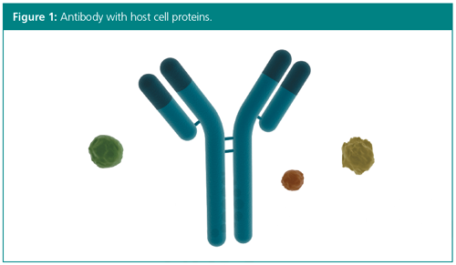
Conventionally, the most common method for the monitoring, detection, and measurement of HCPs during bioprocessing manufacturing has been immunoassays, such as enzyme-linked immunosorbent assays (ELISAs). This is a method based on polyclonal antibodies raised to the host cell (that is, CHO cells or yeast cells) used to synthesize a particular therapeutic product.
ELISA has been the workhorse method for HCP testing because of its high throughput, sensitivity, and selectivity. Typically, an ELISA is established using null host cell line isolates to immunize animals and generate polyclonal antibodies (4). However, the anti-HCP antibodies-which are the critical reagents for HCP ELISA-do not comprehensively recognize all the HCP species, therefore it is especially important to ensure that weak and nonâimmunoreactive HCPs are not overlooked by the ELISA. Moreover, even if the antibodies for a particular HCP are present in the reagent, the corresponding HCP may not be readily detected in the ELISA because of antibody–antigen-binding conditions and availability of HCP epitopes. Another drawback with immunoassays such as ELISA is that they may take up to eight months to develop and require the ethical housing of test animals (5).
Mass Spectrometry Methods for HCP Analysis
The analytical technology that is emerging as the major alternative technology to ELISA is mass spectrometry (MS) (6,7,8). The power of mass spectrometry is the ability to monitor and identify multiple protein analytes in the same sample rapidly and in a high throughput manner. In addition, MS is suitable for this type of analysis because it can detect very low amounts of HCPs, which is important because even very low quantities can adversely affect the drug product or provoke immunogenicity.
Mass spectrometry offers the opportunity to not only monitor and measure the host cell protein and product impurity profile, but also the ability to identify what is, and is not, present in any sample, including low abundance proteins. As such, liquid chromatography coupled with tandem mass spectrometry (LC–MS/MS) has now been applied to the rapid monitoring of HCPs, with or without combining it with HCP enrichment strategies.
However, until recently it was unclear whether available mass spectrometry solutions could provide the sensitivity and dynamic range necessary to detect and quantify trace HCP contaminants amongst an enormous excess of biotherapeutic protein, and at speeds to complete confident analyses in a reasonable time span. Methods are actively being developed, but to date, most have significant liabilities, such as very poor throughput (one sample per day). A commercially viable MS-based solution for HCP analysis must avoid biases, operate without needing to know the contaminant proteins prior to establishing the assay (for example, ELISA), and retain the beneficial aspects of speed, sensitivity, and consistency. A technique that meets these standards, and that is under development for HCP analysis right now, is quadrupole time-of-flight tandem MS combined with SWATH data analysis (9).
Quadrupole Time-of-Flight MS/MS
Typically, mass spectrometry instruments operate in “data dependent” mode, scanning for selected masses and performing MS/MS analysis on the most intense peaks appearing at time points. If the same sample is run multiple times, most of the proteins identified will be the same. However, there will be some variation in the fragmented peptides and host cell proteins identified because the analysis relies on the instrument responding to what it sees, and this cannot be done in a reproducible manner. The process must be repeated many, many times to obtain good statistical data. Alternatively, by using a triple quadrupole time-of-flight MS/MS system that combines fast acquisition speeds with high-resolution detectors, high-resolution quantifiable MS/MS data can be acquired for all detectable analytes in a complex sample, in a single run (10,11,12,13).
Combining mass spectrometry with a “data independent” SWATH analysis allows MS/MS analysis to be performed across small mass windows. A SWATH analysis will collect data for everything, which means that if a sample is run several times the data retrieved will always be the same. This ability to generate reproducible data is vital for drug development studies, and is one of the reasons that it is especially good for HCP analysis. A quadrupole timeâof-flight MS/MS instrument with SWATH acquisition will quantitate and confirm all HCP contaminants in one sample with a digital record of everything in the sample.
When using, for example, a triple quadrupole with multiple reaction monitoring (MRM), it is fundamentally important to know which HCPs to look for, but because it is impossible to predict the identity of contaminants, the use of targeted quantitation should be limited until after HCP profiles are determined. SWATH acquisition, however, captures all ions in a sample, providing information on every component, whether the peak is known or unknown. During SWATH analysis, all peaks within a given mass window undergo fragmentation at the same time, and all resulting fragments travel en masse to the detector to provide comprehensive and high-resolution mass information for the identification and quantitation of each peak.
Summary
HCPs constitute major parts of processârelated impurities and can adversely affect drug safety; therefore, it is critical that they can be identified and quantified accurately. The currently used ELISAs measure total HCP content, but completely lack information about the identity and quantity of individual HCPs in samples. With the improved product quality and time savings resulting from the simultaneous cataloguing and quantifying of all residual background proteins, mass spectrometry has the potential to be the future gold-standard for HCP analysis. The big advantage of SWATH data acquisition is its unbiased identification and quantification of proteins, leading to high reproducibility and sensitivity. Consequently, standardization of the methods to any given set of biotherapeutic samples becomes possible and will have a profound effect on the overall efficiency of biotherapeutic manufacturing.
References
- A.A. Shukla, C. Jiang, J. Ma, M. Rubacha, L. Flansburg, and S.S. Lee, Biotechnol. Prog. 24, 615–622 (2008).
- J.H. Chon and G. Zarbis-Papastoitsis, N. Biotechnol. 28, 458–463 (2011).
- European Medicines Agency http://www.ema.europa.eu/docs/en_GB/document_library/EPAR_-_Scientific_Discussion/human/000607/WC500043692.pdf
- A.L. Tscheliessnig, J. Konrath, R. Bates, and A. Jungbauer, Biotechnol. J.8, 655–670 (2013).
- J. Zhu-Shimoni, C. Yu, J. Nishihara, R.M. Wong, F. Gunawan, M. Lin, D. Krawitz, P. Liu, W. Sandoval, and M. Vanderlaan, Biotechnol. Bioeng.111(12), 2367–2379 (2014).
- C.E. Doneanu, A. Xenopoulos, K. Fadgen, J. Murphy, S.J. Skilton, H. Prentice, M. Stapels, and W. Chen, MAbs4(1), 24–44 (2012).
- G. Joucla, C. Le Sénéchal, M. Bégorre, B. Garbay, X. Santarelli, and C. Cabanne, J. Chromatogr. B Analyt. Technol. Biomed. Life Sci. 30, 942–943, 126–33 (2013).
- Q. Zhang, A.M. Goetze, H. Cui, J. Wylie, S. Trimble, A. Hewig, and G.C. Flynn, MAbs6(3), 659–670 (2014).
- K. Jonakin, https://sciex.com/community/blogs/blogs/host-cell-protein-contaminants-can-t-hide-from-swath-acquisition (2016).
- E. Betgovargez, B. Fonslow, and E. Johansen, 2015. “Ultra-sensitive host cell protein detection using CESI-MS with SWATH®Acquisition.” Poster, accessed April 2017.
- SCIEX Tech Notes: “Developing a routine LC-MS/MS platform for quantitative identification of host cell proteins (HCPs).” Publication no. 7460213-01 (2013). Accessed April 2017.
- J. Blethrow and E. Johansen, 2013. “High-sensitivity host cell protein quantitation in an IgG1 monoclonal antibody preparation via data-independent acquisition with a TripleTOF® System.” Webinar, accessed April 2017.
- E. Johansen and J. Blethrow, 2013b. “Rapid and unbiased quantitation of host cell protein contaminants in biologics by MS/MSALL using a TripleTOF® System Mass Spectrometer.” Webinar, accessed April 2017.
Sibylle Heidelberger is Technical Marketing Manager for Biologics at Sciex.
Kelli Jonakin is Global Marketing Manager for Pharma/CRO at Sciex.
E-mail: biologics@sciex.comWebsite:www.sciex.com

Accelerating Monoclonal Antibody Quality Control: The Role of LC–MS in Upstream Bioprocessing
This study highlights the promising potential of LC–MS as a powerful tool for mAb quality control within the context of upstream processing.
Common Challenges in Nitrosamine Analysis: An LCGC International Peer Exchange
April 15th 2025A recent roundtable discussion featuring Aloka Srinivasan of Raaha, Mayank Bhanti of the United States Pharmacopeia (USP), and Amber Burch of Purisys discussed the challenges surrounding nitrosamine analysis in pharmaceuticals.

.png&w=3840&q=75)

.png&w=3840&q=75)


.png&w=3840&q=75)


.png&w=3840&q=75)


