Advances in Chromatography for Charge Variant Profiling of Biopharmaceuticals
The Column
Biotherapeutic proteins, such as monoclonal antibodies (mAbs), are heterogeneous and exist as variant mixtures of structurally similar molecules. The heterogeneity of monoclonal antibodies is revealed by charge-sensitive methods, such as ion exchange chromatography (IEX). Changes in charge profile can significantly impact the structure, stability, binding affinity, and efficacy of the biotherapeutic drug. It is therefore necessary to understand the profile of the drug so that variants are identified and controlled. This article describes advances in ion exchange column chemistries, elution buffers, and ultrahigh-pressure liquid chromatography (UHPLC) instruments to meet the needs for modern, robust analysis of charge variants in monoclonal antibodies and therapeutic proteins.
Photo Credit: welcomia/Shutterstock.com

Kyle D’Silva, Ken Cook, and Simon Cubbon, Thermo Fisher Scientific, Hemel Hempstead, United Kingdom
Biotherapeutic proteins, such as monoclonal antibodies (mAbs), are heterogeneous and exist as variant mixtures of structurally similar molecules. The heterogeneity of monoclonal antibodies is revealed by charge-sensitive methods, such as ion exchange chromatography (IEX). Changes in charge profile can significantly impact the structure, stability, binding affinity, and efficacy of the biotherapeutic drug. It is therefore necessary to understand the profile of the drug so that variants are identified and controlled. This article describes advances in ion exchange column chemistries, elution buffers, and ultrahigh-pressure liquid chromatography (UHPLC) instruments to meet the needs for modern, robust analysis of charge variants in monoclonal antibodies and therapeutic proteins.
Therapeutic proteins, such as monoclonal antibodies (mAbs), are extremely complex, requiring a multitude of physicochemical tests to validate structure. Once the structure is initially validated, it must be continuously monitored from development through to production and commercialization to ensure safety and drug efficacy. Regulatory guidelines, such as ICH Q6B (1), focus on key structural and physicochemical analyses required for drug sponsors to perform to prove “fingerprint-like” structural similarity of their candidate molecule from clone selection, process development, and final drug product stability. Critical quality attributes (CQAs) of the molecule are those that impact clinical safety or efficacy. CQAs are controlled with tests that are designed to reduce lot-to-lot variability and include, but are not limited to:
- Charge variant profiling
- Aggregate analysis
- Glycan analysis
- Intact mass analysis
- Peptide mapping
Why is Protein Charge Heterogeneity So Important?
Charge variants can occur in therapeutic protein products for a number of reasons, including sequence variation, postâtranslational modification, and chemical degradation. The most common causes of charge variants are deamidation; isomerization; succinimide formation; oxidation; sialylation; N-terminal pyroglutamic acid formation; C-terminal lysine clipping; and aggregation. Depending on the nature of the modification, the resultant species can be more acidic or more basic than the main mAb monomer. As these changes can influence the stability and biological activity of the product, charge heterogeneity represents a challenge to demonstrating comparability.
For candidate innovator biologics, or biosimilars, any differences need to be identified quickly and reduced as early as possible. Process actions could involve further optimization of the cell culture conditions, or further formulation development.
A fast and confident charge variant profiling routine can result in acceleration of candidate development and support efficient ongoing control strategies.
Chromatography of Therapeutic Protein Charge Variants
Ion exchange chromatography (IEX) is the “gold standard” for the analysis of biomolecules, and remains one of the key technologies for charge variant analysis. IEX separates molecules based on the ionic groups on the protein surface, participating in electrostatic interaction with oppositely charged ionic group on the surface of the stationary phase:
- Anion exchange columns have a positively charged stationary phase surface and are operated above the isoelectric point (pI) of the protein.
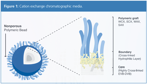
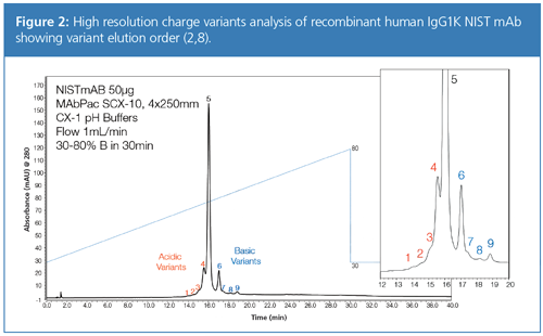
- Cation exchange columns have negatively charged groups grafted to a polymeric stationary phase (Figure 1). They are operated at pH values below the pI of protein. Cation exchange chromatography is typically used for the analysis of mAbs because of their high proportion of basic residues giving rise to basic surface chemistry. Acid variants are less retained by the stationary phase and elute first, followed by neutral then basic variants (Figure 2).
Many common chromatographic techniques, such as reverse phase chromatography, use organic solvents that denature, or unfold, the target protein, changing it from its true native state. An advantage of IEX is that the separation is performed under nondenaturing conditions. Elution is performed using either a salt or pH gradient to disturb electrostatic interactions between the protein and stationary phase.
Salt Versus pH
Historically, protein separation has been most commonly performed using salt gradients. A key challenge of salt-based gradients is that a unique gradient needs to be developed for each individual target protein molecule. Moreover, sensitivity of salt gradients to exact ionic concentrations, including the individual protein, meant that to achieve acceptable reproducibility, long columns and shallow saltâgradients were used. Consequently, method replication and robustness were often poor for fast methods, with method development being time-consuming and repeated for each candidate molecule.
In 2009, Genentech first published the use of pH gradients for charge variant separations instead of salt gradients (3). The advantages of pH-based gradients include global applicability of the method to any mAb, greatly simplifying method development. It was found that mAbs with isoelectric points in the range of around 7–9 could be well separated using cation exchange columns operated in a pHâbased elution mode. The approach was more generic and could easily be used to separate the different charge variants in a range of antibodies using a single method. The analysis time could also be significantly shorter using pH gradients (5 min to 20 min) rather than conventional ionic strength salt gradients (90 min).
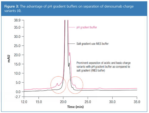
Comparison of the elution order for salt and pH gradient experiments have shown that the mAb variants typically elute in a similar order under both sets of conditions. However, pH gradients typically demonstrate enhanced resolution for mAb variants (Figure 3).
Improved mAb Separations with Generic Platform pH Gradient Methods
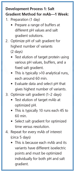
Method optimization for salt-based gradients typically followed the path outlined in Development Process 1. Additional effort is often required to tailor the salt gradient method for individual charge variants. Whilst a well optimized salt gradient can give excellent performance, peak capacity, and resolution, the time required to optimize and transfer methods drains resources. This is especially true for the contract testing organizations who may perform this analysis for multiple clients on dozens of similar mAbs.
This is because each mAb and its variants have different isoelectric points and must be optimized individually for both pH and salt gradient.
Patented pH gradient buffer solutions (5) can generate highly reproducible, linear pH gradients using cation exchange chromatography (Figure 2) (2). This generic LC-based platform approach saves time in method development and facilitates method transfer to QA/QC for a wide range of mAb charge variants. Unlike traditional salt gradients, it is possible to predict the pI from the retention time of the charge variants and use a narrow pH range to obtain high-resolution separations.
Two multi-component zwitterionic buffer concentrates are prepared using a patented formulation. Buffer A is titrated to pH 5.6 and buffer B is titrated to pH 10.2. Good buffers are used as the buffer components, which are zwitterionic and so neutrally charged in this pH range, therefore they will not be retained by the cation exchange column stationary phase.
For pH gradient buffer analysis, a linear gradient from pH 5.6 to pH 10.2 can be generated by running a linear pump gradient from 100% buffer A (pH 5.6) to 100% buffer B (pH 10.2) having general applicability for mAb charge variant analysis (6), and can be easily optimized to improve separation and deliver better charge variant separation than the salt gradient method (4) (Figure 3). The methods described here have already been demonstrated for use in the development of the biosimilars with comparison to the originators of several top-selling mAbs (6).
Ultrafast Separations
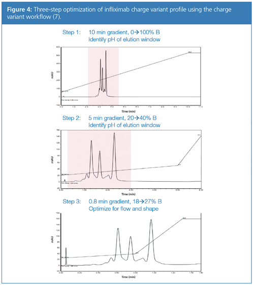
Optimization of methods using pH gradient buffers is fast, and is often performed in just a few hours. The combination of several new technologies into a workflow have enabled scientists to push the bar higher for fast, confident charge variant profiling (7).
The ultrafast charged variant separations shown (Figure 4) are achieved because of several advances in chromatography techniques. The mechanism of pH gradient chromatography lends itself to the use of shorter, faster columns with small particle sizes. The commercial buffer formulations (5) used here form a linear gradient, which allows intelligent optimization of the methods. Finally, UHPLC advances deliver extremely low delay volumes, high precision gradient formation, and a totally inert flow path, resulting in ultrafast separations with a total analysis cycle in the order of 2 min, including column reâequilibration and injection time, enabling users to run more than 1400 samples during 48 h continuous operation (7).
Processing the Data
Sometimes an afterthought, dealing with the data that has been acquired can be one of the biggest challenges for a company. The data obtained has an inherent value; after all, it is the knowledge that is obtained from the samples analyzed. In addition, charge variant analyses are undertaken in GxP compliant environments, requiring any resulting software to be compliance ready.
Ensuring that the knowledge gained can be turned into insights and the relevant actions taken requires any given software system to facilitate this process, from the data capture and processing (typically using a chromatography data system [CDS], which can acquire and process data from multiple vendor systems), through to long-term data storage, trending, and additional compliance tools, such as procedural electronic laboratory notebooks (ELNs), which are contained within laboratory information management systems (LIMS).
Efficiency comes from integrated informatics (connected CDS and LIMS) that allow users to easily interact with their data with minimal manual intervention, through the use of intelligent integration algorithms and ultimately software-driven workflows, guiding users through processes, and simplifying and maintaining data integrity and compliance, a key concern
for the industry.
Acknowledgements
The authors would like to thank Amy Farrell, Craig Jakes, Stefan Mittermayr, Silvia Millan Martin, and Jonathan Bones of the National Institute for Bioprocessing Research and Training (NIBRT), in Dublin, Ireland, for kind provision of some data and methods used in this article. In addition, thanks go to Shanhua Lin, Chris Pohl, Manish Singh, Sachin Pandey, and Mauro De Pra of Thermo Fisher Scientific for the applications and data featured.
References
- www.ich.org/products/guidelines/quality/article/quality-guidelines.html
- Thermo Scientific Apps Lab–3734–Charge Variant Analysis of NIST mAb: https://appslab.thermofisher.com/App/3734/charge-variant-analysis-nist-mab
- D. Farnan and G. Moreno, Anal. Chem.81(21), 8846–8857 (2009).
- Thermo Scientific Application Note 21565–The advantage of pH gradient buffers is demonstrated by the ion exchange separation of charge variants of denosumab: https://tools.thermofisher.com/content/sfs/brochures/AN-21565-LC-pH-Gradient-Buffers-Denosumab-AN21565-EN.pdf
- www.thermofisher.com/chargevariants
- Thermo Scientific Application Note 21092–High-Resolution Charge Variant Analysis for Top-Selling Monoclonal Antibody Therapeutics Using a Linear pH Gradient Separation Platform: https://tools.thermofisher.com/content/sfs/brochures/AN-21092-LC-CX-1-pH-Gradient-Buffer-Kit-mAb-AN21092-EN.pdf
- Thermo Scientific Technical Note 160–Development of Ultra-fast pH-Gradient Ion Exchange Chromatography for the Separation of Monoclonal Antibody Charge Variants: https://tools.thermofisher.com/content/sfs/brochures/TN-160-LC-pH-Gradient-Monoclonal-Antibody-TN71434-EN.pdf
- NIBRT–Webinar, April 2017–Charge Variant Analysis of Monoclonal Antibodies http://bit.ly/NIBRTChargeVariantWebinar
Kyle D’Silva is a Pharma and Biopharma Manager at Thermo Fisher Scientific.Ken Cook is a Biomolecule Separations Specialist at Thermo Fisher Scientific.
Simon Cubbon is a Pharma and Biopharma Manager at Thermo Fisher Scientific.
E-mail: kyle.dsilva@thermofisher.comWebsite: www.thermofisher.com/chargevariants
Accelerating Monoclonal Antibody Quality Control: The Role of LC–MS in Upstream Bioprocessing
This study highlights the promising potential of LC–MS as a powerful tool for mAb quality control within the context of upstream processing.
Common Challenges in Nitrosamine Analysis: An LCGC International Peer Exchange
April 15th 2025A recent roundtable discussion featuring Aloka Srinivasan of Raaha, Mayank Bhanti of the United States Pharmacopeia (USP), and Amber Burch of Purisys discussed the challenges surrounding nitrosamine analysis in pharmaceuticals.

.png&w=3840&q=75)

.png&w=3840&q=75)


.png&w=3840&q=75)


.png&w=3840&q=75)


