Glycosylation Analysis by Hydrophilic Interaction Chromatography (HILIC)–N-Glyco Mapping of The ZP-Domain of Murine TGFR-3
Glycosylation is one of the most common forms of post-translational modification of eukaryotic proteins. Glycosylated proteins (glycoproteins) make up the majority of human proteins.
Kai Darsow1, Sebastian Bartel1, Yves Muller2, and Harald Lange1, 1Chair of Bioprocess Engineering, University of Erlangen-Nuremberg, 2Chair of Biotechnics, University of Erlangen-Nuremberg, and Tosoh Bioscience LLC
Glycosylation is one of the most common forms of post-translational modification of eukaryotic proteins. Glycosylated proteins (glycoproteins) make up the majority of human proteins. The polysaccharide side chains (glycans) play critical roles in physiological and pathological reactions ranging from immunity to cell signaling, development, and death. Besides the interest of researchers in characterizing glycosylation patterns of glycoproteins for structure/function analysis, the thorough characterization of glycosylation is a major quality parameter in the production of biotherapeutics. Hydrophilic interaction chromatography (HILIC) is a well-recognized technique that effectively separates and quantifies isolated glycans.
HILIC
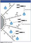
HILIC is used primarily for the separation of polar and hydrophilic compounds. It is commonly believed that in HILIC the aqueous content of the mobile phase creates a water rich layer on the surface of the stationary phase. This allows for partitioning of solutes between the more organic mobile phase and the aqueous layer. Hydrogen bonding and dipole-dipole interactions are supposed to be the dominating retention mechanisms in HILIC mode (see Figure 1 for a schematic representation).

Figure 1
Glycan Analysis
Glycoprotein analysis involves characterizing complex N- and O-linked structures composed of frequently similar and repeating sugar moieties. Several complementary analytical techniques are routinely used to characterize, identify, and quantify oligosaccharides isolated from glycoproteins. Besides mass spectrometric techniques, HILIC using amide-based stationary phases is a well established, robust technique used by many laboratories to obtain high-resolution separation of N-linked glycans released from glycoproteins. Tagging the glycans with a fluorescent label, such as 2-aminobenzamide (2-AB), allows the sugars to be detected at femtomole levels.
HILIC can separate structures with the same composition (isobaric glycoforms such as 2,3- or 2,6-sialic acid) on the basis of both sequence and linkage (1, 2). TSK-GEL Amide-80 column chemistry is ideally suited for the separation of charged and neutral fractions of glycan pools in a single run. The retention of fluorescence labeled (2-aminobenzamide, 2-AB) polysaccharides by a TSKgel Amide-80 column enables the identification of glycan structures by comparison to a labeled dextran ladder. The dextran ladder is used to normalize retention times in order to calculate the number of glucose units (GU values) of the separated glycans. The GU values obtained after separation of sequential exoglycosidase digests can be used to predict the glycan structure by database query (Glycobase, autoGU).

Figure 2
Transforming Growth Factor Receptor
Zona pellucida (ZP) is a glycoprotein matrix surrounding the plasma membrane of oocytes. Many eukaryotic extracellular proteins share a conserved sequence of 260 amino acids, called the ZP domain, which is an integral part of the ZP. The transforming growth factor beta (TGF-β) signaling pathway is involved in many cellular processes including cell growth, cell differentiation, apoptosis, cellular homeostasis, and other cellular functions. The cytokines of the TGF-β family exist in different subtypes (TGF-β1, TGF-β2, TGF-β3). The TGF-β type 3 receptor (TGFR-3) shows a high affinity to these three subtypes of TGF-β and other proteins of the TGF-β super family. Compared to the wide range of cellular processes which are regulated by the TGF signaling pathway, the function of TGFR-3 ZP domain glycans is not fully determined yet. In order to achieve a better understanding of transduction pathways, especially for fibrosis or neurodegenerative diseases, the role of TGFR-3 ZP glycosylation has to be determined (3).

Figure 3
Material & Methods
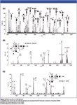
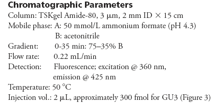
The application of HILIC for the characterization of a complex glycan structure is demonstrated using the example of N-glyco mapping of the ZP-domain of murine transforming growth factor beta type 3 receptor (TGFR-3). Figure 2 shows the protein construct of the ZP domain of murine TGFR-3 expressed in HEK293-EBNA cells which was used for determination of the N- and O-glycosylation of the TGF receptor. Recombinant proteins were purified and submitted to endoglycosidase cleavage. Glycans were fluorescent labeled with 2-aminobenzamide and separated by HILIC. Figure 3 shows the fluorescence chromatograms of HILIC separations of 2-AB labeled N-glycans released from the recombinant ZP domain construct of murine TGFR-3, compared to the dextran ladder (Figure 3A). The structure analysis was completed by high resolution mass spectra acquired on a MALDI QIT ToF MS instrument (Figure 4) (4).

Figure 4

Conclusion
Exoglycosidase sequencing in combination with HILIC separation on a TSKgel Amide-80 column and mass spectrometric analysis enables the determination of different N-glycan structures. Isobaric glycoforms have been identified by retention time (Glycobase) and MS-MS experiments.
References
(1) P. J. Domann, et al.; Proteomics, 1, 70–76 (2007).
(2) L. Royle, et al.; Analytical Biochem., 376, 1–12 (2008).
(3) S. Cheifetz, et al.; J. Biol. Chem., 263, 16984–16991 (1988).
(4) R.L. Martin, et al.; Mass Spectrom., 17, 1358–1365 (2003).

Tosoh Bioscience, LLC
3604 Horizon Drive, Suite 100, King of Prussia, PA 19406
tel. (484)805-1219, (800)366-4875; fax (610)272-3028
Website: www.tosohbioscience.com

Assessing Thorium-Peptide Interactions Using Hydrophilic Interaction Liquid Chromatography
February 4th 2025Paris-Saclay University scientists used hydrophilic interaction liquid chromatography (HILIC) coupled to electrospray ionization mass spectrometry (ESI-MS) and inductively coupled plasma mass spectrometry (ICP-MS) to assess thorium’s interaction with peptides.

.png&w=3840&q=75)

.png&w=3840&q=75)


.png&w=3840&q=75)


.png&w=3840&q=75)


