Characterizing Monoclonal Antibodies and Antibody–Drug Conjugates Using 2D-LC–MS
LCGC Europe
Two-dimensional liquid chromatography (2D-LC) has in recent years seen an enormous evolution, and with the introduction of commercial instrumentation, the technique is no longer considered a specialist tool. One of the fields where 2D-LC is being widely adopted is in the analysis of biopharmaceuticals, including monoclonal antibodies (mAbs) and antibody–drug conjugates (ADCs). These molecules come with a structural complexity that drives state-of-the-art chromatography and mass spectrometry (MS) to its limits. Using practical examples from the authors’ laboratory complemented with background literature, the possibilities of on-line 2D-LC for the characterization of mAbs and ADCs are presented and discussed.
Koen Sandra, Isabel Vandenheede, Mieke Steenbeke, Gerd Vanhoenacker, and Pat Sandra, Research Institute for Chromatography, Kortrijk, Belgium
Two-dimensional liquid chromatography (2D-LC) has in recent years seen an enormous evolution, and with the introduction of commercial instrumentation, the technique is no longer considered a specialist tool. One of the fields where 2D-LC is being widely adopted is in the analysis of biopharmaceuticals, including monoclonal antibodies (mAbs) and antibody–drug conjugates (ADCs). These molecules come with a structural complexity that drives state-of-the-art chromatography and mass spectrometry (MS) to its limits. Using practical examples from the authors’ laboratory complemented with background literature, the possibilities of on-line 2D-LC for the characterization of mAbs and ADCs are presented and discussed.
Monoclonal antibodies (mAbs) have emerged as important therapeutics for the treatment of life-threatening diseases, including cancer and autoimmune diseases (1,2,3). Today, more than 40 mAbs are marketed in the United States and Europe, of which 18 display blockbuster status; six have sales greater than $6 billion (Humira, Remicade, Enbrel, Rituxan, Avastin, and Herceptin) and over 50 are in late-stage clinical development. mAbs are currently considered to be the fastest growing class of therapeutics with sales more than doubled since 2008 (1,2,3).
The successes of mAbs have triggered the development of various next generation formats, such as antibody–drug conjugates (ADC), bispecific mAbs, mAb mixtures, polyclonal antibodies, antibody fragments (Fab, Fc, nanobodies), Fc fusion proteins, brain penetrant mAbs, and glyco-engineered mAbs. In oncology, ADCs are particularly promising because they synergistically combine a specific mAb linked to a biologically active cytotoxic drug using a stable linker (4–6). The promise of ADCs is that highly toxic drugs can be selectively delivered to tumour cells thereby substantially lowering side effects as typically experienced with classical chemotherapy. Two ADCs are currently marketed, namely brentuximab vedotin (Adcetris) and ado-trastuzumab emtansine (Kadcyla), and over 30 are in clinical trials (4–6).
With the top-selling mAbs evolving out of patent there has been a growing interest in the development of biosimilars (7). In 2013, we witnessed the European approval of the first two mAb biosimilars (Remsima and Inflectra), which both contain the same active substance, infliximab (8). In April 2016, Inflectra also reached marketing authorization in the US and a third infliximab biosimilar (Flixabi) was recently approved in Europe. Remicade, infliximab’s blockbuster originator, reached global sales of $8.9 billion in 2013 (3).
Structural Complexity
Together with a huge therapeutic potential, these products come with a structural complexity that drives state-of-the-art chromatography and mass spectrometry to its limits and this is exactly where twoâdimensional liquid chromatography (2D-LC) comes into play. mAbs are tetrameric immunoglobulin G (IgG) molecules with a molecular weight (MW) of 150 kDa (± 1300 amino acids) composed of two light (Lc – 25 kDa) and two heavy (Hc – 50 kDa) polypeptide chains connected through interchain disulphide bridges (Figure 1). Twelve intrachain disulphide bridges, four within each Hc and two within each Lc, furthermore guarantee its structural integrity. The structure can also be divided in the antigen binding fragment (Fab) and the crystallizable fragment (Fc). Antigen binding is mediated by the Fab fragment, while the Fc fragment is responsible for the effector function, that is, antibody dependent cell-mediated cytotoxicity (ADCC) and complement dependent cytotoxicity (CDC). All mAbs are glycoproteins and have two conserved N-glycosylation sites in the Fc region that can be occupied with complex and high mannose type N-glycans.
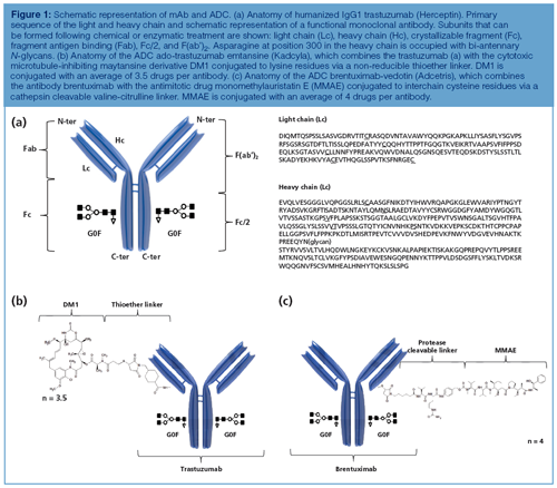
These glycan structures are known to play a role, amongst others, in the effector function and are the subject of extensive research towards the development of glyco-engineered mAbs with improved effector functions. A variety of other chemical and enzymatic modifications (wanted and unwanted) taking place during expression, purification, and longâterm storage further shape the mAb and give rise to a substantial heterogeneity. Modifications and variants often encountered include (but are not limited to) asparagine deamidation, aspartate isomerization, succinimide formation, N-terminal pyroglutamate formation, C-terminal lysine truncation, methionine or tryptophan oxidation, glycation, cysteine variants, and sequence variants (9). Even though only a single molecule is cloned, thousands of possible variant combinations may exist for one given mAb and they all contribute to the safety and efficacy of the product.
Compared to naked mAbs, ADCs further add to the complexity because the heterogeneity of the initial antibody is superimposed with the variability associated with the conjugation strategy. Conjugation typically takes place on the amino groups of lysine residues or on the sulphydryl groups of interchain cysteine residues as is the case in, respectively, ado-trastuzumab emtansine and brentuximab vedotin (Figure 1). With 80–100 lysine and only eight interchain cysteine residues available, lysine conjugation yields a more heterogeneous mixture of species compared to cysteine conjugation.
On top of the described primary structure, mAbs and ADCs also have distinct higher order structures dictating their function, which might be influenced by the above described modifications, and can appear as dimers or aggregates, which have the potential to induce immune responses.
Two-Dimensional Liquid Chromatography
An emerging tool to tackle this complexity is 2D-LC (10–13). In 2D-LC, two different chromatographic separation mechanisms are combined and peaks eluting from the first column are further separated on a second column with an orthogonal separation mechanism. The aim is to substantially increase the separation power for complex samples or to make the firstâdimension separation, in case it makes use of nonâvolatile salts, compatible with mass spectrometry (MS). Orthogonal behaviour is typically governed when combining the following chromatographic modes: affinity chromatography (Protein A), anion or cation exchange chromatography (AEX, CEX), hydrophilic interaction liquid chromatography (HILIC), hydrophobic interaction chromatography (HIC), reversedâphase liquid chromatography (LC), and sizeâexclusion chromatography (SEC).
Peaks can be transferred from one dimension to the other dimension in either an on-line or an off-line approach. Historically, off-line transfer has been the method of choice and is still widely applied. With robust, on-line 2D-LC instrumentation made commercially available in recent years, however, on-line 2D-LC should no longer be considered a specialist tool and is finding its way to mainstream laboratories. This article focuses on on-line 2D-LC.
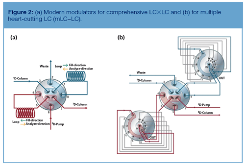
In on-line 2D-LC one makes use of a first-dimension pump, injector, column, and, optionally, a first-dimension detector, which is combined with a second pump, a second injector-now termed modulator, a second column, and a detector. The aim of the modulator is to efficiently transfer peaks from one dimension to the other without compromising the separation (Figure 2). In cases where the modulator transfers all first-dimension peaks to the second dimension, this is known as comprehensive 2D-LC or LC×LC. The modulator is composed of a valve with two loops installed (Figure 2[a]). While one loop is being filled with peaks from the first dimension, the second loop is being analyzed on the second dimension column. This second-dimension separation is typically very fast (in the order of 30 s) to facilitate a high sampling frequency and maintain the first-dimension separation. In cases where the modulator is programmed to transfer one or a couple of peaks from the first dimension to the second dimension, the method is termed (multiple) heartâcutting 2D-LC or (m)LC–LC. A modulator that allows one to store up to 12 first dimension fractions is pictured in Figure 2(b). Loops are then analyzed on the second dimension one by one and the first and second dimension run times are decoupled, which means that there is no requirement to perform fast separations. The following section will highlight how both modes are being applied in the analysis of mAbs and ADCs.
(Multiple) Heart-Cutting LC–LC
(m)LC–LC is typically applied for the analysis of intact mAbs or large fragments thereof (for example, Lc, Hc, Fab, Fc, F[ab’]2, Fc/2, Fd), which can be generated following chemical reduction or enzymatic digestion, with the aim of increasing resolution (for mAbs and ADCs, peaks typically contain many variants) and allowing an in-depth product characterization and to make separations compatible with MS (11,12).
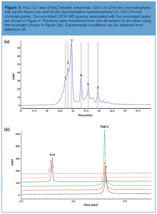
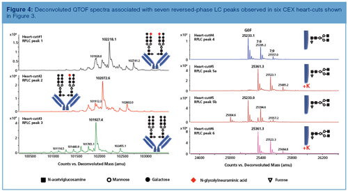
Birdsall et al. showed the potential of an on-line HIC–reversedâphase LC–UV–MS setup for the characterization of interchain cysteine conjugated ADCs (14). HIC separates the ADC based on the number of drugs conjugated for each antibody and as such allows the drug-to-antibody ratio (DAR) and drug distribution to be determined. HIC maintains mAbs in their native state, so it is especially well suited for interchain cysteine conjugated ADCs. The inherent problem of HIC is its incompatibility with MS detection as a result of the use of non-volatile salts. However, applying heart-cutting 2D-LC facilitates the combination of HIC and MS using a reversedâphase LC desalting step prior to MS. The denaturing reversed-phase conditions give rise to dissociation of the ADC into the respective subunits and as such this heart-cutting setup provides unambiguous identification of positional isomers. The downside of the described approach is that the setup only allows the second dimension reversed-phase LC–MS analysis of one heart-cut, even though the HIC chromatogram is heavily populated with peaks. Characterizing all peaks, therefore, requires several re-injections. This issue is alleviated in multiple heartâcutting LC–LC where the modulator design allows the storage of multiple first dimension fractions. Figures 3 and 4 show the mLC–LC analysis of cetuximab (Erbitux). Compared to other mAbs, which have one N-glycosylation site in the conserved region of the Hc, cetuximab contains an additional glycosylation site in the variable region of the heavy chain (15). Both the conserved and second N-glycosylation site are occupied with different types of sugars. While the former contains the typical mAb bi-antennary complex glycans G0F, G1F, and G2F, the latter site is occupied with a diverse mixture of glycans containing a large amount of α-(1γ3) linked galactose residues next to N-glycolylneuraminic (NGNA) acid terminating glycans. This unique glycosylation pattern stems from the fact that these antibodies are produced in murine SP2/0 opposed to CHO cell lines. In this mLC–LC example (Figures 3 and 4), IdeZ digested cetuximab was analyzed on the orthogonal combination CEX–reversed-phase LC hyphenated to UV and QTOF-MS detection. An immunoglobulin-degrading enzyme from Streptococcus equi ssp zooepidemicus (IdeZ) cleaves mAbs underneath the hinge region thereby generating a F(ab’)2 and Fc/2 fragment and as such separates out the two glycosylation sites. Six CEX heart-cuts were transferred to reversed-phase LC–MS. Chromatographic conditions are described in Sandra et al. (16). CEX peaks 1, 2, and 3 correspond to the 100 kDa F(ab’)2 fragment and from the corresponding MS data it is shown that these peaks differ in their NGNA degree with peak 1 carrying NGNA terminating N-glycans on both F(ab’)2 arms, peak 2 carrying one NGNA terminating glycan on one F(ab’)2 arm and a neutral glycan on the other F(ab’)2 arm, and peak 3 carrying neutral glycans on both arms. N-glycolylneuraminic acid renders the protein more acidic, which explains the earlier elution on CEX. Peaks 4, 5, and 6 correspond to the Fc/2 fragments and differ in C-terminal lysine (K) truncation with peak 6 carrying two lysine residues, peak 5 one, and peak 4 being fully truncated. Lysine is a basic amino acid explaining the increased retention time on CEX. Both Fc/2 fragments, which are no longer connected via interchain disulphide bridges, remain associated under the native CEX conditions. Denaturation, however, occurs during reversedâphase LC–MS analysis, which explains the detection of 25 kDa fragments using MS (Figure 4) and the double peaks (5a: Fc/2 + K and 5b: Fc/2 without K) observed on reversed-phase LC (heart-cut 5, Figure 3[b]).
An identical CEX–reversed-phase LC(–UV)–MS setup has recently been used to identify the main isoforms of the mAb rituximab (17) and to characterize the antibody–drug conjugate (ADC) ado-trastuzumab emtansine (16). Instead of storing first dimension fractions in loops, Alvarez et al. designed a setup that allowed six peaks of interest from a CEX or SEC separation to be analyzed and identified by trapping and desalting the fractions onto a series of reversed-phase cartridges with subsequent MS analysis (18). An on-line disulphide reduction step was furthermore incorporated into the workflow, allowing more detailed characterization of mAbs.
The examples described in this section offer a small taste of what can be achieved using (m)LC–LC. In various vendor application notes and non-peer-reviewed journals, more setups and applications can be found (19,20,21); for example, implementing Protein A followed by SEC, CEX, or reversed-phase LC–MS with the aim of selecting the optimal mAbâproducing clones based on various critical attributes, such as titer, charge variants, aggregation, molecular weight, and high level modifications (glycosylation).
LC–LC has not only been applied to characterize the drug substance (mAb or ADC). In a recent study by Li et al., SEC was coupled to reversed-phase LC in a heart-cutting 2D-LC setup for identification and quantification of unconjugated small molecule drugs and related small molecule impurities in ADC samples (22). These free drug species can increase the risk to patients and reduce the efficacy of the ADC. The SEC column in the first dimension provided information on the size variants (dimers, aggregates) of the ADC and separated the small molecular species from the ADC, which were then analyzed by reversed-phase LC. Both small molecule degradation products and aggregation of the ADC were observed in stability samples. The authors subsequently used the same setup to investigate the low recovery of the spiked linker drug in the free drug assay for an ADC (23). It could be concluded that the spiked linker drug reacts with residual reactive groups on the ADC. Birdsall et al. described an alternative approach based on mixed-mode solid-phase extraction (SPE) combined with reversed-phase LC and MS for the measurement of free drug species. The SPE step removes the ADC while it captures the free drug species for transfer to the second dimension. The authors claim that the method is two orders of magnitude more sensitive than UV-based methods (24).
LC–LC combining SEC with mixed-mode chromatography has been used to quantitate mAbs and excipients such as sucrose, Na+, K+, histidine, Cl-, succinate, and polysorbate 80 in drug products including mAbs, ADCs, and vaccines (25). Proteins were directed to a UV detector while excipients were monitored by evaporative light scattering detection (ELSD). Heart cutting 2D-LC with charged aerosol detection (CAD) and MS detection was also implemented to study the molecular heterogeneity and stability of polysorbate 20 in the presence of a mAb formulation sample matrix (26). A mixed-mode column was used in the first dimension to separate the polysorbate esters from the protein in the formulation sample, and the esters were further separated by a reversed-phase LC method in the second dimension to quantify the multiple ester subspecies. A second 2D-LC method using CEX in the first dimension and reversed-phase LC in the second dimension was used for the analysis of degradation products of polysorbate 20.
Comprehensive LC×LC
In biopharmaceutical analysis, comprehensive LC×LC has predominantly been used in peptide mapping studies for the detailed characterization and comparability assessment of innovator and biosimilar mAbs and to assess drug conjugation sites in ADCs (27–29). The use of the technology beyond R&D in a routine environment (QA or QC) has also been suggested (27).
Trypsin digestion of a monoclonal antibody will easily generate over 100 peptides with varying physicochemical properties all present in a wide dynamic concentration range. The complexity associated with these digests demands the best in terms of separation power. Compared to oneâdimensional separations (1DâLC), LC×LC will drastically increase peak capacity if the two dimensions are orthogonal and the separation obtained in the first dimension is maintained upon transfer to the second dimension. Orthogonal combinations for LC×LC-based peptide mapping are strong cation exchange (SCX) × reversed-phase LC, hydrophilic interaction liquid chromatography (HILIC) × reversedâphase LC, and reversedâphase LC × reversedâphase LC at different pHs in the two dimensions (27). Note that in the above combinations, reversed-phase LC is always used in the second dimension. The reasons for that are the commercial availability of highâquality, “fast” reversed-phase LC second dimension columns and their compatibility with MS. The highest orthogonality is obtained with SCX × reversed phase LC and HILIC × reversed phase LC because the separation mechanisms are completely different. However, reversed-phase LC × reversedâphase LC is the preferred approach because (i) it offers the highest ruggedness from the excellent solvent compatibility in both dimensions, (ii) the peak capacity is very high as a result of the high plate numbers in the individual dimensions, and (iii) orthogonality is acceptable as a result of the zwitterionic nature of peptides resulting in major selectivity differences when performing separations using reversed-phase LC at pH extremes (pH 2 versus pH 10).
Vanhoenacker et al. recently described the potential of reversedâphase LC × reversedâphase LC combined with UV and MS detection for the detailed characterization and comparability assessment of a trastuzumab innovator (Herceptin) and candidate biosimilar (27). A wealth of information was provided in the peptide map allowing identity, purity, and comparability to be assessed. Striking similarities were noticed when comparing the peptide maps, but when looking in more detail differences could be found and these were shown to reside in modifications (C-terminal lysine truncation, asparagine deamidation).
To further illustrate the attractiveness of LC×LC for detailed characterization and comparability assessment of mAbs, Figure 5 compares the reversed-phase LC × reversed-phase LC tryptic peptide maps of an infliximab originator and candidate biosimilar for which apparently, the recombinant expression in Chinese Hamster Ovary (CHO) cells was going wrong. Both plots are very similar but some striking differences are noted when the MS data is taken into consideration. The spots SLSLSPG and SLSLSPGK clearly present in the originator are replaced by one spot SLSLSPGI in the biosimilar, which according to the mass spectral data corresponds to the addition of an isoleucine (I) to SLSLSPG or the replacement of lysine (K) by isoleucine in SLSLSPGK at the C-termini of the heavy chain.
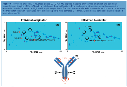
The origin of the two spots, SLSLSPG and SLSLSPGK, in the originator mAb can be explained by the knowledge that heavy chains are historically cloned with a C-terminal lysine but during cell culture production, host cell carboxypeptidases act on the antibody resulting in the partial removal of these lysine residues. A very small retention shift was also noted in another spot that could be attributed to a threonine to serine substitution in heavy chain peptide NYYGS(TγS)YDYWGQGTTLTVSSASTK. Two point mutations are at the origin of this wrong recombinant expression (Figure 5). These mutations were confirmed with reverse transcription polymerase chain reaction (RT-PCR) and DNA sequencing. According to US and European regulatory authorities, identical primary sequence is primordial to similarity, thereby ruling out this candidate biosimilar from further development.
Very recently, the possibilities of LC×LC-based peptide mapping in the characterization of ADCs have been demonstrated (16). The methodology was shown to be particularly valuable to assess drug conjugation sites. Figures 6(a) and 6(b) display the reversedâphase LC × reversedâphase LC–MS tryptic peptide maps of naked trastuzumab (Herceptin) and adoâtrastuzumab emtansine (Kadcyla). The latter combines trastuzumab with the cytotoxic microtubule-inhibiting maytansine derivative DM1 conjugated to lysine residues via a non-reducible thioether linker. Since Herceptin and Kadcyla have the same amino acid sequence, most of the peptide map is identical. Differentiating spots are, nevertheless, observed and are in the upper right corner of the plot of Kadcyla, which corresponds to the most hydrophobic part. Since the conjugation of the drugs makes the peptides more hydrophobic, these spots are expected to correspond to the conjugated peptides. The fragmentation spectra associated with DM1 conjugated peptides are heavily populated by fragments originating from the drug, for example, at m/z 547.2206. This ion can be used to recognize conjugated peptides in LC×LC–MS maps. One can operate the QTOF-MS system in the all ions MS/MS mode, which means that the quadrupole is operated in the RF only mode thereby transferring all peptides to the collision cell where collisionâinduced dissociation takes place.
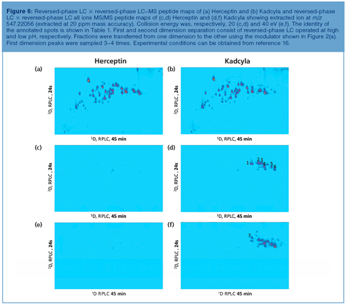
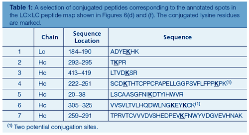
As illustrated in Figures 6(c–f), when extracting from the data the DM1 specific fragment ion at m/z 547.2206 at high mass accuracy, the upper right corner of the 2D peptide map lights up for Kadcyla in comparison to Herceptin, making this LC×LC methodology extremely powerful to highlight conjugated peptides. The latter plot is virtually empty illustrating the selectivity that is offered by this all ions MS/MS functionality. Figures 6(c) and (d) display the all ions MS/MS data obtained at a collision energy of 20 eV, Figures 6(e) and (f) the data at a higher collision energy of 40 eV. Complementary information is obtained when acquiring the data at two different collision energy values. A selection of identified conjugated peptides is shown in Table 1. Since the cytotoxic drug has a maximum UV absorbance at 252 nm, this wavelength can also be used to confirm the positioning of conjugated peptides in the peptide map as an alternative to the all ions MS/MS approach-evidently with a lower specificity.
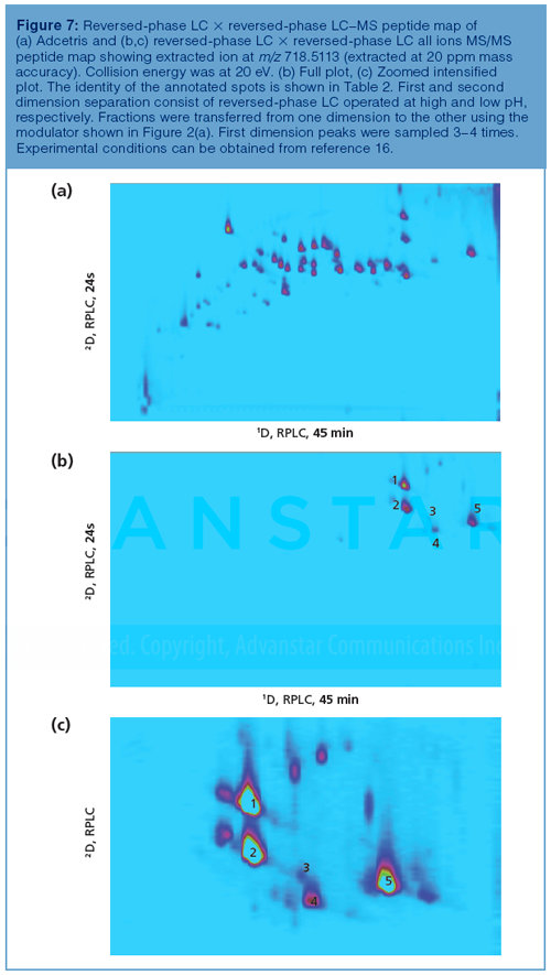
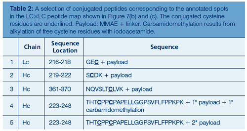
A similar strategy was applied to reveal the conjugation sites on brentuximab-vedotin (Adcetris), the interchain cysteine conjugated ADC. With only eight residues (four on each half antibody) available for conjugation, Adcetris is much simpler compared to Kadcyla. Figure 7(a) displays the reversedâphase LC × reversed-phase LC–MS tryptic peptide map of Adcetris. In analogy to DM1 conjugated peptides, MMAE conjugated peptides also contain specific ions originating from the cytotoxic molecule, that is, at m/z 718.5113. As shown in Figures 7(b) and (c), these ions can be extracted from all ions MS/MS data allowing conjugated peptides in the 2D plots to be recognized. A selection of identified conjugated peptides is shown in Table 2. Of particular interest is the detection of conjugated intrachain cysteine residues here exemplified with heavy chain peptide NQVSLTCLVK (peak 3 in Figure 7[c]). These intrachain residues are not targeted during ADC manufacturing. Apparently the TCEP reduction step applied to the mAb prior to conjugation results in collateral intrachain disulphide bridge reduction next to interchain reduction (the latter links are more labile). The resulting free thiol groups can then react with the protease cleavable linker-MMAE intermediate. These unwanted side products are only detected at trace levels and illustrate the sensitivity of the presented methodology.
It is clear that LC×LC holds great potential in peptide mapping. Recently, this 2D-LC mode has been used at the protein level to resolve intact mAbs or large fragments thereof. Sorensen et al. demonstrated the use of CEX × reversed-phase LC coupled to MS to compare mAb originators and biosimilars following IdeS digestion thereby generating F(ab’)2 and Fc/2 fragments and subsequent reduction generating Lc, Fd’, and Fc/2 (30). 2D-LC enabled direct identification of charge variants separated by the CEX column by indirect coupling of the separation to MS detection through the second dimension reversedâphase LC separation. The authors concluded that the richness of the data enables facile assessment of the degree of similarity between mAbs. In a series of papers, Sarrut et al. described the analysis of the interchain cysteine conjugated ADC brentuximabâvedotin using HIC × reversed-phase LC combined with UV and MS detection (31,32). This setup simultaneously allowed the DAR and the predominant positional isomers to be determined. The authors furthermore suggested the use of this methodology in quality control when operated in UV-only mode.
Acknowledgement
The authors acknowledge Sonja Schneider and Udo Huber (Agilent Technologies, Waldbronn, Germany), Maureen Joseph (Agilent Technologies, Wilmington, USA), and our colleagues from the biopharmaceutical industry.
References
- K. Sandra, I. Vandenheede, and P. Sandra, J. Chromatogr. A 1335, 81–103 (2014).
- S. Fekete, D. Guillarme, P. Sandra, and K. Sandra, Anal. Chem. 88, 480–507 (2016).
- G. Walsh, Nat. Biotechnol.32, 992–1000 (2014).
- S. Panowski, S. Bhakta, H. Raab, P. Polakis, and J.R. Junutula, mAbs6, 34–45 (2014).
- A. Wakankar, Y. Chen, Y. Gokarn, and F.S. Jacobson, mAbs3, 161–172 (2011).
- A. Beck and J.M. Reichert, mAbs6, 15–17 (2014).
- K. Sandra, I. Vandenheede, E. Dumont, and P. Sandra, “Advances in Biopharmaceutical Analysis” supplement to LCGC Europe, Volume 28, Number s10, October 2015.
- A. Beck and J.M. Reichert, mAbs5, 621–623 (2013).
- A. Beck, E. Wagner-Rousset, D. Ayoub, A. Van Dorsselaer, and S. SanglierâCianférani, Anal. Chem.85, 715–736 (2013).
- I. François, K. Sandra, and P. Sandra, Anal. Chim. Acta641, 14–31 (2009).
- K. Sandra and P. Sandra, Bioanalysis7, 2843–2847 (2015).
- D. Stoll, J. Danforth, K. Zhang, and A. Beck, J. Chromatogr. B 1032, 51–60 (2016).
- D. Stoll and P.W. Carr, Anal. Chem.89, 519–531 (2017).
- R.E. Birdsall, H. Shion, F.W. Kotch, A. Xu, T.J. Porter, and W. Chen, mAbs 7, 1036–1044 (2015).
- J. Qian, T. Liu, L. Yang, A. Daus, R. Crowley, and Q. Zhou, Anal. Biochem.364, 8–18 (2007).
- K. Sandra, G. Vanhoenacker, I. Vandenheede, M. Steenbeke, M. Joseph, and P. Sandra, J. Chromatogr. B1032, 119–130 (2016).
- D.R. Stoll, D.C. Harmes, J. Danforth, E. Wagner, D. Guillarme, S. Fekete, and A. Beck, Anal. Chem. 87, 8307–8315 (2015).
- M. Alvarez, G. Tremintin, J. Wang, M. Eng, Y.H. Kao, J. Jeong, V.T. Ling, and O.V. Borisov, Anal. Biochem.419, 17–25 (2011).
- S.M. McCarthy, T.E. Wheat, Y.Q. Yu, and J.R. Mazzeo, Waters Application Note; 720004304EN; (2012).
- S. Schneider, Agilent Technologies Application Note; 5991-6673EN; (2016).
- E. Largy, A. Catrain, G. Van Vyncht, and A. Delobel, Current Trends in Mass Spectroscopy 14(2), 29–35 (2016).
- Y. Li, C. Gu, J. Gruenhagen, K. Zhang, P. Yehl, N.P. Chetwyn, and C.D. Medley, J. Chromatogr. A1393, 81–88 (2015).
- Y. Li, C. Stella, L. Zheng, C. Bechtel, J. Gruenhagen, F. Jacobson, and C.D. Medley, J. Chromatogr. B1032, 112–118 (2016).
- R.E. Birdsall, S.M. McCarthy, M.C. JaninâBussat, M. Perez, J.F. Haeuw, W. Chen, and A. Beck, mAbs8, 306–317 (2016).
- Y. He, O.V. Friese, M.R. Schlittler, Q. Wang, X. Yang, L.A. Bass, and M.T. Jones, J. Chromatogr. A1262, 122–129 (2012).
- Y. Li, D. Hewitt, Y. Lentz, J. Ji, T. Zhang, and K. Zhang, Anal. Chem.86, 5150–5157 (2014).
- G. Vanhoenacker, I. Vandenheede, F. David, P. Sandra, and K. Sandra, Anal. Bioanal. Chem. 407, 355–366 (2015).
- G. Vanhoenacker, K. Sandra, I. Vandenheede, F. David, P. Sandra, U. Huber, and E. Naegele, Agilent Technologies Application Note; 5991â2880EN; (2013).
- G. Vanhoenacker, K. Sandra , I. Vandenheede, F. David, P. Sandra, and U. Huber, Agilent Technologies Application Note; 5991-4530EN; (2014).
- M. Sorensen, D.C. Harmes, D.R. Stoll, G.O. Staples, S. Fekete, D. Guillarme, and A. Beck, mAbs8, 1224–1234 (2016).
- M. Sarrut, A. Corgier, S. Fekete, D. Guillarme, D. Lascoux, M.C. JaninâBussat, A. Beck, and S. Heinisch, J. Chromatogr. B1032, 103–111 (2016).
- M. Sarrut, S. Fekete, M.C. Janin-Bussat, O. Colas, D. Guillarme, A. Beck, and S. Heinisch, J. Chromatogr. B1032, 91–102 (2016).
Koen Sandra is Scientific Director at the Research Institute for Chromatography (RIC, Kortrijk, Belgium).
Isabel Vandenheede is a Protein Analyst at RIC.
Mieke Steenbeke is LC Lab Technician at RIC.
Gerd Vanhoenacker is LC Product Manager at RIC.
Pat Sandra is President of the RIC and Emiritus Professor at Ghent University (Ghent, Belgium).

Regulatory Deadlines and Supply Chain Challenges Take Center Stage in Nitrosamine Discussion
April 10th 2025During an LCGC International peer exchange, Aloka Srinivasan, Mayank Bhanti, and Amber Burch discussed the regulatory deadlines and supply chain challenges that come with nitrosamine analysis.
Polysorbate Quantification and Degradation Analysis via LC and Charged Aerosol Detection
April 9th 2025Scientists from ThermoFisher Scientific published a review article in the Journal of Chromatography A that provided an overview of HPLC analysis using charged aerosol detection can help with polysorbate quantification.
Removing Double-Stranded RNA Impurities Using Chromatography
April 8th 2025Researchers from Agency for Science, Technology and Research in Singapore recently published a review article exploring how chromatography can be used to remove double-stranded RNA impurities during mRNA therapeutics production.


