Characterizing Biological Processes in Single Cells Using Capillary Electrophoresis
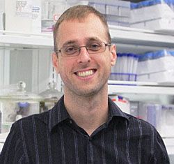
The characterization of transcripts, proteins, peptides, and metabolites in cells is important to study disease mechanisms and develop novel therapeutics. Peter Nemes - from the George Washington University, Washington, USA - spoke to The Column about the important role of capillary electrophoresis–electrospray ionization (CE–ESI) and time-of-flight mass spectrometry (TOF-MS) in this area of research.
Photo Credit: SCIEPRO/Getty Images

The characterization of transcripts, proteins, peptides, and metabolites in cells is important to study disease mechanisms and develop novel therapeutics. Peter Nemes - from the George Washington University, Washington, USA - spoke to The Column about the important role of capillary electrophoresis–electrospray ionization (CE–ESI) and time-of-flight mass spectrometry (TOF-MS) in this area of research.
Q. One strand of your research group’s work focuses on the development of analytical techniques to characterize biological processes that lead to disease in model organisms. Can you explain why this is an important area of research?
A: Our understanding of disease and the development of next-generation therapeutics requires a basic knowledge of the biomolecules that are produced in cells as well as interactions that exist between these molecules, but this hinges on our instrumental ability to characterize transcripts, proteins, peptides, and metabolites in cells. What is particularly needed are new instruments that can measure proteins, peptides, and metabolites in high sensitivity and deep information content to determine molecular processes in volume- and mass-limited samples such as single cells. The availability of this information will help better understand rare events underlying normal development and disease states.
Q. In 2013, you published a protocol outlining the investigation of single neurons by capillary electrophoresis–electrospray ionization and time-of-flight mass spectrometry (CE–ESI-TOF-MS).1 Can you talk about why you developed this method?
A: As a post-doctorate research associate working in Dr Jonathan Sweedler’s laboratory (University of Illinois-Urbana-Champaign, IL, USA), I had the opportunity to use my instrument developmental skills rooted in analytical chemistry to approach basic questions in neuroscience. One interesting aspect of the nervous system that Jonathan, my colleagues, and I have been interested in was whether (or how) neurons that carry out different physiological functions develop specific molecular contents depending on their neuron types. The metabolome, the collection of all the small molecules produced in a cell, was promising to help address this question because metabolites are sensitive to intrinsic and extrinsic events and dynamically reflect the state of the cell, potentially informing us of differences between neuron types. To measure metabolites in single neurons, I established microanalytical workflows and made technical improvements with my colleagues to use a capillary electrophoresis system that Theodore Lapainis, a then-graduate student, designed in Jonathan’s group in 2009, to measure metabolite production between a large number of single neurons. The data that resulted from these measurements allowed us to find reproducible metabolic differences between different neuron types and different levels of heterogeneity within neurons of the same type.
Q. What do analysts need to consider to ensure a successful experiment when using your developed protocol?
A: When measuring trace-level molecules, particularly in single cells, every step of the analytical workflow is critical. We have developed microscale approaches to isolate single cells with minimal damage to the cell, efficiently extract their molecules (metabolites and now proteins), treat these molecules if needed, and separate them to minimize chemical complexity, before analyzing metabolites, peptides, and proteins using our carefully characterized and validated single-cell CE–ESI-TOF-MS instrument. By systematically evaluating each step of the analytical workflow, we have been able to detect and quantify a large number of molecules between single neurons and embryonic cells.
Q. In a recent study, you applied capillary electrophoresis–electrospray ionization mass spectrometry (CE–ESI-MS) to analyze the metabolome of embryonic cells from the South African clawed frog (Xenopus laevis).2 What led you to begin this project?
A: The short answer is fantastic collaborators at GW (George Washington University, Washington DC, USA)! Shortly after joining GW as an Assistant Professor, I met Sally A. Moody and M. Chiara Manzini in the School of Medicine and Health Sciences who research neurodevelopmental and muscular diseases using the Xenopus laevis, zebrafish, and mouse models. Talking to Sally and Chiara, it did not take long for me to become fascinated by how the embryo, literally a ball of just a few cells, knows how to orchestrate a fully functioning organism and how this process can be disrupted by disease. I recognized that the single-cell mass spectrometry tools that I was developing in my laboratory just across the street from their laboratories could be used to determine spatial and temporal molecular processes that unfold in the developing embryo, raising an opportunity to better understand normal and diseased development.
Q. Why did you choose to perform CE–ESI–MS?
A: Having developed single-cell CE–ESI-MS platforms and protocols, I knew that this technology had the analytical metrics I needed to address our basic questions surrounding normal and diseased development of the embryo; CE–ESI-MS is sensitive, specific, label-free, and is able to quantify molecules. Of course, because embryos and embryonic cells are very different to mature neurons, we developed specialized ways to microsample single embryonic cells in the developing embryo, extract small and large molecules (metabolites to proteins) from these single cells, efficiently separate the molecules, and then to identify and quantify them using high-resolution tandem MS. After completing this analytical and instrument development task, we were able to design experiments to answer basic questions in embryonic development.
Q. What were your key findings? What are you working on next?
A: Our single-cell CE–ESI-MS platform and protocols have made it possible to identify and quantify a large number of small (and now also large) molecules between embryonic cells that reproducibly give rise to different tissue types, ranging from the nervous system and skin to the gut. Essentially, we found major metabolic differences between these embryonic cells at a surprisingly early stage of development, and we also discovered metabolites that are capable of altering the tissue fate of embryonic cells in Xenopus. We are now using this knowledge and the single-cell analysis technology we have developed to probe deeper into the molecular underpinnings of normal and diseased development. As we are learning more, we are also asking more specific questions, which require further advances to our technology, mutually fuelling each other.
Q. Are there any challenges specific to researchers working at the intersection of analytical chemistry and biological sciences?
A: Working at the intersection of instrumental analytical chemistry, biology, and health research has been a tremendously exciting and rewarding experience. Of course working in this setting also means that experimental designs, measurements, and data analyses must fulfill the rigorous standards of three different disciplines. Personally, I have found this challenge exceptionally motivating, and I look forward to taking our interdisciplinary approach further to help better human health.
References
1. P. Nemes, S.S. Rubakhin, J.T. Aerts, and J.V. Sweedler, Nature Protocols8(4), 783–799 (2013).
2. R.M. Onjiko, S.A. Moody, and P. Nemes, PNAS112(21), 6545–6550 (2015).

Peter Nemes, Ph.D., is an Assistant Professor of Chemistry (2013–Present) at the George Washington University, Washington DC, USA, whence he obtained his Ph.D. in Chemistry in Professor Akos Vertes’ laboratory in 2009. He completed his postdoctoral research in Bioanalytical Chemistry for Neurobiology in Professor Jonathan Sweedler’s laboratory at the University of Illinois-Urbana-Champaign, IL. In 2011, he assumed a Laboratory Leader position with the US Food and Drug Administration (Silver Spring, MD), where he established a mass spectrometry facility and regulatory research program with the US Public Health Emergency Medical Countermeasures Enterprise to develop a scientific framework permitting to combat present and future threats (chemicals and biologics) to the public. Professor Nemes’ current research at the George Washington University is focused on the development of next-generation mass spectrometry platforms to assess the metabolome and proteome of volume-limited samples, specifically targeting single cells during early development of the embryo and the central nervous system. Professor Nemes has authored and co-authored 4 book chapters and 28 peer-reviewed publications, and has (co-)presented at 60+ national or international conferences. He has received the 2008 International Research Fellowship Award by the Dimitris N. Chorafas Foundation (Switzerland), the 2009 The American Institute of Chemists prize in Chemistry by The American Institute of Chemists (Washington, DC), the 2010 Science and Technology Innovation Award by Baxter Healthcare Corporation (Chicago, IL), and the 2011 Special Recognition Award by the US Food and Drug Administration. In 2015, the Arnold and Mabel Beckman Foundation appointed him a Beckman Young Investigator. Professor Nemes is the co-inventor of laser ablation electrospray ionization (LAESI) mass spectrometry and holds four shared patents for this now-commercialized technology.

Common Challenges in Nitrosamine Analysis: An LCGC International Peer Exchange
April 15th 2025A recent roundtable discussion featuring Aloka Srinivasan of Raaha, Mayank Bhanti of the United States Pharmacopeia (USP), and Amber Burch of Purisys discussed the challenges surrounding nitrosamine analysis in pharmaceuticals.
Extracting Estrogenic Hormones Using Rotating Disk and Modified Clays
April 14th 2025University of Caldas and University of Chile researchers extracted estrogenic hormones from wastewater samples using rotating disk sorption extraction. After extraction, the concentrated analytes were measured using liquid chromatography coupled with photodiode array detection (HPLC-PDA).
Silvia Radenkovic on Building Connections in the Scientific Community
April 11th 2025In the second part of our conversation with Silvia Radenkovic, she shares insights into her involvement in scientific organizations and offers advice for young scientists looking to engage more in scientific organizations.
Regulatory Deadlines and Supply Chain Challenges Take Center Stage in Nitrosamine Discussion
April 10th 2025During an LCGC International peer exchange, Aloka Srinivasan, Mayank Bhanti, and Amber Burch discussed the regulatory deadlines and supply chain challenges that come with nitrosamine analysis.


