Analytical Tools for Enabling Process Analytical Technology (PAT) Applications in Biotechnology
LCGC North America
For PAT, you need to choose the right analytical methods. Here's how.
The success of process analytical technology (PAT), a recent initiative by the United States Food and Drug Administration (FDA), depends to a large extent on efficient control of manufacturing processes to achieve predefined quality of the final product. In this column, we review the various analytical methods that can enable use of PAT. A critical evaluation of suitability of each analytical method as a PAT tool in terms of sampling (in-line, at-line, or on-line), sample preparation, duration of analysis, and its industrial application is performed.

Gary S Chapman/Getty Images
Process analytical technology (PAT) is a system for designing, analyzing, and controlling manufacturing through timely measurement (that is, during processing) of critical quality and performance attributes of raw and in-process materials and processes, with the goal of ensuring final product quality (1–3). Although the term analytical in PAT is broadly defined to include chemical, physical, microbiological, mathematical, and risk analysis conducted in an integrated manner (see Table I), the emphasis in this article is on analytical techniques that enable the monitoring of critical performance attributes of raw and in-process materials and processes during biotechnology manufacturing. However, it is important to understand that the goal of PAT is not only the use of these analytical techniques for monitoring, but also to control the manufacturing process to consistently yield the desired product quality.
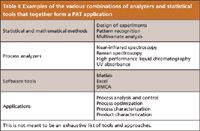
Table I: Examples of the various combinations of analyzers and statistical tools that together form a PAT application
Successful implementation of PAT requires the appropriate selection of a process analyzer. The selection of technique depends on the application and molecule, as well as the capability of the analytical method under consideration. In the biotechnology industry, drug products are manufactured using a series of unit operations. These products have to meet high expectations with respect to product quality, as documented in the pharmacopoeias and other regulatory documents. This is important to ensure the safety and efficacy of the manufactured drug substance and drug product. These requirements may be with respect to identity, content, quality, purity profile, moisture content, particle size, polymorphic form, and other such characteristics of the product. Traditional manufacturing involves the use of extensive analytical testing, most of which is retrospective as the data from analysis is received after the product lot has already advanced to the next process step. This approach results in a waste of manufacturing plant time, product rejects, scraps, and reprocessing (4). In contrast, PAT relies on enhanced process understanding to create controls that can result in continuous verification of product quality through all stages of manufacturing, reducing the chances of product loss.
Process analyzers play a key role in successful implementation of PAT and hence, are the focus of this paper. The analyzers may be used for monitoring of the critical quality attributes of the product, performance attributes of the process, and key characteristics of the various raw and in-process materials used in the process.
Process Analysis Techniques Used in the Biotech Industry
The demand for real-time and near-real-time monitoring in the past two decades has resulted in significant innovation and automation in the field of process analyzers. In the following subsections, we briefly review some of the commonly used process analysis techniques in the biotech industry (5).
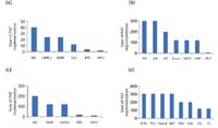
Figure 1: Comparison of analyzers with respect to their ease of implementation in microbial fermentations and mammalian cell culture unit operations for various quality attributes: (a) misincorporation; (b) nutrients; (c) glycosylation; and (d) cell growth.
Near-infrared (NIR) spectroscopy is one of the most commonly used analyzers for PAT applications. It is based on molecular overtone and combination vibrations. This analyzer typically utilizes a frequency range of 4000–12,500 cm-1 (800–2500 nm) to cover overtones and combinations of the lower energy fundamental molecular vibrations that include at least one X–H bond vibration. The functional groups involved in NIR (almost exclusively) are those involving the hydrogen atom: C-H, N-H, and O-H. A key advantage that NIR has is the possibility of direct measurement of the sample (6,7) either in situ, or after extraction of the sample from the process in a fast loop or bypass. The data from NIR measurements require multivariate analysis to extract the desired chemical information (8). NIR spectroscopy and multivariate data analysis (MVDA) has been successfully used for screening basal medium powders used in a mammalian cell culture in the biopharmaceutical industry (9) and also for at-line control and fault analysis of high cell-density fermentations (10). NIR probes also are used in crystallization processes to detect the particle size, shape, and the polymorphic form. This enables monitoring during routine production and determination of the crystallization endpoint (11).
Raman spectroscopy is a spectroscopic technique used to study vibrational, rotational, and other low-frequency modes in a system (12). A monochromatic light, usually from a laser in the visible, near-infrared, or near-ultraviolet range, interacts with molecular vibrations and phonons, resulting in the energy of the laser photons being shifted up or down. The shift in energy gives information about the compound in the system. Samples for analysis can be solids, liquids, gases, or any form in between, such as slurries, gels, and gas inclusions in solids. The Raman spectrum of water is extremely weak so direct measurements of aqueous systems are easy to do, giving this technique an advantage compared to infrared spectroscopy in which water has a very strong absorption. There is no inherent sample size restriction because it is fixed by the optic probe (13). Measurements can be made noninvasively or in direct contact with the targeted material (14). Applications in biotechnology processing include the monitoring of moisture content during lyophilization (15).
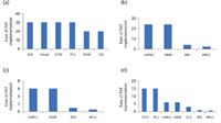
Figure 2: Comparison of analyzers with respect to their ease of implementation in isolation and purification of biotech products; (a) removal of cell mass; (b) misfolds; (c) charge variants; and (d) mass variants.
Nuclear magnetic resonance (NMR) spectroscopy exploits the magnetic properties of atomic nuclei to determine physical and chemical properties of atoms or the molecules in which they are contained. It can provide detailed information about the structure, reaction state, dynamics, and chemical environment of molecules which can be an essential tool for PAT (16). Duarte and colleagues identified and characterized 30 compounds in beer through high resolution (HR)-NMR (17). Two-dimensional (2D) NMR spectroscopy has been used for metabolic flux analysis of high-density perfusion cultures of Chinese hamster ovary (CHO) cells lines (18). Bench-top (BT)-NMR has been used in characterization of emulsions and lipid ingredients and monitoring adsorption as a noninvasive tool in drug delivery research (19).
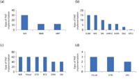
Figure 3: Comparison of analyzers with respect to their ease of implementation in formulation and packaging; (a) moisture content; (b) product content; (c) foreign particles; and (d) labeling.
Mass spectrometry (MS) uses the difference in mass-to-charge ratio (m/z) of ionized atoms or molecules to separate them from each other. It is useful for quantitation of atoms or molecules and also for determining chemical and structural information about them. Because of its inherent sensitivity, speed, and molecular selectivity, MS has been used for process analysis, including in-process monitoring of exhaust gases of fermentation processes, monitoring of drying processes, and environmental monitoring (20). Other applications include on-line, real-time deuterium abundance measurements in water vapor in aqueous liquids, including urine and serum (21); real-time quantification of trace gases in food products (22); and obtaining structural information for identification or structural elucidation of pharmaceutical drug products (23).
Acoustic resonance spectroscopy (ARS) involves spectroscopic measurements in the acoustic region, primarily the sonic and ultrasonic regions. It is typically much more rapid than high performance liquid chromatography (HPLC) and NIR, and it is nondestructive, requiring no sample preparation because the sampling waveguide can simply be pushed into a sample powder, liquid, or solid (24). Applications include a high-throughput, nondestructive method of online analysis and label comparison before shipping to obviate the need for recall or disposal of a batch of mislabeled drugs (25).
Calorimetry involves direct measurement of any endothermic or exothermic change in any process in order to better monitor or control all chemical, physical, and biological processes by providing the ability to measure enthalpy, power, and the heat coefficient (26). It is an on-line, nonintrusive technique for monitoring and optimizing a process. Applications include control of cooling profiles or rates during crystallization, real time measurement of heat transfer coefficient, monitoring and control of bioreactors, and determination of precise specific thermal profiles or signatures (27).
Dielectric spectroscopy is also known as impedance spectroscopy and measures the dielectric properties of a material. It offers the ability to measure bulk physical properties of materials based on the interaction of an external field with the electric dipole moment of the sample. It has an advantage compared to other spectroscopic techniques because it is not an optical spectroscopy or a noncontact technique. This allows for measurement without disturbing a sample in process. This tool has been used as a real-time and in-line monitoring tool for process optimization, automation, consistent product quality, and cost reduction in mammalian and insect cell cultures (28).
Fluorescence spectroscopy is a type of electromagnetic spectroscopy that analyzes fluorescence by using a beam of light, usually ultraviolet light, that excites the electrons in molecules of certain compounds and causes them to emit light at a lower energy (29). Fluorophores such as proteins, coenzymes, and vitamins can simultaneously be detected qualitatively and quantitatively inside and outside the cells. Cell metabolism and cell growth also can be measured (30). Teixeira and colleagues monitored on-line cell viability of recombinant baby hamster kidney (BHK) cell lines, as well as the concentration of the expressed glycoprotein IgG1-IL2 with 2D fluorometry and multivariate chemometric models (31). Other applications include real time monitoring of cell density and antibody titer in bioreactors containing CHO cell lines for production of monoclonal antibodies (32) and detection of antigen-antibody complexes for measuring immunoglobulin G (IgG) concentration in mammalian cell cultures (33).
On-Line HPLC: Rathore and colleagues have published a series of case studies that examine the use of on-line HPLC, at pilot and manufacturing scales, for performing analyses to facilitate real-time decisions for column pooling based on product quality attributes (34,35). HPLC also has been used for the monitoring of protein purity and levels of the different protein-related variants and impurities to enable the timely ending of refold on the basis of product quality data (36). The metabolism in a mammalian cell culture was examined by monitoring the concentration of 17 amino acids and glucose with online HPLC (37). Other applications include separation of recombinant human insulin-like growth factor-I (IGF) from IGF aggregate to allow for real-time control of column pooling (38) and using a combination of on-line size-exclusion HPLC, differential refractometry, and multiangle laser light scattering analysis (MALLS) for real-time estimation of product quality of a mutant form of the human immunodeficiency virus (HIV) vaccine protein antigen (39).
In situ microscopy (ISM) involves the use of a direct-light microscope with a measuring chamber, integrated in a 25-mm stainless steel tube, as well as two CCD-cameras and two frame-grabbers. The data obtained are processed by an automatic image analysis system. The microscopic examination of the liquid is performed in the measuring chamber, which is situated near the front end of the sensor head. ISM mainly allows for on-line microscopic observation of microorganisms during fermentations in bioreactors (40). Applications include in-line monitoring of Hansenula anomala culture in a batch fermentation process (41); real-time monitoring of hybridoma cells concentration in a stirred bioreactor (42); measurement of the level of colonization of fibroblasts (murine cell line NIH-3T3) on microcarrier surfaces during cultivation (43); detection of cell viability without conventional staining techniques (44); and on-line monitoring of enzyme carriers to determine their mechanical stability in the bioreactor (45).
Microwave resonance technology (MRT) is a noninvasive and nondestructive method of evaluating the moisture content of analytes using a microstrip waveguide with a resonance in the 300 MHz to 3 GHz range. It has been used for continuous moisture measurement in pharmaceutical granules and for determining moisture, temperature, and density of the granules to improve process understanding in fluid bed granulation (46).
Terahertz technology has a frequency gap from 100 GHz to 30 THz in the electromagnetic spectrum, which is in between microwave and infrared and has found a variety of applications in biology, medical science, quality control of food, pharmaceutical, and agriculture products, as well as global environment monitoring. Terahertz time domain spectroscopy (THz-TDS) also has been applied to many materials, including biomolecules, medicines, cancer tissue, DNA, proteins, and bacteria (47). It enables 3D imaging of structures and materials, and the measurement of the unique spectral fingerprints of chemical and physical forms, which is especially relevant in 3D imaging of tablets (48) and estimation of concentrations of the various excipients (49).
Photoacoustic spectroscopy (PAS) is based on the absorption of electromagnetic radiation by the molecules in the sample. The nonirradiative collisions of molecules in sample lead to warming of the sample matrix, which gives a pressure fluctuation because of thermal expansion that can be detected in the form of acoustic waves (50). These waves propagate through the volume of the gas to the detector (microphone, piezoelectric transducers, or optical method) where a signal is produced. This detector or microphone signal, when plotted as a function of wavelength, will give a spectrum proportional to the absorption spectrum of the sample (51). Applications include on-line and nondestructive monitoring of growth, thickness, and detachment of biofilms in waste water treatment plants (52) and unraveling the influence of various process parameters (such as pH, flow conditions) on the structure and stability of a biofilm (52).
Suitability of a Process Analyzer for PAT Applications in Biotechnology Processes
There are several challenges in designing a PAT application that can be successfully implemented on the floor of the manufacturing plant (53,54). One of the most significant challenges is the time it takes to analyze compared to the time that is available for making decisions for a step before proceeding to the next unit operation (55,56). In a recent publication, this ratio of times has been proposed as an indicator of the ease of implementation of PAT for a given unit operation (57). Table II presents an analysis of this ratio for a variety of unit operations and attributes. Ratios greater than one indicate that using the analytical method is feasible with the feasibility improving as the number increases. Ratios of one or less indicate that the analytical method cannot be used for PAT applications. It is shown in Table II that for each unit operation there are several methods that can be used to create a successful PAT application. There are a few trends that can be observed. First, while HPLC is one of the most important assays for characterization of biotechnology products, because of the large analysis time it is not well suited for most PAT applications. The emergence of ultrahigh-pressure liquid chromatography (UHPLC) as an alternative has somewhat alleviated this issue (34,35). Second, spectroscopy-based methods such as NIR are well suited for PAT applications because of the rapid analysis that they offer. However, their use is limited by the quality and process attributes that they can monitor. Third, while techniques such as NMR and MS can be useful for several PAT applications, especially in the upstream part of the process, the instrumentation may not be easy to install in a manufacturing plant and also the samples in the upstream process may need to undergo a significant amount of sample preparation before they can be analyzed. Fourth and final, for any given unit operation there are several options available with respect to analytical methods that can be used for creating successful PAT applications. More case studies need to be performed to promote the widespread application of PAT concepts in the biotechnology industry.

Table II: Suitable process analyzer for biopharmaceutical manufacturing process
Summary
Success of PAT depends to a large extent on efficient control of manufacturing processes to achieve predefined quality of the final product. This requires accurate and timely measurement of the critical attributes of the raw materials and process parameters and implies that capability of the analytical methods and their selection are critical for successful implementation of PAT. In this article, we reviewed the various analytical methods that enable the use of PAT. A critical evaluation of suitability of each analytical method as a PAT tool in terms of sampling (in-line, at-line, on-line), sample preparation, duration of analysis, and its industrial application was performed. It is our observation that while significant advancements have been accomplished with respect to our ability to analyze and monitor key process and quality attributes in the biotechnology industry, a lot more needs to be done with respect to utilizing the collected data for subsequent control of the process to achieve optimal yield and product quality. Only then shall we be able to achieve the benefits that will result from true PAT implementation.
Acknowledgments
We would like to acknowledge support of Mr. Muralikrishnan T, Dr. Amarnath Chatterjee, Mr. Samuel Mathew, and Dr. Nitin Patil, all from Biocon Ltd. (Bangalore, India), for critical review of the manuscript.
References
(1) PAT Guidance for Industry — A Framework for Innovative Pharmaceutical Development, Manufacturing and Quality Assurance, US Department of Health and Human Services, Food and Drug Administration (FDA), Center for Drug Evaluation and Research (CDER), Center for Veterinary Medicine (CVM), Office of Regulatory Affairs (ORA). September, (2004).
(2) E.K. Read, J.T. Park, R.B. Shah, B.S. Riley, K.A. Brorson, and A.S. Rathore, Biotechnol. Bioengin. 105, 276–284 (2010).
(3) E.K. Read, J.T. Park, R.B. Shah, B.S. Riley, K.A. Brorson, and A.S. Rathore, Biotechnol. Bioengin. 105, 285–295 (2010).
(4) A.S. Rathore and H. Winkle, Nat. Biotechnol. 27, 26–34 (2009).
(5) A.S. Rathore, R. Bhambure, and V. Ghare, Anal. Bioanal. Chem. 398, 137–154 (2010).
(6) W.F. McClure et al., Near-Infrared Technology in the Agricultural and Food Industries, 2, 145–169 (2001).
(7) Handbook of Near-Infrared Analysis, D.A. Burns and E.W. Ciurczak, Eds. (CRC Press, Taylor & Francis Group, Boca Raton, Florida, 2001) 22, 573–575.
(8) M. Roman Balabin, Z. Ravilya Safieva, and I. Ekaterina Lomakina, Chemometr. Intell. Lab 2, 183–188 (2007).
(9) A.O. Kirdar, G. Chen, and A.S. Rathore, Biotechnol. Prog. 26, 527–531 (2010).
(10) S. Gnoth, M. Jenzsch, R. Simutis, and A. Lubbert, J. Biotechnol. 132, 180–186 (2007).
(11) X. Lawrence Yu, A. Robert Lionberger, S. Andre Raw, R. D'Costa, H. Wu, and S. Ajaz Hussain, Advan. Drug Del. Rev. 56, 349–369 (2004).
(12) C. Dirk Hinz, Anal. and Bioanal. Chem. 384, 1036–1042 (2006).
(13) P.J. Treado and M.D. Morris, Prac. Spectros. Series 16, (1993).
(14) P. Matousek and A.W. Parker, Raman Spectrosc. 38, 563–567 (2007).
(15) T.R.M. De Beer et al., Anal. Chem. 79, 7992–8003 (2007).
(16) http://www.brukeroptics.com/minispec.html.
(17) I. Duarte, A. Nio Barros, P.S. Belton, R. Righelato, M. Spraul, E. Humpfer, and A.M. Gil, Agric. Food Chem. 50, 2475–2481 (2002).
(18) C. Goudar, R. Biener, C. Boisart, R. Heidemann, J. Piret, A. de Graaf, and K. Konstantinov, Metabolic Engin. 12, 138–149 (2010).
(19) H. Metz and K. Mader, Inter. J. of Pharmaceutics 364, 170–175 (2008).
(20) E. Heinzle and A. Oeggerli., Anal. Chim. Acta. 238, 101–115 (1990).
(21) S. Chauvatcharins, T. Seki, K. Fujiyama, and T. Yoshida, J. Ferment Bioeng. 79, 264–269 (1995)
(22) D. Smith and P. Španělm, Mass Spectrom. Rev. 24, 661–700, (2005).
(23) F. Erni, J. of Chrom. 251, 141–151 (1982).
(24) E.P.C. Lai, B.L.C., and S. Chen, App. Spec. 42, 381–529 (1988).
(25) J. Medendorp and R.A. Lodder, PharmSciTech 7, 175–183 (2006).
(26) G. Robert Buice, Jr., P. Pinkston, and A. Robert, Soc. for App. Spec. 48, 517–524 (1994).
(27) K. Hipkins, Pharm. Tech. Eur. 2, 157–163 (2007).
(28) C. Justice, A. Brix, D. Freimark, M. Kraume, P. Pfromm, B. Eichenmueller, and P. Czermak, Biotechnol. Adv. 29, 391–401 (2011).
(29) A. Sharma, and S.G. Schulman, Wiley InterScience 2, 172–176 (1999).
(30) S. Marose, C. Lindemann, and T. Scheper, Biotech. Progress 14, 63–74, (1998).
(31) P. Ana Teixeira, A.M. Carla Portugal, N. Carinhas, M.L. Joá o Dias, P. Joá o Crespo, M. Paula Alves, M.J.T. Carrondo, and R. Oliveira, Wiley InterScience 102, 1108–1116 (2008).
(32) P. Ana Teixeira, M. Tiago Duarte, M.J.T. Carrondo, and M. Paula Alves, Biotechnol.Bioeng. 102, 1852–1861 (2011).
(33) M. Reinecke and T. Scheper, J. Biotechnol. 17, 145–53 (1997).
(34) A.S. Rathore, L. Parr, S. Dermawan, K. Lawson, and Y. Lu, Biotechnol. Prog. 26, 448–57 (2010).
(35) A.S. Rathore, R. Wood, A. Sharma, and S. Dermawan, Biotechnol. Bioeng. 101, 1366–1374 (2008).
(36) A.S. Rathore, A. Sharma, and D. Chilin, BioPharm Int. 19, 48–57 (2006).
(37) M. Tina Larson, M. Gawlitzek, H. Evans, U. Albers, and J. Cacia, Biotech. and Bioengin. 77, 553–563 (2002).
(38) R.L. Fahrner, P.M. Lester, G.S. Blank, and D.H. Reifsnyder, J. Chromatogr. A. 827, 37–43 (1998).
(39) J. Barackman, I.Prado, C. Karunatilake, and K. Furuya, J. Chromatogr. A. 16, 57–64 (2004).
(40) C. Bittner, G. Wehnert, and T. Scheper, Biotechnol. Bioeng. 60, 24–35 (1998).
(41) V. Camisard, J.P. Brienne, H. Baussart, J. Hammann, and H. Suhr, Biotechnol. Bioeng. 78, 73–80 (2002).
(42) J.S. Guez, J.P. Cassar, F. Wartelle, P. Dhulster, and H. Suhr, J. Biotechnol. 111, 335–343 (2004).
(43) G. Rudolph, P. Lindner, A. Gierse, A. Bluma, G. Martinez, B. Hitzmann, and T. Scheper, Biotechnol. Bioeng. 99, 136–145 (2008).
(44) N. Wei, J. You, K. Friehs, E. Flaschel, and T.W. Nattkemper, Biotechnol. Lett. 29, 373–378 (2007).
(45) A. Prediger et al., Chem. Engin. & Tech. 34, 837–840 (2011).
(46) C. Buschmüller, W. Wiedey, C. Doscher, J. Dressler, and J. Breitkreutz, Eur. J. Pharm. Biopharm. 69, 380–387 (2008).
(47) www.nature.com/naturephotonics.
(48) M.P. Wagh, Y.H. Sonawane, and O.U. Joshi., Indian J. Pharm. Sci. 71, 235–241 (2009).
(49) H. Wu, E.J. Heilweil, A.S, Hussain, and M.A. Khan, J. Pharm. Sci. 97, 970–984 (2008).
(50) T. Schmid, Anal. Bioanal. Chem. 384, 1071–1086 (2006).
(51) V. Deepak Bageshwar, S. Avinash Pawar, V. Vineeta Khanvilkar, and J. Vilasrao Kadam, Eurasian J. Anal. Chem. 5, 187–203 (2010).
(52) T. Schmid, U. Panne, C. Haisch, M. Hausner, and R. Niessner, Environ. Sci. Technol. 36, 4135–4141 (2002).
(53) http://pharma.metrohm.com/pdfdownload/Prosp_Pharma_Analytik_e_web.pdf.
(54) W. Chew and P. Sharratt, Anal. Methods 2, 1412–1438 (2010).
(55) P. Werle, R. Mücke, and F. Slemr, App. Phys. B: Lasers and Optics 57, 131–139 (1993).
(56) S. Armenta, S. Garrigues, M. Gauradia, and P. Rondeau, Anal. Chem. Acta. 545, 99–106 (2005).
(57) A.S. Rathore, R. Bhambure, and V. Ghare, Bioanal. Chem. 398, 137–154 (2010).
Rakesh Mendhe is a graduate student in the department of chemical engineering at the Indian Institute of Technology, Hauz Khas, New Delhi, India and an employee of Biocon Ltd., India.

Rakesh Mendhe
Anurag S. Rathore is a biotech CMC consultant and an associate professor with the Department of Chemical Engineering at the Indian Institute of Delhi, India.

Anurag S. Rathore
Ira S. Krull is Professor Emeritus of Chemistry and Chemical Biology at Northeastern University, Boston, Massachusetts, and a member of LCGC's editorial advisory board.

Ira S. Krull

Thermodynamic Insights into Organic Solvent Extraction for Chemical Analysis of Medical Devices
April 16th 2025A new study, published by a researcher from Chemical Characterization Solutions in Minnesota, explored a new approach for sample preparation for the chemical characterization of medical devices.


