Analysis of Phenazepan in Whole Blood Using Solid-Phase Extraction and LC-Tandem Mass Spectrometry
LCGC North America
Phenazepam is becoming a drug of interest in forensic laboratories. This new procedure will enable forensic toxicologists to analyze phenazepam in biological fluids quickly and easily.
In this study, a solid-phase extraction (SPE) procedure is described for the analysis of phenazepam in whole blood. Extraction was performed using a mixed-mode SPE column. Samples of whole blood were diluted with aqueous phosphate buffer (pH 6). After loading the diluted sample onto the SPE column, the sorbent was washed with deionized water, acetic acid, and methanol. After drying the SPE columns, the analytes were eluted from the SPE column with 3 mL of an elution solvent consisting of methylene chloride, isopropanol, and ammonium hydroxide. The eluates were collected, evaporated to dryness, and dissolved in mobile phase (100 µL) for analysis by liquid chromatography–tandem mass spectrometry (LC–MS-MS). Chromatography was performed in gradient mode using a C18 column and a mobile phase consisting of acetonitrile and 0.1% aqueous formic acid. The total run time for each analysis was 5 min. The limits of quantitation and detection for this method were determined to be 1.0 ng/mL and 0.5 ng/mL, respectively. The method was found to be linear from 1.0 ng/mL to 100 ng/mL (r2 > 0.995). Recoveries of the phenazepam were found to be greater than 90%.
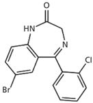
Phenazepam (7-bromo-5-[2-chlorophenyl]-1,3-dihydro- 2H-1,4-benzodiazepin-2-one) (Figure 1) is a benzodiazepine-type drug that was developed in the former Soviet Union and is now produced in Russia and some other countries (1). Phenazepam is used in the treatment of neurological disorders such as epilepsy, alcohol withdrawal, and insomnia (2), but it is now becoming a drug of interest to the forensic community because of its reported misuse (3). It can be used as a premedication before surgery because it augments the effects of anesthetics and reduces anxiety. Phenazepam is available as a 0.5-mg tablet, and the maximum daily dosage should not exceed 10 mg (2). The possible side effects of using phenazepam include dizziness, loss of coordination, and drowsiness, along with anterograde amnesia that can be quite pronounced in high doses (4). As with other benzodiazepines, in case of abrupt discontinuation following prolonged use, severe withdrawal symptoms may occur including restlessness, anxiety, insomnia, and convulsions (5). The metabolism of phenazepam in several species of mammals including humans has been known since the 1980s, when it was reported (6) that after oral administration (human) peak blood concentrations of the parent drug were achieved in 4 h and had a half life (t½) of 60 h. The authors of the study observed that the conversion of phenazepam to the metabolite 3-hydroxyphenazepm is not significant in humans; thus phenazepam is the main analyte of interest for forensic toxicologists because its use and misuse is becoming prevalent (3). Phenazepam has been determined in biological fluids by gas chromatography–mass spectrometry (GC–MS) (7) and GC using nitrogen specific detection (NPD) (8) as well as liquid chromatography–tandem mass spectrometry (LC–MS-MS) (9), following liquid–liquid extraction (LLE).

Figure 1: The structure of phenazepam.
This article is (to our knowledge) the first report on the continent of North America of phenazepam in a drugs-and-driving case employing mixed-mode solid-phase extraction (SPE) and LC–MS-MS. A recent report has been published in Europe for the analysis of this drug in Finland (10).
Experimental
Chemicals and Reagents
Phenazepam was obtained from Lipomed (Cambridge, Massachusetts) as a 1-mg/ mL methanolic solution. The internal standard (diazepam-d5) was purchased from Cerilliant (Round Rock, Texas) as a 100-µg/mL methanol solution. Acetonitrile, acetic acid (glacial), concentrated ammonium hydroxide solution (32% by volume), formic acid, isopropanol, methanol, and methylene chloride were obtained from Fisher Scientific (Pittsburgh, Pennsylvania). The SPE columns (CSDAU206) were obtained from UCT Inc. (Bristol, Pennsylvania). Deionized (DI) water was laboratory grade and was generated in the Massachusetts State Police Crime Laboratory (MSPCL). The water was produced by passing water through mixed-bed ion-exchange filters followed by ultraviolet light radiation; the resulting deionized water had 18-MΩ resistance. All chemicals were of ACS grade.
Acetic acid was prepared as a 1.0 M solution by diluting glacial acetic acid (58.0 mL to 500 mL), making it up to 1 L with DI water, and mixing well. Formic acid was prepared as a 0.1% (v/v) solution by adding 1 mL of the acid to 900 mL of DI water and diluting to 1 L. Acetonitrile containing 0.1% formic acid (v/v) was prepared by adding 1 mL of formic acid to 900 mL of acetonitrile and diluting to 1 L. Phosphate buffer (pH 6, 0.1 M) was purchased from Fisher Scientific as a ready-to-use solution.
Chromatographic Analysis
Analysis was performed using an API 3200 Q-Trap instrument supplied by Applied Biosystems (Foster City, California). The chromatographic system consisted of a Shimadzu CBM 20 A controller, two Shimadzu LC 20 AD pumps including degasser, a Shimadzu SIL 20 AC autosampler, and a Shimadzu CTO AC oven (set at 10 °C) (Shimadzu Scientific Instruments, Columbia, Maryland). The instrument was fitted with a 50 mm × 2 mm, 5 µm Imtakt US-C18 column from Silvertone Sciences (Philadelphia, Pennsylvania), which was attached to a Unison US-C18 guard column (5 mm × 2 mm) obtained from the same supplier. The LC system's column oven was maintained at 40 °C throughout the analyses. The injection volume was 10 µL. The mobile phase consisted of solvent A, DI water containing 0.1% formic acid, and solvent B, acetonitrile containing 0.1% formic acid. The mobile phase was delivered at a flow rate of 0.5 mL/min. The mobile-phase gradient was programmed as follows: 5–90% B in 4.0 min, then the proportion of solvent B was returned to 5.0%. The instrument was ready for reinjection after 5.1 min.
The mass spectrometry was performed on an API 3200 QTRAP system using multiple reaction monitoring mode (MRM). The following transitions were monitored (quantification ions underlined): m/z 350.8 → 206.3, 104.4, for phenazepam. The internal standard (diazepam-d5 ) was monitored at the following transitions: m/z 290.1 →198.3, 154.3. Tandem mass spectrometry was performed under the following conditions: curtain gas setting, 15; collision gas setting, medium; ion spray voltage setting, 5000 V; temperature setting, 650 °C; ion source gas 1 setting, 50; ion source gas 2 setting, 50. Tandem mass spectrometer conditions are shown in Table I. The analytical data were collected using Analyst Software Version 1.5 supplied by Applied Biosystems.

Table I: Tandem mass spectrometry conditions
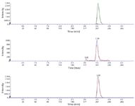
The retention times for phenazepam and the internal standard (diazepam-d5) were 3.49 and 3.54 min, respectively (Figure 2).

Figure 2: Chromatogram of a blood extract containing phenazepam at LOQ (1.0 ng/ mL) showing total ion chromatogram (TIC) (upper), phenazepam (middle), and internal standard (lower).
Sample Preparation for Analysis
Calibrators and Controls
A solution of phenazepam was prepared at a concentration of 1 µg/mL by the dilution of 10 µL of stock solution with acetonitrile to 10 mL in a volumetric flask. A solution of the internal standard (diazepam-d5) was prepared by diluting 100 µL of the stock solution (100 µg/mL) to 10 mL with acetonitrile in a volumetric flask. The choice of internal standard was based on the fact that deuterated analogs of phenazepam are not currently available and that an isotopically labeled analog of a benzodiazepine (which shares structural similarities to phenazepam) would not be observed in a case sample.
Calibrators were prepared by the addition of 0.5, 1.0, 10.0, 50, and 100 ng of phenazepam into 1.0-mL samples of drug-free whole blood. Then, 50 ng of the internal standard was added to these samples. Control samples were repared by the addition of 4 ng of phenazepam to 1.0 mL samples of drug-free whole blood in addition to 50 ng of the internal standard. A negative control sample was prepared by adding only the internal standard (50 ng) to a sample of drug-free whole blood (1.0 mL). To each of the calibrators, control, and test samples was added 5 mL of pH 6 buffer. These were then well mixed on a vortex mixer (1 min) and centrifuged at 3000 rpm for 10 min before application on individual SPE columns. All determinations were performed in duplicate.
To assess the performance of the procedure, calibration curves were constructed twice daily over five consecutive days using the spiked controls; we obtained intraday and interday values from these data.
Solid-Phase Extraction
Solid-phase extraction columns were conditioned by the sequential addition of 1 × 3 mL of methanol, 1 × 3 mL of DI water, and 1 × 1 mL of 0.1 M phosphate buffer (pH 6). Each liquid was allowed to percolate through the sorbent using gravity without allowing the sorbent to dry out between steps.
Following the passage of the methanol, DI water, and 0.1 M phosphate buffer (pH 6) through the SPE columns, each diluted sample (that is, calibrator, control, and case item) was loaded on to an individually marked SPE tube, and allowed to pass through the sorbent using gravitational flow. The columns were then washed with 1 × 3 mL of DI water, 1 × 3 mL of 1.0 M acetic acid, and 1 × 3 mL of methanol, respectively. The SPE columns were then dried by applying a vacuum to the SPE manifold at 15 in. of mercury pressure with the aid of an electric vacuum pump connected to the vacuum manifold.
The analytes were eluted from the SPE columns by the addition of 1 × 3 mL of a 78:20:2 methylene chloride–isopropanol–ammonium hydroxide solution. This solution was prepared daily by adding 2 mL of concentrated ammonium hydroxide solution to 20 mL of isopropanol and mixing well. Finally, 78 mL of methylene chloride was added to this solution and the resultant solution was transferred to a clean screw-top glass bottle for use. A screw-top bottle ensures that the basicity of the solution remains high by eliminating any loss of ammonia from the bottle. The elution solvent was allowed to flow through the SPE sorbent with the aid of gravity and was collected in separate glass tubes (75 mm × 125 mm). Glass tubes were chosen because they are standard laboratory materials within this toxicology laboratory.
The eluate from each SPE column was evaporated to dryness using a gentle stream of nitrogen at 35 °C, after which the samples were dissolved in 100 µL of a solution consisting of 95% mobile-phase A and 5% mobile-phase B for LC–MS-MS analysis.
Recovery Studies
To determine the recovery values across the dynamic range of the analysis, the results of the SPE extractions of the whole blood extracts (as duplicate analyses) were compared to the values obtained from unextracted standards at corresponding concentrations. The unextracted standards were prepared by evaporation of acetonitrile solutions containing phenazepam (including 50 ng of the internal standard). The dried residues were dissolved in mobile phase (100 µL) before analysis by LC–MS-MS.
Matrix Effects
Studies into the matrix effects were performed according to procedures described by Matuszewski and colleagues (11). In this process, samples of drug-free whole blood (1 mL) were spiked with phenazepam before analysis using the SPE methodology. A second set of drug-free whole extracts was analyzed according to the SPE method. Following elution from the SPE columns, the extracts were spiked with phenazepam. Both sets of samples were evaporated to dryness under a gentle stream of nitrogen at 35 °C, and the residues were dissolved in 100 µL of a solution consisting of 95% mobile-phase A and 5% mobile-phase B, the samples were combined for analysis by LC–MS-MS.
Phenazepam solutions (each with a concentration of 50 ng/mL) were infused into the tandem mass spectrometer using the on-board syringe pump (controlled by Analyst 1.5 software) via a 1-mL Hamilton syringe (model 1001TLL, supplied by Fisher Scientific) at a flow rate of 5 µL/min. At the same time as the phenazepam solution was flowing into the mass spectrometer, a 10-µL aliquot of the SPE-extracted whole blood matrix (drug-free whole blood, free of phenazepam) was injected using the autosampler syringe on the Shimadzu liquid chromatograph using Analyst 1.5 software. The liquid chromatograph and mass spectrometer were arranged so that samples from the liquid chromatograph were mixed into the flow of phenazepam via a three-port T-section before the total flow entered the mass spectrometer. Any suppression effects on the phenazepam could be monitored at the MRMSs for the noted drugs.
Selectivity
When analyzing samples of biofluids such as blood via SPE and LC–MS-MS, it is essential to ensure that the interfering effects of other drug compounds can be eliminated. In this procedure, samples of drug-free whole blood (1 mL) were spiked with 49 drugs at a concentration of 100 ng/mL (bupropion, lidocaine, methadone, amitriptyline, nortriptyline, thioridazine, trazodone, mesoridazine, pethidine, diphenhydramine, phenyltoloxamine, imipramine, desipramine, benztropine, trimethoprim, diltiazem, haloperidol, strychnine, morphine, codeine, 6-acetylmorphine, oxycodone, oxymorphone, hydrocodone, noroxycodone, hydromorphone, diazepam, nordiazepam, oxazepam, temazepam, alprazolam, α-hydroxyalprazolam, lorazepam, triazolam, α-hydroxytriazolam, flunitrazepam, 7-amino-flunitrazepam, chlordiazepoxide, midazolam, α-hydroxymidazolam, flurazepam, desalkyl-flurazepam, cocaine, ecgonine methyl ester, ecgonine ethyl ester, benzoylecgonine, cocaethylene, clonazepam, and 7-amino-clonazepam) and were extracted according to the SPE method. The interfering effect of these compounds was not found to be significant.
Results and Discussion
Recovery
The recovery of phenazepam from drug-free whole blood was 98% (±2%). This result is an excellent indicator for the efficiency of the extraction procedure of phenazepam using whole blood as a matrix. The procedure was performed twice daily during a period of five days.
Imprecision of Analysis
The spiked control samples (4 ng/mL) were determined to have concentrations of 3.9 ng/mL (±0.2 ng/mL). This value was determined during a period of five days.
Intraday variation and interday variation for the analysis of phenazepam were found to be less than 5% and less than 8%, respectively. Ion suppression studies revealed that suppression of monitored ions was less than 2%. This method was found to be linear (r2 > 0.995) throughout the 1.0–100 ng/mL dynamic range for phenazepam.
LOD and LOQ
The limit of detection (LOD) of a particular method can be defined as the level at which the signal-to-noise ratio for the particular analyte is greater than or equal than 3:1. The limit of quantification (LOQ) for the method is the level at which the signal-to-noise ratio for a particular analyte is greater than or equal to 10:1. In this study, LOD values were determined empirically by analyzing extracted samples of drug-free whole blood fortified with phenazepam by LC–MS-MS according to the SPE method. This analysis was performed until the lowest level at which each of the respective analytes just failed the signal-to-noise ratio of 3:1. This was observed to be 0.5 ng/mL. In terms of LOQ, samples of drug-free blood were spiked with phenazepam at concentrations below 10 ng/mL and extracted according to the SPE procedure until the analytes just failed a signal-to-noise ratio of 10:1; this value was found to be 1.0 ng/mL.
Solid-Phase Extraction
As noted earlier, phenazepam is a relatively new compound of interest to forensic toxicologists. The use of mixed-mode SPE offers toxicologists in forensic laboratories a very clean sample to analyze. The sample is loaded onto the sorbent as a diluted solution at pH 6, and it is cleaned and concentrated on the SPE column. The use of an ion-exchange moiety allows coextracted materials to rinse off the sorbent while the drug of interest is retained in a clean condition. In this situation, the drug can be eluted using a mid-polarity solvent mixture that is easily evaporated for further analysis. This combination of hydrophobic and ion-exchange chemistries is a powerful tool for producing clean samples for chromatographic analyses.
Tandem Mass Spectrometry
This project was aimed at introducing new methodology to the forensic community involved in the analysis of phenazepam in biological samples, with selectivity and sensitivity in mind. In other words, the ability to detect, confirm, and quantify a compound such as phenazepam in a complex mixture at low levels is a highly desired quality in a new procedure, especially if it can lead to a fast turnaround time and an increase in laboratory efficiency.
Conclusion
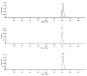
Phenazepam is quickly becoming a drug of interest in forensic laboratories in the United States, the United Kingdom, and Europe (3,12), and analysts will be asked to test for it on a routine basis. With that in mind, this new procedure using SPE and LC–MS-MS will offer forensic toxicology laboratories the ability to perform the analysis of phenazepam in biological fluids, such as blood, quickly and efficiently. When this new method was applied to a genuine case sample taken from a driver operating a motor vehicle, the whole blood sample was found to contain 9 ng/mL of phenazepam (Figure 3).

Figure 3: Chromatogram of an actual blood extract containing phenazepam showing total ion chromatogram (TIC) (upper), phenazepam (middle), and internal standard (lower).
Albert A. Elian is with the Massachusetts State Police Crime Laboratory in Sudbury, Massachusetts.
Jeffery Hackett and Michael J. Telepchak are with UCT Inc. in Bristol, Pennsylvania. Direct correspondence to: jhackett@unitedchem.com
References
(1) V.V. Zakusov, Pharm. Chem. J. 13, 1094–1097 (1979).
(2) R.C Baselt, Dispostion of Toxic Drugs and Chemicals in Man 8th (Biomedical Publications, Foster City, California, 8th ed., 2008) p. 1221.
(3) P.D. Maskell, G.D. Paoli, L.N. Seetohul, and D.J. Pounder, B.M.J. 343, d4207 (2011).
(4) T.A. Vorinina and T.L. Garibova, Bull. Exp. Biol. Med. 95, 65–68 (1983).
(5) V.P. Zherdev, T.A. Voronina, T.L. Garibova, G.B. Kolyvanov, A.A. Litvin, A.K. Sariev, V.N. Tokhmakhchi, and A.E. Vasil'ev, Eksp Klin Farmakol. 66, 50–53 (2003).
(6) V.P. Zherdev, S. Caccia, S. Garattini, and A.L. Ekonomov, Eur. J. Drug Metab. Pharmacokinet. 7, 191–196 (1982).
(7) I. Rasenen, M. Neuvonen, I. Ojanpera, and E. Vuori. Forensic Sci. Int. 112, 191–200 (2000).
(8) W. Johnson, Toxtalk. 34, 17–18 (2010).
(9) K. Bailey, L. Richards-Waugh, D. Clay, M. Gebhardt, H. Mamoud, and J.C.Kraner, J. Anal. Toxicol. 34, 527–32 (2010).
(10) L. Wilhelm, S. Jenckel, P. Kriikkuu, J. Rintatalo, J. Hukke, and J. Kramer, Toxichem. Krimtech. 78, 302–305 (2011).
(11) B.K. Matuszewski, M. Constanzer, and C.M. Chavez-Eng, Anal. Chem. 75, 3019–3030 (2003).
(12) J. Mrozkowska, E. Vinge, and C. Borna, Lakartidningen. 106, 516–517 (2009).

Extracting Estrogenic Hormones Using Rotating Disk and Modified Clays
April 14th 2025University of Caldas and University of Chile researchers extracted estrogenic hormones from wastewater samples using rotating disk sorption extraction. After extraction, the concentrated analytes were measured using liquid chromatography coupled with photodiode array detection (HPLC-PDA).
Polysorbate Quantification and Degradation Analysis via LC and Charged Aerosol Detection
April 9th 2025Scientists from ThermoFisher Scientific published a review article in the Journal of Chromatography A that provided an overview of HPLC analysis using charged aerosol detection can help with polysorbate quantification.
Removing Double-Stranded RNA Impurities Using Chromatography
April 8th 2025Researchers from Agency for Science, Technology and Research in Singapore recently published a review article exploring how chromatography can be used to remove double-stranded RNA impurities during mRNA therapeutics production.


