2020 HTC Innovation Award – Ryan Kelly
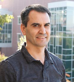
LCGC Europe sponsored the 2020 HTC Innovation Award to highlight innovation in separation science. The winner, Ryan Kelly, from Brigham Young University, in Utah, USA, has introduced an impressive array of innovative approaches to advance proteomics research using nano-liquid chromatography–mass spectrometry (nano‑LC–MS), LC–MS, and two‑dimensional (2D)-LC–MS, including the development and application of nanodroplet processing in one pot for trace samples (nanoPOTS). This platform, when combined with nano-LC–MS/MS, can identify more than 3000 protein groups in 10 cells, a greater level of proteome coverage than was previously possible for samples containing 5000 cells.
LCGC Europe sponsored the 2020 HTC Innovation Award to highlight innovation in separation science. The winner, Ryan Kelly, from Brigham Young University, in Utah, USA, has introduced an impressive array of innovative approaches to advance proteomics research using nano-liquid chromatography–mass spectrometry (nano LC–MS), LC–MS, and two-dimensional (2D)-LC–MS, including the development and application of nanodroplet processing in one pot for trace samples (nanoPOTS). This platform, when combined with nano-LC–MS/MS, can identify more than 3000 protein groups in 10 cells, a greater level of proteome coverage than was previously possible for samples containing 5000 cells.
Q. Congratulations on winning the 2020 HTC Innovation Award, which was sponsored by LCGC Europe. When did you first become involved in hyphenated techniques?
A: Thank you very much. It means a lot to see the work of our team being recognized by the separation science community. I first became involved in hyphenated techniques as a postdoc in Dick Smith’s group at Pacific Northwest National Laboratory. I initially focused on electrospray emitter technology at the interface between liquid chromatography (LC) and mass spectrometry (MS), developing a chemical etching technique to shape fused-silica capillaries into fine tips rather than standard heating and pulling methods. These etched tips have no internal taper, making them far less prone to clogging than conventional nanospray emitters, and because they are fabricated through a purely chemical means, they are highly reproducible. Perhaps most importantly, they can operate stably at far lower flow rates than conventional nanospray emitters (extending to the picolitreâperâminute range), opening the door to much lower flow separations than were previously possible.
Another area of interest in my early involvement with hyphenated techniques was dividing the flow from an LC separation among an array of emitters. There are tradeoffs in miniaturizing LC–MS analyses for ultralow flow rates; compared to micro LC–MS operated at flow rates greater than 1 µL/min, it can be difficult to achieve high run-to-run reproducibility, extreme measures need to be taken to minimize dead volumes, and sample loading and column regeneration times can become excessive. Yet, the sensitivity of an LC–MS analysis is dramatically improved at low flow rates as a result of increased ionization efficiencies, driving the need for column miniaturization.
Multielectrospray sources could potentially provide the best of both worlds, allowing for robust micro-LC separations, but then dividing the flow postcolumn among an array of nano electrospray ionization (ESI) emitters, providing high ionization efficiencies. We were able to demonstrate an approximate 40-fold sensitivity increase for our multispray emitters in combination with a modified MS inlet. A remaining challenge is that the emitter arrays can generate more electrospray current than commercial mass spectrometers are designed to deal with, so this is an area that could definitely benefit from additional R&D.
Q. What are the biggest challenges facing chromatographers in proteomics research from an analytical perspective?
A: Well, I think that we and others are demonstrating significant sensitivity gains by continuing to miniaturize liquid chromatography with ultranarrow-bore columns that operate at low nanolitre per-minute flow rates, but commercial LC systems are not well equipped for these columns. The autosamplers can’t inject samples smaller than several microlitres and the extracolumn tubing can lead to huge delay volumes with long sample loading times and gradient delays. Also, there are limits to the flow rates that current systems can deliver, and flow splitting is typically necessary to get to these really low flow rates. Besides this, almost all commercial columns for proteomics have an internal diameter of 75 µm and operate at 300 nL/min, which is not ideal for proteomic analysis of small samples. This leads to relatively few groups who have expertise in customized column preparation and instrumentation being able to work in ultrasensitive nano LC–MS. Of course, each one of these challenges is also an opportunity for LC instrument and column manufacturers, and as demand for these capabilities continues to grow, the market will respond with new solutions.
Q. You recently published a paper on improved single-cell proteome coverage hyphenating narrow-bore packed nano-LC columns with ultrasensitive mass spectrometry (1). What is innovative about this research?
A: When you are trying to extract as much chemical information as possible from a tiny biological sample, such as a single human cell, you have to look at every possible way to boost signal and reduce noise. One of the ways we are trying to do that is by continuing to miniaturize LC. Early on in this project we demonstrated substantial improvements in proteome coverage using custom packed 30-µm internal diameter (i.d.) columns compared to conventional 75-µm-i.d. columns, and we have used the 30-µm columns ever since. In this most recent study, we wanted to see how much more could be gained by going to even smaller columns, and in this case we packed 20-µm-i.d. columns in-house and evaluated their performance in the context of single-cell proteomics. We found that dropping to 20 µm, which resulted in a flow rate reduction from ~50 nL/min to ~20 nL/min, allowed us to identify about 40% more proteins on average from single cells.
Q. You recently gained recognition for developing nanoPOTS. What is nanoPOTS and what led you to develop this concept?
A: A major challenge in the field of proteomics has been that large amounts of sample comprising thousands or millions of cells were required to achieve sufficient signal for a measurement. This means that we are typically measuring a mixture of different cell types in different states, so we were getting a bit of a blurry picture of the underlying cell biology. Overcoming this limitation has motivated efforts to extend proteomic analyses to the single-cell level.
We saw that the sensitivity of LC–MS analyses for proteomics continued to improve to the point that samples containing about the same protein content as single mammalian cells (around 0.1 ng or 0.2 ng of total protein) could be analyzed, but these demonstrations were typically performed using small aliquots of bulk-prepared samples and there was no way to prepare actual single cells. A biological sample has to undergo a number of processing steps such as cell lysis, protein extraction, reduction of disulphide bonds, digestion with trypsin, etc. to produce ready-to-analyze peptides. Conventional preparation methods are performed in volumes of tens of microlitres, and there are multiple sample transfer steps.
If you take a single cell through a conventional sample preparation workflow, you’re probably diluting the cell’s proteins by a factor of about 108 (from ~1 pL to ~100 µL). Such dilution results in samples being exposed to a lot of adsorptive surfaces, and the reaction kinetics for processing steps such as trypsin digestion become highly unfavourable at these reduced concentrations.
NanoPOTS, which stands for Nanodroplet Processing in One pot for Trace Samples, essentially preserves the standard form factor used for proteomic sample preparation, but miniaturizes volumes by several orders of magnitude; we are still pipetting into a well plate, but the pipettor is now a robotic nanolitre liquid handler, and the well plate is now a micropatterned glass slide. This allows trace proteomic samples such as single cells to be effectively prepared in a volume of just ~200 nL versus ~100 µL, and the sample is exposed to less than 1 mm2 of surface, so sample recovery is greatly improved. In our initial study (2), we were able to use the nanoPOTS platform in combination with highly sensitive nano-LC–MS/MS to identify more than 3000 protein groups from as few as 10 cells. This is a greater level of proteome coverage than was previously achieved for samples containing 5000 cells, so this is an “orders-of-magnitude” improvement.
Q. What benefits does this offer the analyst?
A: NanoPOTS enables us to extract more chemical information from smaller samples than was previously possible. As analytical chemists, we are often focused on improving the separation or the mass spectrometry, but sometimes the battle is won or lost before we even get to the analytical platform. If most of the sample is being lost during the preanalytical steps, which was the case for small proteomic samples, that’s where we need to focus and that’s what drove the development of nanoPOTS. Another benefit is that nanoPOTS preserves the basic form factor of a pipettor interfacing with a well plate. There are a lot of other ways that surface exposure and dilution could be minimized, but, because nanoPOTS is an open platform, it is compatible with widely used sample isolation techniques including fluorescence activated cell sorting and laser microdissection. This gives us options for targeting specific cells or tissues of interest that, for example, a closed microfluidic platform doesn’t have.
Q. Can you describe an application that demonstrates the inventiveness of nanoPOTS in practice?
A: One example of interest is proteome profiling of circulating tumour cells (CTCs), which originate at a solid tumour but make their way into the blood stream. These cells can potentially inform on disease progression-how an individual is responding to a therapy and when resistance to therapy is developing-all without requiring invasive and risky biopsies. So this is a high value target that could have a real impact on treatment, but CTCs are very rare and only a small number are isolated from a blood draw. Conventional proteomics approaches lack the sensitivity to obtain an in-depth profile of protein expression from such small samples, but nanoPOTS sample preparation in combination with highly sensitive LC–MS offers hope that we can begin to extract protein information from these previously inaccessible samples. In fact, in collaboration with George Thomas at Oregon Health and Science University, we have begun to develop a workflow that begins with enriching CTCs from whole blood and then profiling the cells for biomarkers and an initial demonstration was recently published (7).
This is just one example, and really any sample that was too small for proteome profiling using conventional techniques, even single mammalian cells, can now be addressed.
Q. Where else has nanoPOTS been beneficial?
A: Another area that we are very interested in is mapping protein expression across tissues. The amount of biochemical information obtained from a thin tissue section on a microscope slide like those studied by pathologists is generally limited to staining for a few molecules of interest. We used laser microdissection to convert a thin section of mouse uterine tissue into tiny pixels just 100 µm × 100 µm across and then subjected each of these pixels to the nanoPOTS workflow. This allowed us to reconstruct an image in which the protein abundance of each of the 2000 proteins (approximately) that were detected were overlaid over an optical image of the same tissue to determine which proteins were up and downregulated across different regions of the tissue. Proteins have been very difficult to detect using other mass spectrometry imaging approaches, and so nanoPOTS enables us to identify more than 100 times more protein groups from each pixel than was previously possible. We think this will provide biomedical researchers with an important tool to determine the contribution of the microenvironment to give rise to a given phenotype, and to get a better understanding of what is happening in different tissues.
Q. You have also published on multidimensional separations combining nano-LC with structures for lossless ion manipulation ion-mobility mass spectrometry (SLIM IM-MS) (6). What is novel about SLIM IM-MS?
A: Yes, while we were focused on developing nanoPOTS, Dick Smith, Yehia Ibrahim, and colleagues at Pacific Northwest National Laboratory have been developing structures for lossless ion manipulation (SLIM). SLIM devices comprise ion optical components that are typically constructed using printed circuit boards, and these can provide extended pathlengths for ion-mobility (IM) spectrometry separations. SLIM IM makes it possible to achieve high peak capacities akin to those of LC separations, but with a total separation time of ~1 s or less. We wanted to couple LC with SLIM IM to achieve ultrahigh peak capacity separations for trace samples using no more time than a typical LC separation requires. To interface LC with SLIM IM, we used our nanoPOTS robot to fractionate a nano-LC separation into different nanowells in 1-min intervals. The fractions were then dried for storage. To inject each sample, the modified nanoPOTS robot dispensed solvent into each well to reconstitute the sample, then pick up that sample and electrospray it into the SLIM IM-MS platform. Using this multidimensional separation platform, we were able to achieve peak capacities of more than 3600 in about 90 min, which is an order of magnitude increase in peak capacity per unit time compared to LC alone.
Q. What benefits does nanoâLCâSLIM IM-MS offer the analyst?
A: It is difficult to achieve a peak capacity more than 500 for a one dimensional (1D)-LC separation, even when extended (more than10 h) gradients are employed. Nano-LC–SLIM IM-MS offers ultrahigh peak capacity separations of greater than 3600 in a short timeframe of less than 90 min by coupling two orthogonal techniques. We are in the early days of coupling LC separations with high peak capacity IM-MS such as SLIM provides, but I think this is a very promising avenue of research for analyzing difficult-to-separate compounds and for comprehensive analysis of complex mixtures.
Q. You have developed picoelectrospray ionization mass spectrometry using narrowbore chemically etched emitters. What was the rationale behind this research?
A: Since the initial development of ESI MS, it had been observed that a greater portion of solution-phase molecules could be converted to gasâphase ions by operating the electrospray at reduced flow rates, that is, ionization efficiency increases at low flow rates as a result of the formation of smaller, more readily desolvated charged droplets. This led to the development of nano-ESI, where the electrospray emitter is operated at flow rates of less than 20 nanolitres to several hundred nanolitres per minute. Indeed, the vast majority of proteomics separations take place at 300 nL/min to realize some of the benefits of nano-ESI. But we analytical chemists are greedy, and we reasoned that if nano-ESI was good, wouldn’t picoESI, where we operate at less than 1 nL/min, be better?
In other words, we had some unanswered questions about the limits of improved ionization efficiency and reduced ionization suppression at low flow rates, but there were no electrospray emitters that could operate reproducibly at such low flow rates.
I had previously developed a chemical-etching technique to shape fused-silica capillaries into very sharp emitters that are ideal for nano ESI (8). In this study, we applied the same technique to 2-µmâi.d. capillaries and evaluated their spray stability at ultralow flow rates. We were able to spray stably at flow rates as low as 0.4 nL/min, which enabled us to explore the analytical performance of ESI in uncharted territory. We found that ionization efficiency continued to improve in the picoelectrospray range, ionization suppression was not much different than in the nanospray regime, and we observed greatest signalâtoânoise ratios at about 1 nL/min.
Q. Where has this approach been adopted?
A: Until recently, this was more of an academic curiosity rather than of practical utility as it was exceedingly difficult to perform LC separations at approximately 1 nL/min to take advantage of the sensitivity gains that our study indicated should be achieved at such low flow rates. However, in a new study led by Shaorong Liu from the University of Oklahoma with Ying Zhu at Pacific Northwest National Laboratory (currently in review), it has been shown for the first time that pico LC–MS using 2-µm-i.d. open-tubular LC columns can identify ~1000 proteins from just a few pg of protein equivalent to 10% of a typical single cell. While this is not quite ready for prime time, this is definitely an area worth continuing to explore.
Q. What benefits does nanowellâmediated two-dimensional liquid chromatography (2D-LC) provide in proteomics research (4)?
A: Higher peak capacity separations can greatly increase proteome coverage by reducing the number of peptides entering the mass spectrometer at any given time, reducing ionization suppression, and, with typically longer analysis times, providing more time for MS/MS sequencing. Two-dimensional separations are often employed to dig deeper into the proteome, with the first dimension being distributed into fractions that are then analyzed by standard low-pH reversed-phase nano-LC. For 2D-LC employing high pH followed by low pH reversedâphase LC, a relatively high flow high-pH separation is employed and samples are fractionated into multiwell plates. The fractions are dried and then reconstituted in tens of microlitres of aqueous solution for injection into a low-pH reversed-phase LC–MS/MS analysis platform. The problem we run into is that we are again exposing our samples to a lot of surface area with potential for losses in between these two separation dimensions and so large samples are required up front so that we still have sufficient material to measure after the peptide losses and incomplete injection. We thought that just as microfabricated nanowells could replace well plates for reduced sample losses during sample preparation in the nanoPOTS workflow, the same nanowells could also reduce losses in between separation dimensions. So we went ahead and used a nano-LC separation for the first dimension, fractionated into nanowells, and then reconstituted the samples for injection into our custom ultralow-flow nano-LC. From samples comprising just a few hundred cells, we were able to identify around 6000 proteins, which was unprecedented from such small samples. We are currently miniaturizing this to extend 2D-LC to even smaller samples.
Q. What are you currently working on at the moment?
A: We are definitely still interested in increasing proteome coverage for single cells and increasing the throughput with which these can be measured. We are very encouraged that there appears to be no end in sight to the achievable gains and have a couple of significant improvements close to submission for publication. At the same time, we are working to automate sample injection from nanoPOTS chips to nano-LC, which should take a lot of the skill and labour intensiveness from the process (and allow my group members to sleep at night rather than tending to the instruments around the clock). We are also working hard to make the nanoPOTS platform accessible to a wide range of researchers interested in single-cell and nanoscale proteomics. Currently, the robotic platforms that we use are quite expensive and require some expertise to fabricate and use. We are now adapting some low-cost automated liquid handling systems for nanopipetting, which will hopefully broaden access. Finally, it is very important that we not only develop new technologies but that we also put them into practice, and we enjoy working with a number of biomedical researchers to apply our systems to new sample types and help to uncover new biomedical insights.
Q. Can you name one scientist who represents innovation in separation science?
A: I would like to recognize Yufeng Shen, who I think is a bit of an unsung hero in separation science. Yufeng received his Ph.D. at Brigham Young University under the direction of Milton Lee after publishing something like 35(!) papers (~25 first author) before graduating. He then moved to the Biological Separations and Mass Spectrometry group at Pacific Northwest National Laboratory where he was responsible for many innovations in ultrahigh-pressure liquid chromatography (UHPLC), ultrafast separations, and ultrahigh peak capacity separations that made modern proteomics possible. Much of this groundbreaking work was done in the early 2000s, but I think a number of the “world records” that he set in separation science still stand today, and his work certainly inspires a lot of what we do.
References
- Y. Cong, Y. Liang, K. Motamedchaboki, R. Huguet, T. Truong, R. Zhao, Y. Shen, D. Lopez-Ferrer, Y. Zhu, and R.T. Kelly, Anal. Chem. 92, 2665–2671 (2020).
- Y. Zhu, P.D. Piehowski, R. Zhao, J. Chen, Y. Shen, R.J. Moore, A.K. Shukla, V. Petyuk, M. CampbellâThompson, C.E. Mathews, R.D. Smith, W.-J. Qian, and R.T. Kelly, Nat. Commun. 9, 882 (2018).
- Y. Zhu, G. Clair, W.B. Chrisler, Y. Shen, R. Zhao, A.K. Shukla, R.J. Moore, R.S. Misra, G.S. Pryhuber, R.D. Smith, C. Ansong, and R.T. Kelly, Angewandte Chemie 57, 12370–12374 (2018).
- M. Dou, Y. Zhu, A. Liyu, Y. Liang, J. Chen, P.D. Piehowski, K. Xu, R. Zhao, R.J. Moore, M.A. Atkinson, C.E. Mathews, W.-J. Qian, and R.T. Kelly, Chem. Sci. 9, 6944–6951 (2018).
- M. Dou, C.-F. Tsai, P.D. Piehowski, Y. Wang, T.L. Fillmore, R. Zhao, R.J. Moore, P. Zhang, W.-J. Qian, R.D. Smith, T. Liu, R.T. Kelly, T. Shi, and Y. Zhu, Anal. Chem. 91, 9707–9715 (2019).
- M. Dou, C.D. Chouinard, Y. Zhu, G. Nagy, A.V. Liyu, Y.M. Ibrahim, R.D. Smith, and R.T. Kelly, Anal. Bioanal. Chem. 411, 5363–5372 (2019).
- Y. Zhu, J.Podolak, R. Zhao, A.K. Shukla, R.J. Moore, G.V. Thomas, and R.T. Kelly, Anal. Chem. 90, 11756–11759 (2018).
- R.T. Kelly, J.S. Page, Q. Luo, R.J. Moore, D.J. Orton, K. Tang, and R.D. Smith, Anal. Chem. 78, 7796–7801 (2006).

Ryan Kelly is an associate professor in the Department of Chemistry and Biochemistry at Brigham Young University (BYU), in Utah, USA. He received his Ph.D. in 2005 from BYU and then spent 13 years at Pacific Northwest National Laboratory, Washington (USA) before returning to BYU in 2018. A central theme of Kelly’s research is the development of new technological solutions in separations, microfluidics, and MS for improved biochemical analyses, including nanoâLC–MS-based proteomics.
Research Interests
Professor Ryan Kelly develops innovative hyphenated separation techniques including ultrasensitive nano-liquid chromatography mass spectrometry (nanoâLC–MS) using narrow-bore (20 µm) separation columns combined with mass spectrometry (MS) for bioanalysis, comprehensive two-dimensional liquid chromatography–mass spectrometry (2D-LC–MS), and liquid chromatography ion mobility spectrometry (LC–IM-MS) or proteomic analysis of trace samples, including single cells. He also develops microfluidic sample preparation strategies for these trace samples.
Winning Credentials
Ryan Kelly was awarded the 2020 HTC Innovation Award because of his outstanding contributions to the field of microcolumn separations involving hyphenation. A central theme of Kelly’s research is the development of new technological solutions in separations, microfluidics, and mass spectrometry (MS) for improved biochemical analyses. To this end, he has developed novel techniques based on one-dimensional (1D)- and 2D-LC, hyphenated LC-ion mobility spectrometry-MS, and novel preconcentration/injection strategies for capillary LC–MS and electrophoresis MS. He is the author of more than 90 publications with more than 3500 total citations and inventor of 11 issued and pending patents, several of which have been licensed and commercialized.
Kelly has an extensive background in microfluidics that allows him to address preanalytical bottlenecks to overcome the inefficiencies associated with sample isolation and processing of biological samples for proteomic analysis. His research group recently developed nanodroplet processing in one pot for trace samples (nanoPOTS) a breakthrough microfluidic platform based on robotic nanopipetting from microfabricated well plates, which greatly reduces sample losses and enhances reaction kinetics. When combined with ultrasensitive LC–MS, nanoPOTS has enabled more than1000 proteins to be quantified from single mammalian cells. This is the first example of in-depth proteome profiling from such trace samples, and it has enormous implications for advancing biomedical research. A summary of Kelly’s recent achievements include:
NanoPOTS in combination with 30-µm-i.d. nano-LC–MS enables highly quantitative profiling of more than 3000 proteins from as few as ten mammalian cells. This level of proteome coverage has not been achieved previously for fewer than 10,000 cells, indicating a three-order-of-magnitude advance. Approximately 700 proteins were subsequently identified from single cells (3), which has since been increased to more than1000 proteins/cells by further miniaturization of LC columns to a 20-µm i.d.
He developed nanowell-mediated 2D-LC, where high-pH reversed-phase nano-LC separations are fractionated into microfabricated nanowells for subsequent low-pH nano-LC. This method was used to identify more than 6000 proteins from low-nanogram biological samples (4). A similar, fully automated platform was subsequently developed for 2D-LC of phosphopeptides (5).
In another hyphenated separation technique, he coupled nano-LC with high-resolution ion mobility spectrometry to achieve a total peak capacity of approximately 3600 in 2 h (6).
Kelly focuses on applying his technologies to biological systems to maximize societal impact. For example, the nanoPOTS/nano-LC platform has been applied to a wide variety of biological tissues and problems, including comparing individual pancreatic islets of type 1 diabetic and non-diabetic donors, brain tissues, hair cells involved in auditory signal transduction, lung, liver, circulating tumour cells, and plants.
How Does This Research Benefit Society?
Healthy and diseased tissues contain diverse cell types and subtypes with distinct functions, and understanding the heterogeneity of cellular sub-populations at the single cell level is of great importance for biomedical research. Kelly’s ultrasensitive separations and sample processing enable biological systems to be probed with unprecedented molecular depth and granularity, extending to the single cell level. The biological insights that these analytical tools provide are expected to lead to advances in understanding and therapies for cancer, diabetes, neurological disorders, and other pathologies.

Detecting Hyper-Fast Chromatographic Peaks Using Ion Mobility Spectrometry
May 6th 2025Ion mobility spectrometers can detect trace compounds quickly, though they can face various issues with detecting certain peaks. University of Hannover scientists created a new system for resolving hyper-fast gas chromatography (GC) peaks.
Altering Capillary Gas Chromatography Systems Using Silicon Pneumatic Microvalves
May 5th 2025Many multi-column gas chromatography systems use two-position multi-port switching valves, which can suffer from delays in valve switching. Shimadzu researchers aimed to create a new sampling and switching module for these systems.
New Study Reviews Chromatography Methods for Flavonoid Analysis
April 21st 2025Flavonoids are widely used metabolites that carry out various functions in different industries, such as food and cosmetics. Detecting, separating, and quantifying them in fruit species can be a complicated process.

.png&w=3840&q=75)

.png&w=3840&q=75)


.png&w=3840&q=75)


.png&w=3840&q=75)


