Utilization of CESI Technology for Comprehensive Characterization of Biologics
Monoclonal antibodies (mAbs) form a major class of biologics and recently biosimilars and biobetters are being added to the growing inventory of therapeutics.
Monoclonal antibodies (mAbs) form a major class of biologics and recently biosimilars and biobetters are being added to the growing inventory of therapeutics. In-depth characterization of mAbs at various stages of development and manufacturing is essential to maintain product safety and efficacy. However, analysis of mAbs is challenging due to their high molecular weight, the microheterogeneity presented by the glycans, and degradative modifications that occur during production. Any analytical technique that provides greater depth of information without a time penalty is an advantage. A recent advancement to meet this need was the introduction of CESI–MS. CESI is the integration of capillary electrophoresis (CE) and electrospray ionization (ESI) in a dynamic process, within the same device. In this technology, the analytes are separated inside an open nontapered capillary based on their electrophoretic mobility, and electrosprayed directly into the MS (2). At operating flow rates less than 30 nL/min, very efficient desolvation and, thus, ionization is achieved.
Though high speed CESI separations reduce analysis time, it also necessitates the use of high speed MS to preserve the separation efficiency. The TripleTOF® 5600+ system (AB Sciex) offers the necessary high acquisition speed, high resolution, and high mass accuracy, in both MS and MS-MS modes. CESI performance was evaluated by analyzing a tryptic digest of trastuzumab using the SCIEX CESI 8000 - TripleTOF® 5600+ MS platform.
Experimental Conditions
Trastuzumab was reduced, alkylated, and digested with trypsin. After drying, it was resuspended in the leading electrolyte (100 mM ammonium acetate at pH 4) and 50 nL (100 fmol) was injected into the separation capillary. The background electrolyte used was 10% acetic acid and a separation voltage of 20 kV (normal polarity) was applied for 60 min. Information dependent acquisition (IDA) was utilized to trigger MS-MS. IDA parameters were optimized so that the duty cycle of the MS was matched to the high speed CE separation. Data analysis was performed using BioPharmaView™ software (AB Sciex, Massachusetts).
Results and Discussion
Primary Sequence Coverage: 100% primary sequence coverage of both the light and heavy chains of the antibody were obtained. Peptides ranging from 4 to 63 amino acids in length without any missed cleavages were detected. Electrophoretic separation is based on the charge-to-mass ratio of the peptides and is not dependent on relative hydrophobicity. Thus, small hydrophilic peptides often lost in the LC void volume, and large hydrophobic peptides, which tend to be retained on the column, can be identified by CESI–MS, resulting in the high sequence coverage observed.

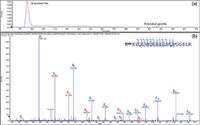
Figure 1: (a) Extracted ion electropherogram of N-terminal peptide with Glu and pyroGlu separated by CESI and (b) MS-MS identification of N-terminal peptide with pyroGlu.
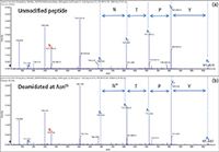
PTM Characterization: Data from CESI-MS analysis showed the presence of several PTM hotspots such as N-terminal pyroGlu formation, methionine oxidation, and asparagine deamidation. Pyroglutamination leads to loss of a positive charge which results in the electrophoretic mobility of the modified peptide being lower than the unmodified one. This is advantageous since the modified and unmodified forms can be separated by CESI–MS and the MS-MS spectra confirmed the presence of the pyroGlutamate residue (Figure 1). Oxidative degradations at Met255 and Met431 and deamidations at Asn55 and Asn387 in the heavy chain and at Asn30 in the light chain were also identified. A typical identification from the CESI–MS data is shown in Figure 2.

Figure 2: MS-MS identification of asparagine deamidation with (a) showing unmodified peptide and (b) deamidation at Asn55 in the heavy chain of trastuzumab.
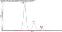
Glycosylation Heterogeneity: Trastuzumab possesses one N-glycosylation site at Asn300 in the HC where different glycoforms such as a-fucosylated or fucosylated glycans can be present (3). By using CESI-MS, the G0F, G1F, and G2F forms of the peptide TKPREEQYNSTYR were separated well as shown in Figure 3. Furthermore, the identification of G0F, G1F, and G2F forms of peptide EEQYNSTYR without the missed cleavage (at the arginine residue in the peptide TKPREEQYNSTYR) also confirmed the presence of these glycoforms. In addition, the a-fucosylated forms of this peptide, such as G0 and G1, were identified, but it has to be further confirmed that the a-fucosylated forms were not generated due to source fragmentation of fucosylated counterparts.

Figure 3: Extracted ion electropherograms of peptide TKPREEQYNSTYR with G0F, G1F, and G2F modifications.
Conclusions
We have presented CESI–MS, a robust ultra-low flow and highly efficient separation technology in combination with TripleTOF MS, a high resolution accurate mass measurement system for qualitative analysis of biopharmaceuticals. CESI–MS is attractive for simultaneous analysis of primary sequence coverage and glycopeptide profiling, without carry-over concerns. The combination of high separation efficiency and high sensitivity allows the analysis of all peptides including modified and low abundant species, in addition to confirming the amino acid sequence of the antibody.
References
(1) J.M. Busnel, B. Schoenmaker, R. Ramautar, A. Carrasco-Pancorbo, C. Ratnayake, J. Feitelson, J. Chapman, A. Deelder, and O. Mayboroda, Anal. Chem. 82, 9476–9483 (2010).
(2) A. Beck, S. Sanglier-Cianferani, and A. Van Dorsselaer, Anal. Chem. 84, 4637–4646 (2012).

AB Sciex
500 Old Connecticut Path, Framingham, MA 01701
tel. (877) 740-2129, fax (800)343-1346
Website: www.sciex.com/cesi

SEC-MALS of Antibody Therapeutics—A Robust Method for In-Depth Sample Characterization
June 1st 2022Monoclonal antibodies (mAbs) are effective therapeutics for cancers, auto-immune diseases, viral infections, and other diseases. Recent developments in antibody therapeutics aim to add more specific binding regions (bi- and multi-specificity) to increase their effectiveness and/or to downsize the molecule to the specific binding regions (for example, scFv or Fab fragment) to achieve better penetration of the tissue. As the molecule gets more complex, the possible high and low molecular weight (H/LMW) impurities become more complex, too. In order to accurately analyze the various species, more advanced detection than ultraviolet (UV) is required to characterize a mAb sample.


