Size-Exclusion Chromatography–Mass Spectrometry Analysis of mAbs on a Biocompatible Flow Path
Size-exclusion chromatography (SEC) is an essential analytical tool in discovering and developing recombinant therapeutic proteins, such as monoclonal antibodies (mAbs). SEC is commonly carried out using nonvolatile phosphate buffers, which makes the separation incompatible with mass spectrometry (MS). The use of volatile MS-compatible buffers, such as ammonium acetate, has been shown to be significantly less effective compared to phosphate buffers when applied on classic stainless-steel instrument/column configurations due to nonspecific interactions. This article details a recent experiment that demonstrates how these interactions can be prevented by using metal-free or low adsorption flow paths.
Over the past three decades, monoclonal antibodies (mAbs) have emerged as promising treatments for a range of illnesses, from cancer to autoimmune diseases, COVID-19, and beyond. Regulatory agencies in the U.S. and Europe have approved more than 100 mAbs (1), and more are on the way (2).
Size-exclusion chromatography (SEC) and mass spectrometry (MS) are both essential tools in characterizing mAbs. In recent years, there has been a trend to combine these techniques to simultaneously assess multiple structural attributes. Doing so successfully requires fully metal-free or low adsorption, flow paths that prevent unwanted secondary interactions. This articles describe such a flow path for native SEC–MS analysis of mAbs.
Performance Hurdles in SEC and MS for mAbs
SEC is widely used for studying aggregation and fragmentation associated with mAbs (3–6). The technique can detect the presence of size variants in mAbs that can have adverse effects on safety and efficacy. SEC is conducted under nondenaturing aqueous conditions, often using a phosphate buffer at neutral pH and high ionic strength.
Because SEC does not rely on interactions with the stationary phase, it does not require the application of a mobile phase gradient. Instead, molecules travel through the column at a speed dependent on particle pore accessibility. Very large molecules are excluded first, while smaller molecules penetrate pores to various degrees depending on size.
However, unwanted secondary interactions with the stationary phase can take place, drastically impacting separation by causing tailing peaks, poor recoveries, and shifted elution times. These interactions, commonly electrostatic or hydrophobic, can be suppressed by changing the mobile phase composition by adding salts or organic solvent (7). More recently, however, novel and more inert SEC materials have been introduced that limit or prevent secondary interactions while using common mobile phase compositions. In this way, potential active sites on the stationary phase are shielded (8–9).
The nonvolatile buffers and salts used in SEC interfere with MS performance, rendering the two techniques incompatible. For this reason, a desalting step (online or offline) is required for peak identification. Typically, this is done with two-dimensional liquid chromatography (2D-LC)–MS, where first‑dimension SEC peaks are collected in loops installed on a multiple heart-cutting valve and then desalted using reversed-phase LC or SEC in the second dimension (10–11).
SEC can be directly combined with MS by using volatile mobile phases such as ammonium acetate (12). However, this practice has shed light on another type of nonspecific interaction: that of mAbs with stainless-steel surfaces, which are omnipresent in the LC system, capillary tubing, and column hardware (13–14). This problem is often masked when using a phosphate buffer at high ionic strength. Successful native SEC–MS measurements of mAbs, therefore, demand fully biocompatible flow paths.
Uniting Two Seemingly Incompatible Methods
The combination of a biocompatible LC instrument with a polyether ether ketone (PEEK)-lined column was evaluated for native SEC–MS analysis of three mAbs: trastuzumab, NISTmAb, and the antibody‑drug conjugate (ADC) brentuximab vedotin. The results confirmed the importance of using a biocompatible LC system and columns for mAbs analysis.
Experimental:
The Agilent 1290 Infinity II Bio LC System, with an iron-free flow path that reduces the risk of unwanted interactions, was used (15). To enable chromatography at very high pressures, the LC system employed the metal alloy MP35N instead of stainless steel. This was combined with a 4.6 × 150 mm, 1.9-μm AdvanceBio SEC column (Agilent), in which PEEK covers the stainless-steel surfaces. The combination of this system and column demonstrated superior biocompatibility, chemical resistance, and mechanical and thermal stability.
Results:
Figure 1 shows the SEC UV 280 nm chromatograms of several mAbs in overlay with the gel filtration standard obtained on the biocompatible system and column using 150 mM sodium phosphate (the golden standard mobile phase in SEC) and 100 mM ammonium acetate as the MS-compatible alternative. The successful use of the latter mobile phase opens opportunities for directly combining SEC with MS without the need for a desalting step.
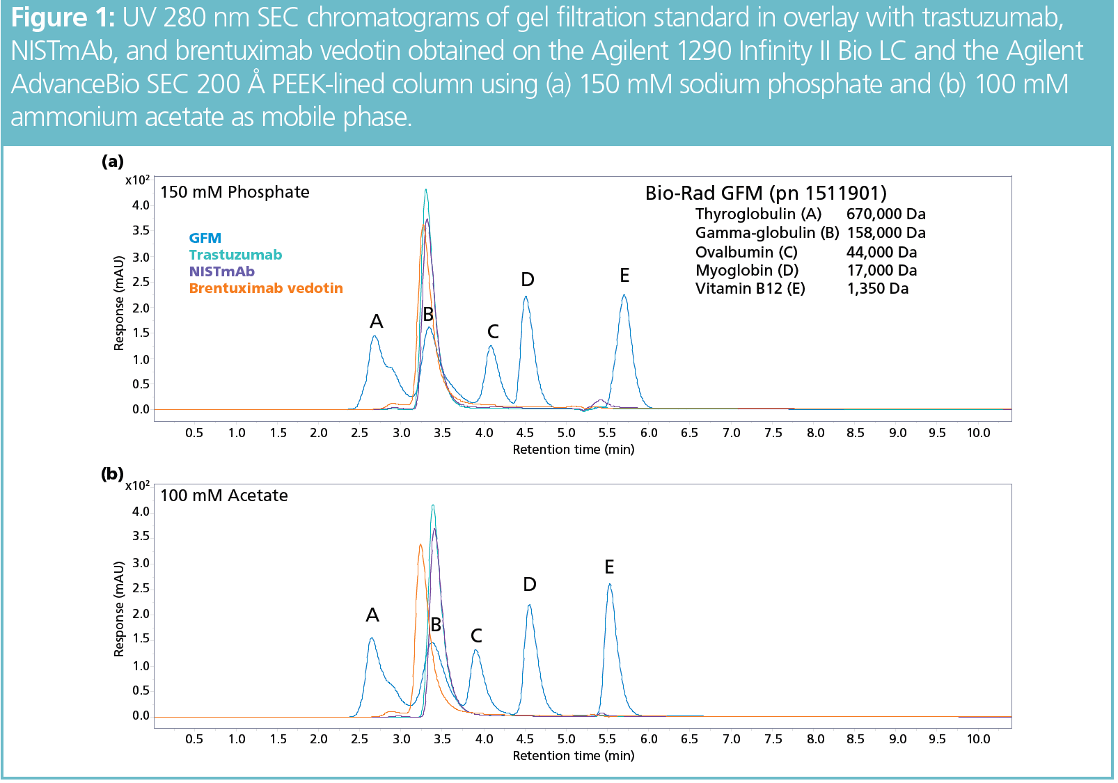
Note the satisfactory separation performance for all mAbs, including the ADC, which was facilitated by the hydrophilic polymeric coating of the particles. The hydrophobic nature of ADCs made their analysis challenging on earlier-generation SEC particles due to secondary interactions with the stationary phase.
To evaluate the impact of the absence or presence of stainless-steel components in the SEC flow path, mAb samples were analyzed on several system or column combinations. Figures 2 and 3 show the analysis of, respectively, trastuzumab and NISTmAb on a classic stainless-steel system and column (left), a stainless-steel system and PEEK-lined column (middle), and a bio-inert, biocompatible, metal-free LC system and PEEK-lined column (right).
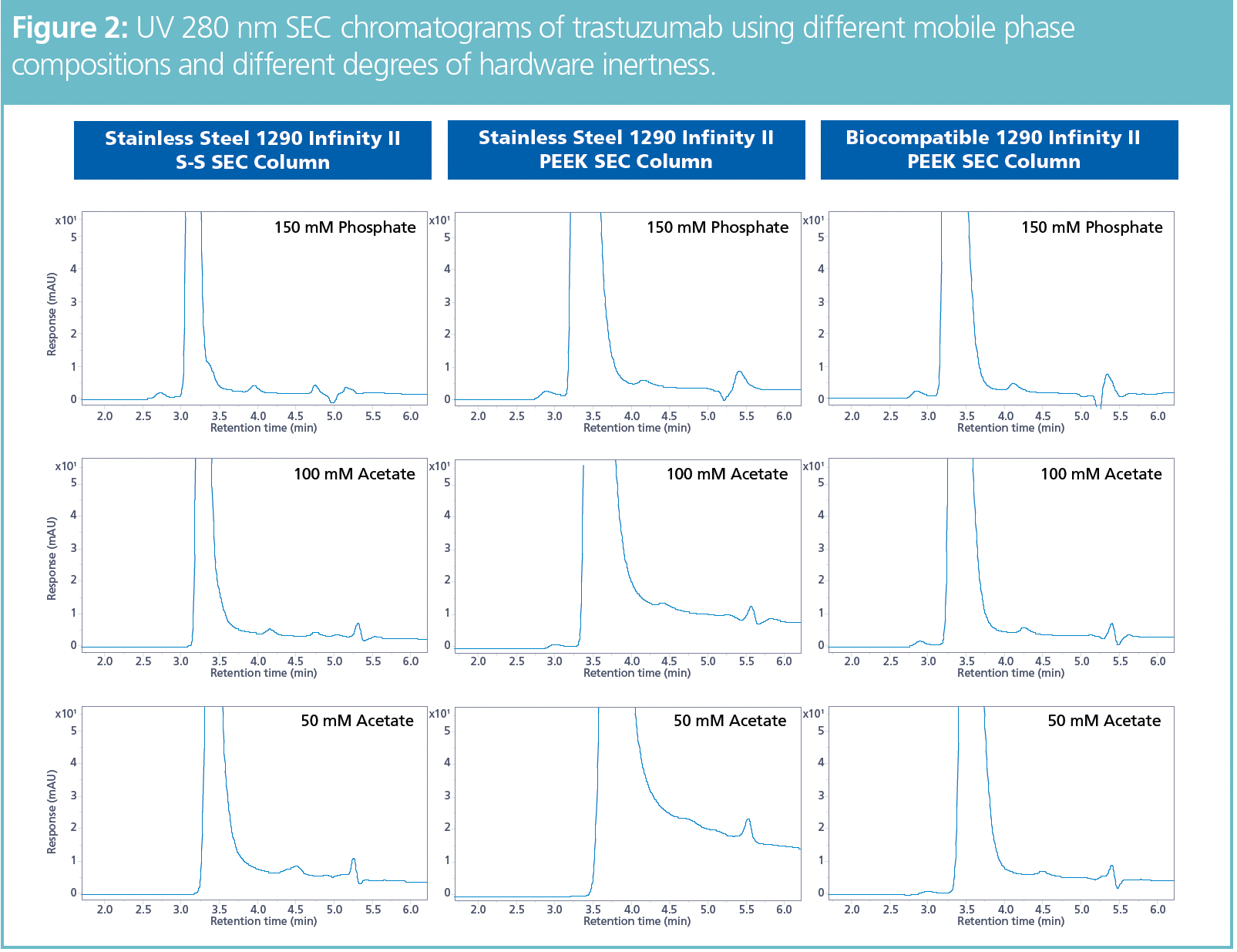
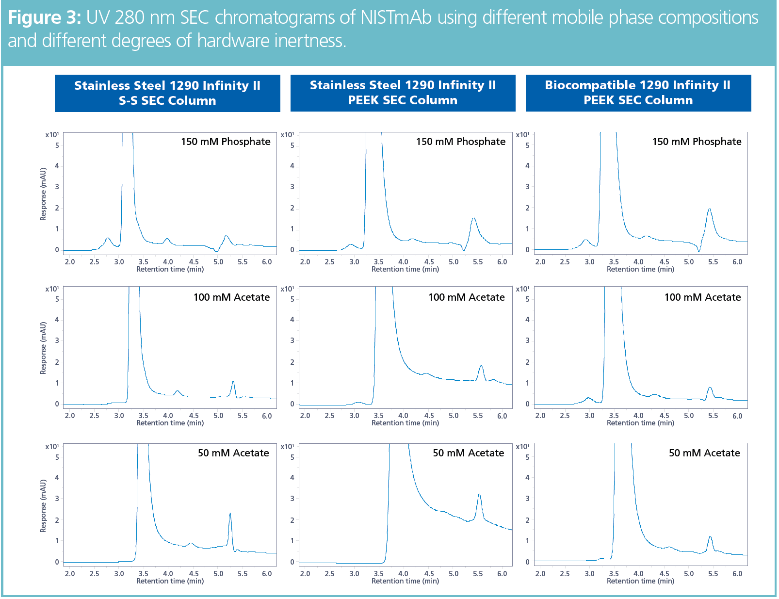
All combinations were evaluated with a 150 mM phosphate mobile phase and with 50 mM and 100 mM ammonium acetate phases as MS-compatible alternatives. Sodium phosphate resulted in satisfactory separation performance on all combinations tested, with good recovery of high-molecular-weight (HMW) species.
When sodium phosphate was replaced with ammonium acetate and the mobile phase ionic strength was reduced, the quality of the separation was adversely affected where there were stainless-steel components in the flow path. The impact of the column hardware was such that HMW species were scarcely detected, if at all, when a stainless-steel column was used with ammonium acetate as mobile phase.
HMW species reappeared when a PEEK‑lined column was installed and run with 100 mM ammonium acetate. With the 50 mM ammonium acetate mobile phase—which was the most beneficial in terms of MS sensitivity—only the biocompatible LC system and PEEK-lined column combination (bottom‑right chromatograms of Figures 2 and 3) provided a useful result, with detection of the HMW impurities.
Peak widths in SEC chromatograms were consistently smaller when the biocompatible LC system was applied. Peak tailing also did not increase upon replacing the phosphate mobile phase with MS-compatible acetate. Both peak width and tailing deteriorated significantly when the mobile phase was altered on a stainless-steel LC system. Reducing the ionic strength of the mobile phase amplified this effect (Figures 2 and 3).
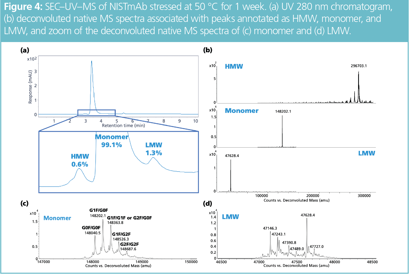
SEC–UV–MS analysis of NISTmAb is presented in Figure 4. The UV 280 nm chromatogram shows HMW and low-molecular-weight (LMW) variants that could be identified as mAb dimer and Fab fragments, respectively, based on the deconvoluted MS spectra. The spectrum associated with the main peak highlights several glycoforms decorated with the bi‑antennary complex N-glycans G0, G0F, G1F, and G2F. The spectrum also reveals the existence of mAb variants carrying C-terminal lysine.
Conclusion
LC instrument and column inertness are both essential when analyzing mAbs by native SEC–MS. The reduction of nonspecific interactions with stainless-steel components in the flow path will help guarantee adequate chromatographic performance and recovery of HMW species. In addition, conventional phosphate mobile phases can successfully be exchanged with MS-compatible ammonium acetate with limited impact on separation performance, resulting in excellent native MS measurements.
References
(1) Lyu, X.; Zhao, Q.; Hui, J.; et al. The Global Landscape of Approved Antibody Therapies. Antib. Ther. 2022, 5 (4), 233–257. DOI: 10.1093/abt/tbac021
(2) The Antibody Society, Antibody Therapeutics Approved or in Regulatory Review in the EU or US. https://www.antibodysociety.org/resources/approved-antibodies/ (accessed 2023-04-17).
(3) Sandra, K.; Vandenheede, I.; Sandra, P. Modern Chromatographic and Mass Spectrometric Techniques for Protein Biopharmaceutical Characterization. J. Chromatogr. A 2014, 1335, 81–103. DOI: 10.1016/j.chroma.2013.11.057
(4) Fekete, S.; Beck, A.; Veuthey, J.-L.; Guillarme, D. Theory and Practice of Size Exclusion Chromatography for the Analysis of Protein Aggregates. J. Pharm. Biomed. Anal. 2014, 101, 161–173. DOI: 10.1016/j.jpba.2014.04.011
(5) Fekete, S.; Guillarme, D.; Sandra, P.; Sandra, K. Chromatographic, Electrophoretic, and Mass Spectrometric Methods for the Analytical Characterization of Protein Biopharmaceuticals. Anal. Chem. 2016, 88, 480–507. DOI: 10.1021/acs.analchem.5b04561
(6) Goyon, A.; Fekete, S.; Beck, A.; Veuthey, J.-L.; Guillarme, D. Unravelling the Mysteries of Modern Size Exclusion Chromatography – the Way to Achieve Confident Characterization of Therapeutic Proteins. J. Chromatogr. B 2018, 1092, 368–378. DOI: 10.1016/j.jchromb.2018.06.029
(7) Arakawa, T.; Ejima, D.; Li, T.; Philo, J.S. The Critical Role of Mobile Phase Composition in Size Exclusion Chromatography of Protein Pharmaceuticals. J. Pharm. Sci. 2010, 99, 1674–1692. DOI: 10.1002/jps.21974
(8) Goyon, A.; Beck, A.; Colas, O.; et al. Evaluation of Size Exclusion Chromatography Columns Packed with Sub-3 μm Particles for the Analysis of Biopharmaceutical Proteins. J. Chromatogr. A 2017, 1498, 80–89. DOI: 10.1016/j.chroma.2016.11.056
(9) Schneider, S.; Jegle, U.; Dickhut, C. Size Exclusion Chromatography Analysis of Antibody Drug Conjugates. Agilent Technologies Application Note, 5991-8244EN, 2017.
(10) Vanhoenacker, G.; Vandenheede, I.; Sandra, P.; Sandra, K.; Steenbeke, M. Characterizing Monoclonal Antibodies and Antibody-Drug Conjugates Using 2D-LC–MS. LCGC Eur. 2017, 30, 149–157.
(11) Vanhoenacker, G.; Sandra, P.; Sandra, K. Minimizing the Risk of Missing Critical Sample Information by Using 2D-LC. LCGC Eur. 2022, 35, 348–353. DOI: 10.56530/lcgc.na.vg2884v4
(12) Vandenheede, I.; Sandra, P.; Sandra, K. Denaturing and Native Size-Exclusion Chromatography Coupled to High-Resolution Mass Spectrometry for Detailed Characterization of Monoclonal Antibodies and Antibody–Drug Conjugates. LCGC Eur. 2019, 32, 304–311.
(13) Goyon, A.; D’Atri, V.; Colas, O.; et al. Characterization of 30 Therapeutic Antibodies and Related Products by Size Exclusion Chromatography: Feasibility Assessment for Future Mass Spectrometry Hyphenation. J. Chromatogr. B 2017, 1065–1066, 35–43. DOI: 10.1016/j.jchromb.2017.09.027
(14) Murisier, M.; Fekete, S.; Guillarme, D.; D’Atri, V. The Importance of Being Metal-Free: The Critical Choice of Column Hardware for Size Exclusion Chromatography Coupled to High Resolution Mass Spectrometry. Anal. Chim. Acta 2021, 1183, 338987. DOI: 10.1016/j.aca.2021.338987
(15) Nägele, E. Elevate Your mAb Aggregate Analysis. Agilent Technologies Application Note, 5994-2709EN, 2020.
Liesa Verscheure is a scientist at RIC group (Kortrijk, Belgium).
Gerd Vanhoenacker is senior scientist at RIC group.
Pat Sandra is founder and advisor of the RIC group and emeritus professor at Ghent University (Ghent, Belgium).
Koen Sandra is CEO and scientific director at RIC group and visiting professor at Ghent University.
Sonja Schneider is manager, Applications Development at Agilent Technologies. She has led the application development team in Waldbronn, Germany, since 2016.
Udo Huber is the director of LC Application Solutions at Agilent Technologies.

New Method Explored for the Detection of CECs in Crops Irrigated with Contaminated Water
April 30th 2025This new study presents a validated QuEChERS–LC-MS/MS method for detecting eight persistent, mobile, and toxic substances in escarole, tomatoes, and tomato leaves irrigated with contaminated water.

.png&w=3840&q=75)

.png&w=3840&q=75)


.png&w=3840&q=75)


.png&w=3840&q=75)


