SEC-MALS of Antibody Therapeutics—A Robust Method for In-Depth Sample Characterization
Monoclonal antibodies (mAbs) are effective therapeutics for cancers, auto-immune diseases, viral infections, and other diseases. Recent developments in antibody therapeutics aim to add more specific binding regions (bi- and multi-specificity) to increase their effectiveness and/or to downsize the molecule to the specific binding regions (for example, scFv or Fab fragment) to achieve better penetration of the tissue. As the molecule gets more complex, the possible high and low molecular weight (H/LMW) impurities become more complex, too. In order to accurately analyze the various species, more advanced detection than ultraviolet (UV) is required to characterize a mAb sample.
Multi-angle light scattering (MALS) is suited to determine the molecular weight of sample components to better understand molecule aggregation as well as fragmentation. MALS detects the light a molecule scatters if hit by a laser beam. The intensity of scattered light depends on its concentration and on its molecular weight. In this application note, we coupled the TSKgel® UP-SW3000-LS column and the LenS3® MALS detector for the analysis of a mAb standard. We demonstrate which data can be derived from adding MALS detection, how robust the method is, and which equipment is suited to easily integrate MALS detection into the mAb analysis workflow.
Experimental HPLC Conditions
To demonstrate the benefits of MALS detection combined with size-exclusion analysis of a monoclonal antibody, we employed a reference mAb and a universal method for separation as described below. Adaptations were required to optimize MALS data quality because light scattering is more sensitive for particles such as salt crystals, microbes, or column shedding than UV detection. The mobile phase was filtered twice, and a light-scattering dedicated UHPLC column (TSKgel UP-SW3000-LS) was employed. The TSKgel UP-SW3000-LS column had previously proven the lowest noise levels in comparison to two other columns. The LenS3 MALS detector was selected for its high sensitivity to achieve characterization of
low-abundance impurities.
Column: TSKgel UP-SW3000-LS, 2 μm, 4.6 mm ID × 30 cm
Mobile phase: 100 mmol/L phosphate buffer, pH 6.7, 100 mmol/L, Na2SO4 (filtered twice using a 0.1 μm pore size PES vacuum filter)
Flow rate: 0.35 mL/min
Detection: UV-absorbance @ 280 nm, multi-angle lightscattering with LenS3 (RALS for MW)
Injection vol.: 10 μL
Sample: SILu™ Lite SigmaMAb (1 mg/mL), IgG1 mAb standard (Sigma #MSQC4)
Instrument: Thermo Scientific™ Vanquish™ UHPLC
Results
Molecular Weight Determination of High- and Low-Molecular Weight Impurities of mAbs
To analyze the molecular weight (MW) profile of the antibody sample, the system was first calibrated with BSA. A solution of 1 mg/mL was analyzed according to the method described above and the monomer peak was selected to calibrate the system. Calibration corrects for signal offset and band broadening due to dead volume between the detectors, and it determines the UV and MALS response factors, as well as the normalization factors of the MALS detector.
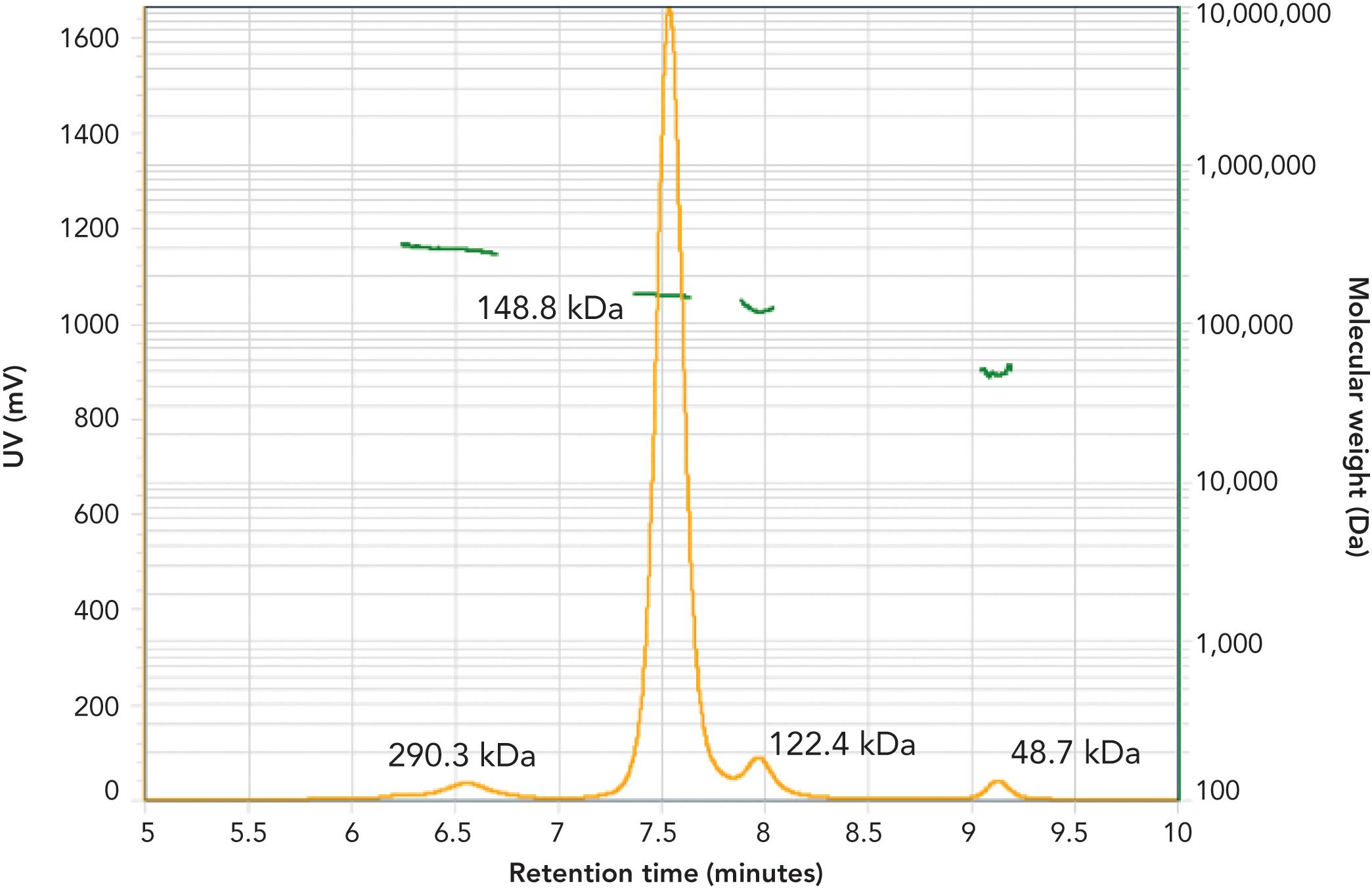
The mAb sample was analyzed with UV absorbance at 280 nm and MALS. The UV trace (Figure 1, yellow) shows a monomer peak eluted at 7.6 min, high MW impurities eluted around 6.6 min, and two low MW impurities, one eluted at 7.9 min, another at 9.2 min.
Figure 1: Molecular weight determination of high- and low-molecular weight impurities of mAbs.

Employing the concentration calculated from the UV signal (extinction coefficient = 1.5 mL/mg) and the right angle light scattering signal (RALS), the MW (green) for the identified species was calculated in the SECview® software. The MW of the monomer was determined to be 148.8 kDa, which is in line with the weight of the antibody without modifications (146 kDa) plus glycosylation modifications. The HMW species eluted at 6.6 min was calculated to have 290.3 kDa, indicative of a mAb dimer. One of the LMW impurities was 122.4 kDa which may be assigned to a mAb missing a light chain, and the smallest peak is 48.7 kDa, the weight of the Fab fragment or two aggregated light chains.
Sensitivity and Robustness of SEC-MALS Analysis of mAbs
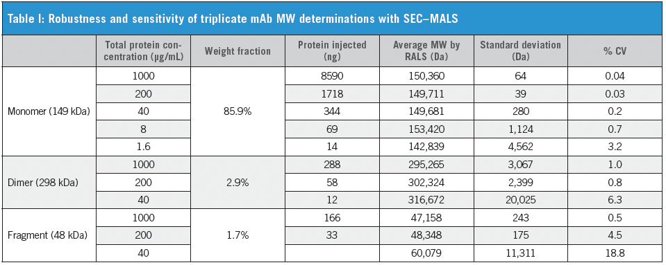
Next, the stability and sensitivity of SEC–MALS analysis of mAbs were determined by injecting triplicates of a 1:5 serial dilution of the mAb sample. Table I shows the MW determination of monomer, dimer, and fragment at the different concentrations that were injected. As these molecules are present in the sample at different weight ratios, the injected amounts of each of them at the given total protein concentration were calculated and are outlined as well.

For the monomer, we find a MW determination within +/-5% of the expected MW (149 kDa) for a sample as small as 14 ng. However, the accuracy decreases, and the standard deviation increases with decreasing amounts of the analyte. A similar trend is seen for the MW determination of the dimer. The lowest amount injected was 12 ng which led to a calculated MW of 317 kDa (+6 %) and a high variation of 6.3%. In contrast, at higher concentrations, the determination for the dimer MW was accurate(< 2% deviation from expected value) with a low coefficient of variation(% CV < 1.1 %). Similarly, the small Fab fragment (48 kDa) shows reliable MW determination down to 33 ng, whereas the lowest injected amount of 7 ng results in poor MW determination and high variations in triplicate measurements. This is explained not only by the low analyte amount but also by the lower amount of scattered light of smaller molecules.
Conclusion
Analyzing monoclonal antibody samples with SEC-MALS directly determines the MW of sample components. This facilitates the assignment of peaks to the structures of impurities. Employing the sensitive LenS3 MALS detector, the method is sensitive down to less than 50 ng of dimerized antibody. MW determination of monomer and impurities are possible at concentrations down to 15 ng but with less accuracy and higher variation.
TSKgel and Tosoh Bioscience are registered trademarks of Tosoh Corporation.
LenS is a registered trademark of Tosoh Bioscience LLC in the USA, India, and Japan.
SECview is a registered trademark of Tosoh Bioscience LLC in the USA, EU, and India; and of Tosoh Corporation in Japan.
SILu is a trademark of Sigma-Aldrich.
Thermo Fisher and Vanquish are trademarks of Thermo Fisher Scientific.

Tosoh Bioscience LLC
3604 Horizon Drive, Suite 100, King of Prussia, PA 19406
Tel. (484) 805-1219, fax (484) 805-1277
Website: www.tosohbioscience.com

.png&w=3840&q=75)

.png&w=3840&q=75)


.png&w=3840&q=75)


.png&w=3840&q=75)

