Practical Electromigration Techniques to Separate Microorganisms in Medical Analysis
LCGC Europe
This article looks at the use of electromigration techniques to determine microorganisms, such as viruses, bacteria and other biologically important macromolecules (erythrocytes), in medical analyses. It was found that electromigration techniques could be used for the identification of several viruses, including the identification of a specific marker for the Hepatitis C virus infection and another for a urinary tract infection. The determination of cell viability and the quality control of probiotics and consumer products that contain active bacteria is also possible using electromigration. Special attention is paid to the modification of capillary wall surfaces using different monomers and the application of monolithic columns to determine active bacteria in pharmaceutical products using capillary electrochromatography (CEC) conditions. This approach represents a new frontier for separation science and the possibility to apply it in medical diagnosis.
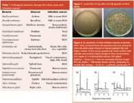
Microorganisms, such as bacteria, viruses and fungi, are widespread throughout nature and the environment. Some microbes are included in health products, medicines and supplements as the active ingredients and they generate the useful components for human health. The majority of microorganisms perform essential activities in nature and many are beneficial to the animals they interact with. However, potentially harmful microorganisms can have profound effects on humans and may be the cause of different dangerous diseases (Table 1).1
For example, Escherichia coli can easily contaminate beef and milk and is the pathogen most frequently linked to urinary tract infections. Salmonella causes a dangerous, infectious disease that continues to plague human populations in developed and developing countries. Another example of pathogenic bacteria is Helicobacter pylori, which causes severe diseases of the gastric tract ranging from chronic gastric ulcer to gastric cancer.2
Some pathogenic bacteria are the major cause of death in many countries (Bacillus anthracis, Rickettsia rickettsi, Salmonella thypi).1 Thus, identification, characterization and monitoring of pathogenic bacteria and other microorganisms or living cells are very important in clinical diagnosis, analysis in the food industry and quality control of several processes.1
Conventional bacterial identification methods usually require time consuming and laborious procedures so are not capable of fast diagnosis in emergency instances. Traditional methods to determine bacteria involve following basic steps: preenrichment, selective enrichment, biochemical screening, isolation of pure culture and serological confirmation. Hence, a series of tests is required and the results of such tests are often very difficult to interpret (Figure 1).

Time and sensitivity of analysis are the most important limitations related to the usefulness of microbiological testing. Therefore, in order to increase the speed, selectivity and sensitivity of microbial assays, many different techniques have been used, such as polymerase chain reaction detection (PCR),3 flow cytometry,4,5 differential staining,6 serological methods and recently dielectrophoresis,7–21 field-flow fractionation (FFF), a combined dielectrophoretic and field-flow microsystems,24,25 microchip-based technology,26 and matrix-assisted laser desorption/ionization (MALDI) mass spectrometry.27 In recent years electromigration techniques, in particular, capillary electrophoresis (CE) has shown increasing potential in the determination of charged particles such as erythrocytes, haemoglobin and its variants, serum proteins, urine proteins, lipoproteins, molecular diagnosis and microorganisms that cause infectious diseases.28 Molecular diagnosis by electromigration techniques is the newest and most rapidly growing area in laboratory medicine. These assays give very important information that could not be obtained using standard methodologies. In this study the detection, characterization and quantification of cells are used to diagnose and manage several diseases. Fully automated fast diagnosis of microbial diseases without isolation of pure culture is of obvious importance.
History of Electromigration Techniques to Separate Microorganisms
Early work in microbial assays was performed by Hjerten and co-workers in 1987.29 They showed that Tabacco mosaic virus (TMV) and Lactobacillus casei migrated through a methylcellulose-coated 100 µm i.d. capillary and proved that the orientation of the TMV virus affected its electrophoretic mobility.29 Kenndler et al.30 performed the determination of the isoelectric point of Human Rhinovirus by capillary isoelectric focusing (CIEF) using fused silica capillaries dynamically coated with hydroxypropylmethyl cellulose (Figure 2).
Three years later the same group demonstrated that the extremely large common cold virus (HRV) can be successfully analysed by capillary electrophoresis.31,32 They describe a novel high-resolution technique for using a ternary mixture of detergents as a background electrolyte. The detergents were very important for the prevention of aggregation of subviral particles and adsorption to the capillary wall. Affinity capillary electrophoresis was performed by Okun et al.33 This is a relatively new technique that gives some information about the interaction of a wide variety of molecules and macromolecules. They describe some interaction between HRV and certain monoclonal antibodies. Mann et al. demonstrated a capillary zone electrophoresis-based method for the analysis of intact adenovirus.34 They used polyvinyl alcohol (PVA)-coated capillary that eliminates the need for detergent additives in the running buffer. This system allows for the observation of new viral peaks appearing after a freeze-thaw cycle indicating discrete modifications to the viral surface. G.E. Shiha and co-workers determined Hepatitis C Virus (HCV) in urine samples from infected individuals.35 The characteristic peak (migration time 2.72 min) was collected from urine samples using CZE and was subjected to the nested PCR technique. The amplified product of the fractionally collected peak was detected by agarose gel electrophoresis (Figure 3).35 Detection of HRV RNA using CZE may be very suitable for use in the screening of blood units to identify infecting samples that do not contain specific antibodies.
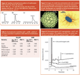
It was known that the human HBO-type system is a polymorphism of complex carbohydrate structures at the surfaces of erythrocytes and that the differences between these form types are very subtle. Wan-Hua Lu and co-workers used CE for cell studies and compared erythrocytes from different sources.36 Erythrocyte sample from sheep, duck and human sources showed characteristic migration times and the migration times of A-,B-, AB- and 0-type erythrocytes from human blood were different (Figure 4). It was found that CE is a powerful and practical alternative for characterizing and quantifying erythrocytes or other cells, thereby providing additional information for clinical diagnosis and therapy.
Small viruses have diameters in the range of several tens of nanometres. Bacteria are larger by a factor of approximately 100 and the increasing size leads to increased complexity. Viruses exist mainly in one of two forms (helical and/or icosahedral) and they typically contain only a few types. Viral capsids are composed entirely of protein and they typically contain only a few types. By their very nature viruses are highly uniform and simplistic. The outer membrane of bacteria has a large number of lipids, proeins and techoic acids. Therefore, analysis of bacteria is more complicated. Bacteria can adopt an enormous variety of shapes and sizes, both among and within species. This physiological difference makes characterization and identification of bacterial cells by electromigration techniques more difficult (Figure 5).

As with other colloidal particles bacteria have a surface charge that originates from the ionization of surface molecules and of the adsorption of ions from solution. Bacterial cell wall and membranes contain numerous proteins, lipid molecules, teichoic acids and lipopolysaccharides, which give them characteristic charge (Table 3). Therefore, bacterial cells undergo electrophoresis in a free solution with their own mobility depending on ionic strength and the pH of the buffer solution.
Many of the bacteria can aggregate to form long chains or clusters. They can easily attach to other microbes and surfaces such as the fused silica capillary wall. Some microorganisms can secrete substances that result in cell lysis, altered migration times or unwanted peaks.39,40
Armstrong and co-workers applied two electromigration techniques for the separation of several species of bacteria.40,41 They used a direct injection capillary electrophoresis system for the rapid identification of the bacterial pathogens, responsible for urinary tract infections (UTI).42 Because urinary tract infections are caused by strains of E. coli or by Staphylococcus saprophyticus and since urine is a water matrix with macromolecular or colloidal contaminants, capillary electrophoresis is an ideal technique for high-efficiency separation. They obtained very good electropherograms showing the different migration times of the pathogen bacteria and showed that urea and various inorganic salts in urine sample migrated with electroosmotic flow (Figure 6). Of course, the amount and the composition of dissolved solids in urine sample depends on an individual's liquid intake and food intake.
A number of health consumer products contain active ingredients that are microbes because specific bacteria are beneficial to human health. However, there are no effective methods for the determination of the active bacterial ingredients. Armstrong at al. reported the first microbial assays of tablets (pills) and powder-based commercial products for identifying Lactobacillus and Bifidobacterium in a powdered formula supplement.43 They showed that both the bacteria and the maltodextrin from Schiff tablets can be identified and quantified in the same run.43 Another example is in the analysis of L. acidophilus from Schiff tablets. The electropherograms showed that microorganisms can be determined in widely different matrices and the migration times can vary slightly in the presence of certain matrices or contamination (Figure 7).

All of the microbes which are found in consumer products are helpful only if they are alive. Armstrong and co-workers used fluorescent dyes and capillary electrophoresis coupled with laser-induced fluorescence (LIF) detection for the determination of bacterial viability.37,38,42,43 SYTO 9 stains all bacteria while propidium iodide stains only bacteria with damaged membranes. Viable bacterial cells produce an enhanced green fluorescence while nonviable (dead) bacterial cells produce a red fluorescence. By simultaneously monitoring and by the comparing the ratio of viable to nonviable cells they showed that only about 60% of L. acidophilus is viable.
Shintani and co-workers used the same system (CE-LIF) for the selective identification of Salmonella enteritidis.45 They used two fluorescent staining methods: a cell-permeable nucleic acid stain and a salmonellae-specific polyclonal antibody. The CE-LIF system successfully detected as few as three cells per injection from a pure culture of S. entritidis.
A very important application of microbial assays is the analysis of health-related microbes called probiotics. Moon and Kim described the rapid determination of probiotics such as Saccharomyces cerevisiae and Enterococcus faecalis using uncoated fused silica capillaries with a polyethylene glycol (PEG) additive to the running buffer.46 They showed the possibility of using capillary electrophoresis as a potential for the quality control (QC) and quality assurance (QA) of the production of a medicine containing the probiotics.
It was reported that the addition of other components, such as PEG in the running buffer, could enhance the resolution and has been used by many research groups.39–42,47,48 Without PEG addition the electroosmotic flow was too fast and all species of bacteria migrated near the EOF resulting in the broad bandwidths. When the small amount of PEG (Mw = 600000) was used, bacteria migrated as narrow zones. Nevertheless, PEG addition prevents adsorption of bacteria to the capillary wall and clusters formation (Figure 8).

The Influence of Capillary Surface Chemistry
In order to increase the speed and sensitivity of bacterial analysis Buszewski et al.47,48 investigated modification of the internal capillary surface using three different monomers: acrylamide, trimethylchlorosilane and divinylbenzene (DVB). Such an approach should result in suppressed electroosmotic flow and prevent the adsorption of bacteria to the capillary wall. The picture demonstrates the scanning electron microscope (SEM) micrograph of the capillary wall modified with DVB and corresponding separation of four species of bacteria.
Modification of the capillary wall with trimethylchlorosilane (CH3SiCl) gave similar results and we obtained a successful separation of five species of bacteria on an 8.5 cm distance in 8?min (Figure 10). All of the microbes migrated as narrow zones and the bacteria exhibited negative electrophoretic mobilities, which reflected their surface charge. As the bacteria were separated in the same buffer in modified capillaries (trimethylchlorosilane and divinylbenzene) it is likely that weak adhesion to the capillary wall may have some effects on the separation, which resulted in slightly greater values of mobilities in DVB modified capillary (Table 4).
Recently, together with miniaturization of separation systems, a lot of attention has been paid to the use of monolithic columns that are prepared by the polymerization of various monomers (hydrophobic — responsible for the separation, polar — responsible for the presence of the electroosmotic flow, and crosslinking agents) inside the capillary.49 The presence of the porogenic solvent provides the formation of a continuous, porous monolith (rod). In our recent study we used lauryl methacrylate capillary columns (monolith) described previously by Buszewski et al. for the determination of Lactobacillus rhamnosus in pharmaceutical products using capillary electrochromatography (CEC) conditions.49 The picture demonstrates the SEM micrograph of lauryl monoliths and typical electropherogram of bacteria (Figure 11).

Conclusions
All of these publications suggested that it is possible to separate and characterize different species of bacteria and other microorganisms and these assays could be used for the rapid identification of living cells, which plays a very important role in medical diagnosis and treatment. Fast diagnosis of microbial-based diseases without isolation of pure cultures is obviously needed. Because of the high efficiencies and short analysis times these techniques seem to be very promising in modern clinical laboratories and the impact on this selective characterization of living microorganisms will continue to grow.
Ewa Klodzi´nska is a PhD student of Professor Buszewski at Nicolas Copernicus University (NCU), Toru´n, Poland. Her main research focuses on miniaturization and the application of electromigration techniques and separation of microorganisms.
Boguslaw Buszewski is Head of the Department of Environmental Chemistry and Ecoanalytics, NCU. His main research area focuses on analytical chemistry, physicochemistry of surface processes, development of new stationary phases and columns, miniaturization, separation mechanisms (HPLC, TLC, GC, CZE), hyphenated techniques, sample preparation, adsorption and chemometry.
Hanna Dahm is Head of Department of Microbiology, Faculty of Biology and Earth Sciences, NCU. Her main research focuses on the production of growth substances (e.g., auxins, gibberellins, cytokinins, vitamins), biology of endophytic organisms and the identification of bacteria.
Marek Jackowski is Head of Department of Surgery, Collegium Medicum, NCU. He works as a surgeon at clinics in Toru´n. His main research focuses on oncology and the influence of microorganisms on cancer diseases.
References
1. D. Ivnitski et al., Biosensors & Bioelectronics, 14, 599–624 (1999).
2. H. Sjunnesson et al., Current Microbiol., 47, 278–285 (2003).
3. L. Jimenez, S. Smalls and R. Ignar, J. Microbiol. Methods, 41, 259–265 (2000).
4. H.B. Steen, J. Microbiol. Methods, 42, 65–74 (2000).
5. T. Katsuragi and Y. Tani, J. Biosci. and Bioeng., 89(3), 217–222 (2000).
6. I.V. Kourkine et al., Electrophoresis, 24, 655–661 (2003).
7. W.B.Betts, Trends in Food Sci. and Tech., 6, 51–58 (1995).
8. R. Pethig and G.H. Markx, TIBTECH, October, 15, 426–432 (1997).
9. G.H. Markx and C.L. Davey, Enzyme and Microbial Tech., 25, 161–171 (1999).
10. A.P. Brown et al., Biosensors & Bioelectronics, 14, 341–351 (1999).
11. Z. Yunus et al., J. Microbiol. Methods, 51, 401–406 (2002).
12. P.R.C. Glascoyne and J.Vykoukal, Electrophoresis, 23, 1973–1983 (2002).
13. J. Suehiro et al., J. Electrostatics, 57, 157–168 (2003).
14. N.G. Green, H. Morgan and J.J. Milner, J. Biochem. Biophys. Methods, 35, 89–102 (1997).
15. S. Fiedler et al., Anal. Chem., 70, 1909–1915 (1998).
16. T. Müller et al., Biosensors & Bioelectronics, 14, 247–256 (1999).
17. T. Schelle et al., Biochimica et Biophysica Acta, 1428, 99–105 (1999).
18. B. Malyan and W. Balachandran, J. Electrostatics, 51–52, 15–19 (2001).
19. H. Li and R. Bashir, Sensors and Actuators B, 86, 215–221 (2002).
20. J. Suehiro et al., Sensors and Actuators B, 96, 144–151 (2003).
21. M. Frénéa et al., Mater. Sci. and Eng. C, 23, 597–603 (2003).
22. R. V. Sharma, R. T. Edwards and R. Beckett, Wat. Res., 32(5), 1508–1514 (1998).
23. T. L. Edwards, B. K. Gale and A.B. Frazier, Anal. Chem., 74, 1211–1216 (2002).
24. J. Yang et al., Anal. Chem., 71, 911–918 (1999).
25. X. Wang et al., Anal. Chem., 72, 832–839 (2000).
26. P.C.H. Li and D.J. Harrison, Anal. Chem., 69, 1564–1568 (1997).
27. J. Bundy and C. Freselau, Anal. Chem., 71, 1460–1463 (1999).
28. R.Petersen et al., Clin. Chim. Act.,330, 1–30. (2003)
29. S. Hjerten et al., J. Chromatogr., 403, 47–67 (1987).
30. U.Schnabel et al., Anal. Chem., 68, 4300–4303 (1996).
31. V.M.Okun, D. Blaas and E.Kenndler, Anal. Chem., 71, 4480–4485 (1999).
32. V.M.Okun et al., Anal. Chem.,71, 2028–2032 (1999).
33. V.M.Okun et al., Anal. Chem., 72, 4634–4639 (2000).
34. B.Mann et al., J.Chromatogr. A, 895, 329–337 (2000).
35. M.Attallah et al., Clin. Chim. Act, 346, 171–179 (2004).
36. W.-H. Lu et al., Anal. Biochem., 314, 194–198 (2003).
37. Dennis Kunkel Microscopy (http: //www.pbrc.hawaii.edu/Vkunkel).
38. A.T. Poortinga et al., Surf. Sci. Rep., 47, 1 (2002).
39. M.J. Desai and D.W. Armstrong, Microbiology and Molecular Biology Reviews, Mar. 38–51 (2003).
40. J.M. Schneiderheize et al., Microbiology Letters, 189, 39–44 (2000).
41. M.A. Rodriguez and D.W. Armstrong, J. Chromatogr. B, 800, 7–25 (2004).
42. D.W. Armstrong and J.M. Schneiderheize, Anal. Chem., 72, 4474–4476 (2000).
43. D.W. Armstrong et al., FEMS Microbiol.Lett., 194, 33–37 (2001).
44. D.W. Armstrong and L. He, Anal. Chem., 73, 4551–4557 (2001).
45. T. Shintani, K. Yamada and M. Torimura, FEMS Microbiology Letters, 210, 245–249 (2002).
46. B. Moon and Y. Kim, Bull. Korean Chem. Soc., 24(8), 1203 (2003).
47. B. Buszewski et al., J. Sep. Sci., 26, 1045–1049 (2003).
48. M. Szumski, E. Klodzi´nska and B. Buszewski, J. Chromatogr. A (in press) 2004.
49. B. Buszewski, M. Szumski and Sz. Sus, LCGC Eur., 15(12), 782–798 (2002).

Miniaturized GC–MS Method for BVOC Analysis of Spanish Trees
April 16th 2025University of Valladolid scientists used a miniaturized method for analyzing biogenic volatile organic compounds (BVOCs) emitted by tree species, using headspace solid-phase microextraction coupled with gas chromatography and quadrupole time-of-flight mass spectrometry (HS-SPME-GC–QTOF-MS) has been developed.
Identifying PFAS in Alligator Plasma with LC–IMS-HRMS
April 15th 2025A combination of liquid chromatography ion mobility spectrometry, and high-resolution mass spectrometry (LC–IMS-HRMS) for non-targeted analysis (NTA) was used to detect and identify per- and polyfluoroalkyl substances (PFAS) in alligator plasma.


