Practical Considerations of Capillary Electrophoresis–Mass Spectrometry for Analysis of Biotherapeutics
LCGC North America
In this article, we discuss the use of CE-MS (sheath flow interface) for analysis of intact proteins as well as of protein digests. We discuss the unique aspects that the user needs to be aware of while testing biotherapeutics versus small molecule drugs. We also highlight that the optimization of CE and MS parameters together result in the creation of a more robust and reproducible protein analysis approach. Finally, we list some of the most common errors that are likely to occur during CE-MS analysis and suggest ways to overcome them.
In this installment of “Biotechnology Today,” we discuss the use of capillary electrophoresis–mass spectrometry (CE–MS) (with a sheath flow interface) for the analysis of intact proteins as well as protein digests. We discuss the unique aspects that the user needs to be aware of while testing biotherapeutics versus small-molecule drugs. We also highlight that the optimization of CE and MS parameters together results in the creation of a more robust and reproducible protein analysis approach. Finally, we list some of the most common errors that are likely to occur during CE–MS analysis and suggest ways to overcome them.
The rapid advancements in the field of biopharma have increased the demand for analytical techniques that can accurately fingerprint the analyte (1). This challenge takes on a whole new dimension when we apply it to analytical characterization of biotherapeutics, which are a set of complex molecules with diversity ranging from cytokines, growth factors, hormones, peptides, and proteins including monoclonal antibodies (mAbs) and fusion proteins (2,3). The recent spurt of patent expiries, together with the high cost of biotherapeutics have fueled the interest in biosimilars (4). The success of biosimilars depends on achieving the optimum trade-off between the need to demonstrate equivalence of quality, safety, and efficacy with the innovator product and achieving a reduction in the extent of clinical examination that the drug has to undergo. The latter is the primary contributor to the lower cost of development and commercialization of a biosimilar. A key component in achieving this balance is that of performing a thorough analytical characterization using a multitude of orthogonal, high resolution, analytical tools (5). Apart from the sophistication and orthogonality of the analytical methods used, it is also important that the analytical method be such that it allows the characterization of biologics while conserving any potentially present modifications and without changing the heterogeneity of the sample (6,7).
As highlighted by several researchers, biotech products, as well as processes, are complex (8). The process complexity essentially pertains to the often uncharacterized feed material and raw materials that are used and the complex interplay of metabolic dynamics in the host organism, that makes it difficult to predict the influence of one, or a combination of variables, on process performance. The presence of such a large number of product related variants, including oxidized, reduced, deamidated, phosphorylated, and glycosylated forms in a typical biotherapeutic, can be attributed to the high complexity of these products. For accurate identification and quantification of these attributes, it is necessary to have high selectivity and sensitivity of the analytical methods used for their characterization (3,5,9). Conventional techniques, such as liquid chromatography (LC), size-exclusion chromatography (SEC), and gel electrophoresis have been quite successful in terms of their utility in characterization of protein therapeutics, and they still continue to be essential components of a typical, characterization armory of proteins. However, these techniques suffer from one or more limitations, with respect to analysis time, sample processing requirements, feasibility of automation, selectivity, and sensitivity for deciphering the minute changes in the overall protein architecture.
During the past several decades, capillary electrophoresis (CE) has emerged as an analytical tool for characterization of large molecules (for example, protein therapeutics) (10). The major reason for this emergence is the high separation efficiency that it offers, due to minimal longitudinal diffusion, which otherwise contributes to the peak broadening during separations in other, contemporary analytical tools like high performance liquid chromatography (HPLC). In addition, since CE analysis is performed in a fused-silica capillary or in a coated capillary, under aqueous conditions (for minimizing protein adsorption), protein analysis can typically be achieved with minimal conformational changes. Finally, since CE separation is based on differences in charge, shape, and size of the analyte, it can be advantageously capitalized for differentiating among various post-translationally modified (PTM) forms of the proteins, which are likely to have different electrophoretic mobilities during separation.
Mass spectrometry (MS) has also gained wide popularity as one of the most useful detection devices in proteomics research. For biotherapeutic characterizations, MS is widely used to gain information about the primary sequence of the protein, often via peptide mapping (also known as bottom up methods). MS is also used to better understand any possible modifications, higher order structure, glycosylation, and host cell protein (HCP) analysis (11,12). However, the real power of MS is truly harnessed once it is used in conjunction with a powerful separation technique such as LC or CE. However, interfacing CE with MS is technically not as straightforward as it is to couple LC to MS. The approaches developed so far to couple CE with MS can be divided into three categories (Figure 1). First, the use of an interface based on direct spray with a metal or metallized needle for electrical contact (Figure 1a). A second approach uses a liquid junction interface, in which the background electrolyte (BGE) in the capillary is hydraulically connected to the reservoir where an electrical contact is made (Figure 1b). The third generation of CE–MS coupling makes use of a coaxial sheath flow interface, whereby a conductive solvent flows around the outside of the capillary, and it provides a hydraulic connection to an electrical contact (Figure 1c). A comprehensive description of the various CE–MS coupling approaches is available in the literature (13–16). It should also be noted here that although the sheathless-low sheath flow interfaces were developed with superior sensitivity as compared to the sheath flow interface, the commercial use of the latter far exceeds the former (17).
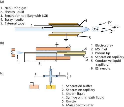
Figure 1: Illustration of the three commonly used interfaces. (a) Coaxial sheath liquid interface, (b) porous tip sheathless interface, and (c) electroosmotic flow driven sheath liquid Interface used for coupling CE to MS.
In this article, we discuss the use of CE–MS with a sheath flow interface for the analysis of intact proteins as well as protein digests. We discuss the unique aspects that users need to be aware of while testing biotherapeutics versus small-molecule drugs. We also highlight that the optimization of CE and MS parameters together results in the creation of a more robust and reproducible protein analysis. Finally, we list some of the most common errors that are likely to occur during CE–MS analysis and suggest ways to overcome them.
Materials and Methods
Protein Samples
The therapeutic protein sample used in this study was an IgG1 mAb (derived from CHO cell culture). The mAbs were donated to us by a major domestic biotech manufacturer. Sample preparation reagents were of reagent grade, and CE–MS buffer reagents were of LC–MS grade.
Instrumentation
An Agilent capillary electrophoresis (G7100) system coupled to an Agilent 6230B time-of-flight (TOF) mass spectrometer through Agilent’s CE–MS adapter kit (G1603A) was used for performing CE–MS analysis. An Agilent 1260 Infinity II binary pump (G4220A) was used for sheath liquid delivery employing a splitter with a 99:1 ratio, effectively producing a sheath flow rate of 4 µL/min.
Sample Preparation
- Intact mAb: mAb sample (2 mg/mL) was buffer exchanged in 50 mM ammonium acetate, pH 4.
- mAb digest: mAb sample (1 mg/mL) was denatured, reduced, and alkylated according to the standard protocol (18). The reduced mAb was digested using trypsin (trypsin–protein = 1:50) and incubated overnight at 37 °C. Before analysis, the digested mAb was buffer exchanged to 50 mM ammonium acetate, pH 4.0. The digested sample was desalted before analysis using ZipTip according to the manufacturer’s protocol.
Analysis Conditions
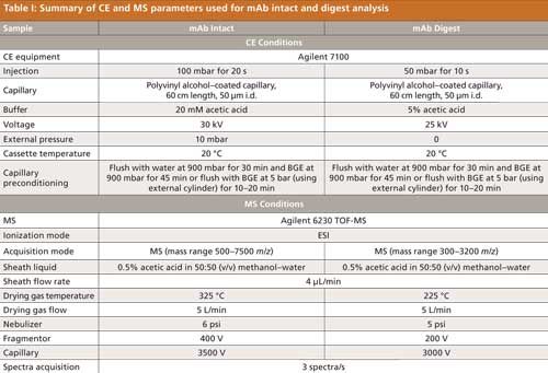
The CE and MS parameters used for the mAb (intact and digest) analysis are summarized in Table I.
Results and Discussion
Analysis of Intact and Digested mAb
CE–MS analysis of mAbs requires more thorough optimization of both separation (CE) as well as ionization (MS) parameters. This more thorough optimization is needed primarily because of the difficulty in creating the gas phase ions using electrospray ionization (ESI) for such a large compound without compromising the separation. Also, as discussed earlier, a reliable analytical method for protein analysis should be such that it preserves all the modifications that are potentially present in the molecule without introducing any artifacts during sample preparation and analysis. Thus, it would be desirable to perform the analysis in the native form of the protein. However, for achieving good electrophoretic separations, the molecule has to be sufficiently charged. The choice of buffer should also take into consideration the compatibility of the buffer and ionization of the analyte in MS.
Sample Preparation
In LC–MS analysis of proteins, the sample is generally reconstituted or buffer exchanged in 0.05–0.1% trifluoroacetic acid or formic acid (5). However, in our experience better results were obtained when using ammonium acetate buffer of suitable strength and pH (depending on target analytes pI). A clean mass spectrum was obtained while maintaining the protein’s native structure. For the present case, the mAb sample reconstituted in 50 mM ammonium acetate (pH 4.0) was found to give satisfactory results.
Choice of Capillary
A large number of publications have highlighted the issue of adsorption of proteins onto the walls of the fused-silica capillary. This adsorption problem has been generally tackled using coated capillaries where interaction between the analytes and the capillary wall is minimal. For basic proteins, positively charged coatings in combination with a low-pH BGE minimize the adsorption on the wall. Similarly, for acidic proteins, negatively charged coatings in combination with a high-pH BGE have been found to minimize electrostatic and hydrophobic interactions of the proteins with the capillary walls (19). Coatings such as polyvinyl alcohol also help in eliminating electroosmotic flow (EOF) independent of the pH of the BGE. This helps in overcoming cumbersome washing procedures and significantly improves reproducibility of the migration time. For our study, we used a polyvinyl alcohol-coated capillary for the analysis of the intact and digested protein.
Capillary Preconditioning
Before analyzing samples by CE–MS, it is essential to properly condition the capillary so as to achieve reproducible migration of charged analytes under the applied field. For uncoated capillaries (for example, bare fused-silica capillaries) a flush of 1 N sodium hydroxide at 900 mbar for 20 min, followed by flushing with water and the method’s BGE for 10 and 30 min, respectively, at 900 mbar. For most of the coated capillaries-coated with polyvinyl alcohol, for example-a flush with sodium hydroxide is not required. It should be noted that the nebulizer pressure should be set at 0 psig and sheath liquid flow kept on while flushing the capillaries with sodium hydroxide because of the incompatibility of MS toward nonvolatile buffers. Alternatively, the flushing can be done without inserting the capillary into the mass spectrometer’s main body and setting the capillary voltage to 0 V. In either case, the frequency of MS source cleaning (generally with 60% acetonitrile) and MS capillary maintenance should be increased for retaining acceptable performance of the instrument. For mAb analysis, we found that flushing at 5 bar (high-pressure flushing) for 15–20 min greatly enhanced the results obtained. A stable current profile was maintained during the entire analysis period, as shown in Figure 2.
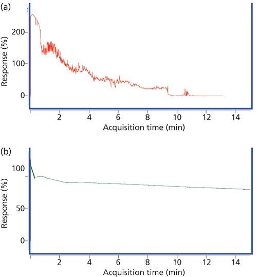
Figure 2: Current profile during intact mAb analysis after conditioning the capillary (a) at 900 mbar and (b) 5 bar.
Sheath Liquid Optimization
The results of CE–MS with a sheath liquid interface greatly depend on the optimization of the sheath liquid composition as well as flow rates. Also, an important factor that needs to be kept in mind is that the polyimide coating of the capillary at the sprayer tip (MS system end) should be carefully removed. If the coating is not removed, the sheath liquid causes swelling and expansion of the polyimide coating to the sprayer tip, which can result in improper nebulization, reduced sensitivity, and current abnormalities. The sheath liquid composition that is widely reported in the literature is 50% methanol as the base solution with a suitable acid or base with a volatility comparable to that of the base solution (20). Isopropanol and ethanol are also used as the base solution. Acetonitrile, though well suited for ionization in MS, is known to deteriorate the polyimide coating of the capillaries and is therefore generally not used. Acids of choice are acetic acid and formic acid, and ammonium acetate and ammonium formate of suitable strengths are recommended as the base of choice (20). The optimization of sheath liquid flow rate is aimed at reaching the right balance between sensitivity and stability of measurements. Although high flow rates enhance the stability of the measurements, they also lead to multifold sample dilution, which affects the sensitivity. Keeping these aspects in mind, the sheath liquid flow rate is typically kept between 0.4 µL/min to 0.8 µL/min depending on the demands of the analysis (20). Most sheath liquid interfaces use an external pump with a recycling function that splits the flow in predefined ratios. If the same solution is used for several days, recycling increases the risk of dirt accumulation in the sheath liquid. As a rule of thumb, the sheath liquid should always be prepared fresh daily. Failing to prepare fresh sheath liquid is likely to result in significant difficulties during analysis (Figure 3).
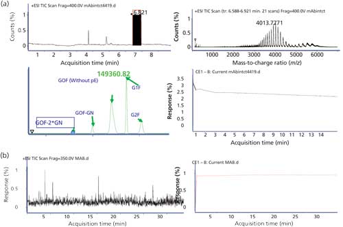
Figure 3: CE–MS results using (a) fresh sheath liquid and (b) recycled sheath liquid on third day. No spectra corresponding to the mAb can be observed in (b) because the analyte signal is suppressed by the relatively high amount of the contaminant in the CE effluent.
Applied Voltage for Separation
The applied voltage for electrophoretic separation varies according to the properties of the analytes. However, the maximum applied voltage should not exceed 300–350 V/cm. Also, when using high-concentration acid BGEs such as 1 M formic acid, the applied voltage should be kept around 250–300 V/cm because such buffers cause sudden application of the voltage and thereby result in the cracking of the capillaries. The developed cracks, in most cases, are not visible to the naked eye but do result in unstable current profiles during analysis.
Optimization of MS Parameters
At the MS level, optimization achieves two objectives. One is to obtain stable spray and the other is optimal ionization and transportation of the analyte into the MS system. For optimization of spray conditions, the nebulizer pressure and drying gas temperature need to be optimized. For ionization of the analyte, ion introduction voltage (capillary voltage) and fragmentation voltage need to be optimized (for TOF-MS). The parameters for obtaining stable spray conditions depend on the effluent from the upstream separation device. It should be noted here that because of the difference in flow characteristics of liquid chromatography (milliliters per minute) and capillary electrophoresis (nanoliters per minute), the parameters of LC cannot be translated to CE for obtaining equivalent response for a particular analyte. It was observed that a nebulizer pressure greater than 10 psig during CE–MS analysis of mAbs resulted in suction pressure at the mass spectrometer end that caused backflow of the analyte to the CE. This problem was overcome by using nebulizer pressure (5–6 psig) with a drying gas flow rate of 5–10 L/min. Also, switching off the nebulizer gas during injection by creating a time segment in the acquisition software prevented entry of the bubble into the capillary during injection and thereby helped achieve a stable current profile throughout the run. Gas temperatures of 150 °C (for digest) and 200 °C (for intact mAb) yielded satisfactory results. Fragmentor and capillary voltages of 400 V and 3500 V, respectively, were found to be optimal for intact mAb, and 200 V and 3000 V, respectively, were found to be optimal for the digest mAb analysis. It should be noted that while the optimal values of capillary and fragmentor voltages depend on the nature of compound (intact or digest), parameters such as drying gas temperature and flow, nebulizer pressure, and so forth, are flow dependent parameters and therefore are linked to the separation conditions used.
Figure 4 illustrates the effect of sheath liquid composition on the analysis of mAb digests.
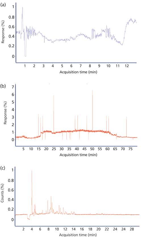
Figure 4: Total ion chromatogram (TIC) corresponding to mAb digest analysis using the conditions specified in Table I. Sheath liquid: (a) 0.1% acetic acid in 50% methanol, (b) 0.3% acetic acid in 50% methanol, and (c) 0.5% acetic acid in 50% methanol. The sequence coverage of 0%, 13%, and 98%, respectively, could be achieved in the three cases.
Conclusions
CE–MS has gradually become a gold standard in mAb analysis and is applied routinely from product development to release testing. Nevertheless, the robustness of CE–MS continues to be a concern in the minds of analytical chemists. However, over the past two decades, researchers have published a number of interlaboratory studies that have belied these speculations and have provided ample evidence of the robustness, efficacy, specificity, and reliability of CE–MS. As demonstrated in this column installment, adapting an existing analytical method (for example, LC–MS) to CE–MS is nontrivial and requires optimization of the individual parameters that may impact separation as well as detection. Another major issue is the limitations in the availability of CE–MS interfaces, particularly across vendors. Despite these challenges, the superiority of CE–MS for testing the various attributes of proteins including charge heterogeneity, size distribution, and glycan profiles will ensure that it remains a potent tool in the analytical arsenal.
References
- S.A. Berkowitz, J.R. Engen, J.R. Mazzeo, and G.B. Jones, Nat. Rev. Drug Discovery11(7), 527–540 (2012).
- G. Narula, V. Hebbi, S.K. Singh, A.S. Rathore, and I.S. Krull, LCGC North Am.34(1), 34–46 (2016).
- A. Guttman, LCGC North Am.30(5), 412–421 (2012).
- A.S. Rathore, Trends Biotechnol. 27(12), 698–705 (2009).
- N. Nupur, S.K. Singh, G. Narula, and A.S. Rathore, J. Chromatogr. B: Biomed. Sci. Appl. DOI: 10.1016/j.jchromb.2016.05.027 (2016).
- I.S. Krull, and A.S. Rathore, LCGC North Am.33(1), 42–49 (2015).
- S.A. Berkowitz, I.S. Krull, A.S. Rathore, LCGC North Am. 33(7), 478–489 (2015).
- A.S. Rathore and S.K. Singh in Topics in Medicinal Chemistry (Springer Berlin Heidelberg, 2016), pp. 1–27. DOI: 10.1007/7355_2015_5004
- A.S. Rathore and R. Bhambure, Anal. Bioanal. Chem. 406(26), 6569–6576 (2014).
- E. Tamizi and A. Jouyban, Electrophoresis36(6), 831–858 (2015).
- A. Beck, H. Diemer, D. Ayoub, F. Debaene, E. Wagner-Rousset, C. Carapito, A.V. Dorsselaer, and S. Sanglier-Cianférani, TrAC, Trends Anal. Chem.48, 81–95 (2013).
- A. Beck, S. Sanglier-CianfeÌrani, and A.V. Dorsselaer, Anal. Chem. 84(11), 4637–4646 (2012).
- R.D. Smith, J.A. Olivares, N.T. Nguyen, and H.R. Udseth, Anal. Chem. 60(5), 436–441 (1988).
- J.F. Banks, Electrophoresis18(12–13), 2255–2266 (1997).
- J. Ding and P. Vouros, Anal. Chem. 71(11), 378A–385A (1999).
- E.J. Maxwell and D.D.Y. Chen, Anal. Chim. Acta627(1), 25–33 (2008).
- G. Rozing blog, https://www.rozing.com/.
- M.D. Jones, L.A. Merewether, C.L. Clogston, and H.S. Lu, Analytical Biochemistry216(1), 135–146 (1994).
- R. Haselberg, G.J. De Jong, and G.W. Somsen, LCGC North Am. 30(6), 504–518 (2012).
- H.H. Lauer and G.P. Rozing, Agilent Technologies Primer on CE-MS, P/N 5990_3777EN. (http://www.agilent.com/cs/library/primers/Public/5990_3777EN.pdf).

Sumit Kumar Singh is a graduate student in the Department of Chemical Engineering at the Indian Institute of Technology.

Vishwanath Hebbi is a graduate student in the Department of Chemical Engineering at the Indian Institute of Technology.

Anurag S. Rathore is a professor in the Department of Chemical Engineering at the Indian Institute of Technology in Delhi, India.

Ira S. Krull is a Professor Emeritus with the Department of Chemistry and Chemical Biology at Northeastern University in Boston, Massachusetts, and a member of LCGC’s editorial advisory board.

Silvia Radenkovic on Building Connections in the Scientific Community
April 11th 2025In the second part of our conversation with Silvia Radenkovic, she shares insights into her involvement in scientific organizations and offers advice for young scientists looking to engage more in scientific organizations.


