Multidimensional LC Approaches for Intact Protein Biopharmaceutical Characterization
LCGC Europe
This instalment provides a brief overview of some multidimensional approaches targeted at intact protein analysis for life science applications, particularly for biopharmaceutical analysis.
This instalment provides a brief overview of some multidimensional approaches targeted at intact protein analysis for life science applications, particularly for biopharmaceutical analysis.
Since the 1944 publication of a paper by Consden, Gordona and Martin (1) describing multidimensional separations, the prospect of using this technique to address complex analytical challenges has sustained the interest of the analytical chemistry community. Analysts often use multidimensional techniques when they require higher separation capacity than single-dimension analyses can afford. By using dimensions of various selectivity for complex analytes such as food, polymers and hydrocarbons, maximum resolution can be achieved among components that are generally difficult to separate using a single-dimension technique.
The multidimensional approach for separations has been — and continues to be — highly utilized. Not surprisingly, its use as an analytical technique has been expanded. It now includes various applications that do not necessarily result in greater resolution. In those applications, the technique serves as the means for conducting separate analyses of the same analyte without the need for multiple instruments. It can also reduce steps for sample preparation and provide a convenient way to perform automated assays.
In the past few years, an emerging area of focus in the pharmaceutical industry is the biopharmaceuticals. The compounds in this class are very complex and require a high degree of characterization. Indeed, the level of characterization necessary is increasing, largely because of improved analytical technologies and regulatory requirements. What's also at play is the emergence of biosimilar approval pathways, in which protecting intellectual property for innovators or proving similarity for biosimilar producers is increasingly important (2).
Developing and manufacturing biopharmaceuticals requires amassing a variety of chromatographic and mass spectral data. The different chromatographic modes probe particular characteristics of the analyte. Some of these characteristics include size, charge and mass profile. As such, they are related to, or are themselves, critical quality attributes.
For some biopharmaceutical applications, a multidimensional separation can reduce a practitioner's analytical burden, yielding greater assay reproducibility by dispensing with hands-on sample preparation. Note, however, that although adding chromatographic dimensions can prove useful, to do so without realizing additional benefit, such as increased resolution, sensitivity,or automation, introduces unnecessary complexity.
This column instalment provides a brief overview of some multidimensional approaches targeted at intact protein analysis for life science applications, particularly for biopharmaceutical analysis. For the applications described here, adding a liquid chromatography (LC) dimension automates sample preparation and provides a convenient means of analysing an analyte by different methods that require different chromatographic eluents. As such, these applications are meant for targeted analyses where sample complexity does not demand adding a dimension. Rather, the criterion for needing a multidimensional separation is the primary chromatographic method's incompatibility with the desired detection method. For example, because of high mobile-phase salt concentrations, ion-exchange chromatography (IEC) is incompatible with mass spectrometry (MS) detection directly. Capturing a peak of interest to a second-dimension reversed-phase column, however, allows for desalting the analyte and collecting MS data.
Off-Line Multidimensional Approach
A convenient way to perform a multiple-dimension analysis is to collect fractions from one dimension, and then reinject those fractions onto the next dimension. Often, the first-dimension separation serves as a sample-preparation step, for example, an affinity purification using protein A. Yet fraction collection of a sample separated by IEC or sizeexclusion chromatography (SEC) is also commonplace (3). Moreover, fraction collection provides a convenient means to comprehensive analysis, regardless of the chromatographic run time for each dimension because the fractions can be stored in a temperaturecontrolled environment.
One type of first-dimension separation based on affinity purification serves to enrich the collected fraction in the analyte of interest in an automated fashion. The method used is often similar, if not identical, to the method that will be used when purifying the molecule during the production process. During an affinity purification using protein-A, a UV detector monitors the column eluent, and the resultant peak area can be used to assess a protein titer. Following UV detection, the purified material is collected using a fraction collector. The fraction collector is usually temperature controlled to prevent sample degradation. A significant benefit of an affinity purification step lies in its highly reproducible peak elution, allowing the generation of a generic method suitable for a variety of analytes (4).
After the first-dimension purification, the collected fractions (or fraction) are reinjected for subsequent analyses, such as IEC (to access charge heterogeneity) or SEC (often to access aggregation and uniformity of the molecular size). With limited handling, a single sample can be purified and analysed using various second-dimension methods, without the need for multiple chromatographic systems. Commercial instruments are available for these types of analyses in an automated fashion; they enable users to define the purification conditions and methods for each of the secondary analyses for each sample.
Another type of first-dimension separation for intact proteins relies on the first dimensions to be more highly resolving, such as IEC or SEC. Fractions can be collected in two ways: by a timed event, with equal fractions collected across either part or all of the chromatographic separation, or by detection thresholds for collection, which limits the number of fractions collected. In either collection regime, each collected fraction undergoes a single second-dimension analysis or, potentially, multiple second-dimension analyses.
This type of protocol is useful to identify and characterize low-level impurities present in a sample. For example, an SEC analysis for a sample clearly indicates that the sample contains aggregated species, evidenced by a peak correlating to a molecule roughly twice the size of the desired product. One can easily adopt a multidimensional approach by collecting the suspected aggregated species. The collected fraction could then be analysed by an orthogonal LC method to obtain the distribution of components in the aggregated species. The second-dimension separation can be done with IEC or a reversed-phase-MS analysis. The IEC analysis can help determine whether the species is enriched or deficient in charged species. The reversed-phaseMS analysis can help determine the presence of species with different masses in their monomeric forms.
A potential problem associated with the fraction-collection technique arises when it is used for lowabundance species, often the target of this type of analysis. It is common for low concentrations of protein species to adsorb to the walls of collection vessels, which can impede the ability to conduct a second-dimension analysis. Nevertheless, one can avoid this phenomenon by prudently choosing a collection vessel, assuming a suitable vessel exists for the particular application (5). Alternatively, you can conduct multiple first-dimension separations and collections to ensure the retention of sufficient analyte for subsequent analysis.
On-Line Multidimensional Separations
An alternative multidimensional approach adopts an on-line collection scheme where fractions, or "heart cuts," collected from the first dimension are captured directly onto a second chromatographic column for the second-dimension analysis. Life science applications have used multidimensional approaches for some time; for example, Washburn and colleagues (6) conducted proteomics studies using multidimensional analysis. Recently, multidimensional separations have also helped to identify host-cell proteins during biopharmaceutical development (7).
As with the off-line fraction-collection techniques, on-line multidimensional separation proves useful when investigating impurities that are insufficiently resolved from other components in a complex mixture in a single-dimension LC analysis. In addition, on-line approaches are useful for investigating low-level impurities, because analytes are less susceptible to loss upon collection. In either case, a multidimensional approach allows analysts to either reduce sample complexity or enhance an assay's dynamic range. One particular advantage of the on-line approach is that analytes can be stored at physiologic conditions until just before an analysis.
A common application of an on-line, multidimensional approach that is similar to off-line fraction-collection techniques involves purifying an antibody using protein A followed by an orthogonal approach. Emphasized here is the coupling of a first-dimension, protein-A purification followed by reversed-phase-MS analysis of the heart cut. Each of these analyses are commonly performed, often separately, during the development and production of antibodies, but with modern instrumentation they can be accomplished sequentially on a single LC–MS system. Other second-dimension analyses exist, and their utility in a heart-cut scheme equals that of the off-line fractioncollection methods.
In the application example discussed below, the second-dimension separation is a low-resolution separation conducted on a short column — one generally used for trapping and eluting analytes. Although higher-resolution separations can be performed in the first dimension, second dimension or both, one must consider overall system-pressure restrictions when both columns are placed in-line.
The production and formulation of antibodies intrinsically results in mixtures that contain excipients that are not compatible with MS analysis. Particularly in the case of MS analysis, efficient removal of these components is imperative for achieving sufficient MS sensitivity. Hence the attractiveness of affinity purification, which efficiently removes excipients by selectively binding only the analyte of interest while the remaining components are eluted.
The remainder of this article discusses in detail a specific application example that focuses on the challenge of coupling purification with mass profiling of antibodies using an on-line, multidimensional LC system. The methods were developed on an LC system with 2D technology coupled to a quadrupole time-of-flight (Q-TOF) mass spectrometer.
Figure 1 depicts a plumbing diagram with an affinity purification column configured for conducting the heart cut. The configuration provides two valve positions. In position 1, the loading position, the first-dimension column is loaded. Eluent flows through a UV detector and then to a waste vessel. Diverting the eluent to waste is necessary to minimize unwanted analytes (such as components from cell culture media) from collecting on the reversed-phase column. During the loading step, the 2D LC pump delivers initial gradient conditions to the second-dimension column. After removing unbound components, the antibody elutes from the affinity column. During the first-dimension elution phase, in an actuation sequence known as "heart cutting," the valve switches to position 2, diverting a portion of the first-dimension eluent to the second-dimension column.

Figure 1: Example plumbing diagram for on-line heart-cutting analyis.
After collection from the first dimension, the valve returns to position 1, and the second-dimension gradient is initiated. Because each dimension is used in series for analysis, the overall run times for each dimension (either protein A or mass measurement by reversed-phase-MS analysis) is reduced compared to running either dimension individually off-line. The run time is decreased because equilibration time for each dimension can mainly be accomplished while the other dimension is conducting an analysis.
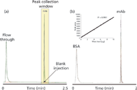
Figure 2(a) shows that the on-line affinity purification is highly reproducible, as evidenced by an overlay of 12 replicate injections with a relative standard deviation (RSD) of 0.078%. In addition, these methods provide a linear response based on mass load, which is shown in Figure 2(b). One would expect similar results using an off-line fractioncollection system.

Figure 2: Representative UV chromatograms for purification of mAb by protein A.
An important consideration, besides chromatographic reproducibility, is heart cutting through an on-line method. Making appropriate choices in valves, valve event timing and ensuring optimal plumbing connections are all important factors to consider when designing the experiment. Because collection is generally based on a timed event or by a specified threshold, such as UV intensity, the valve trigger events must be reproducible. One must also account for any delay time in the system between the point at which a desired peak is detected and its subsequent collection. Particularly, note that the flow rate, tubing length and the internal diameter all affect valve timing.
A first-dimension protein A purification for a monoclonal antibody (mAb) is shown in Figure 2(a). The highlighted area represents the time selected for peak collection. The area was defined by timed events to actuate the left-hand valve (shown in Figure 1) to move to position 2 at the start of collection and then back to position 1 at the end of collection. The collected peak is focused on the second-dimension column and quickly desalted before elution. A valve on the mass spectrometer diverts the remaining eluent stream to waste. After sufficient removal of salt and buffer, primary components of the protein-A effluent, the desalting valve diverts the flow to the mass spectrometer's source, a steep gradient runs and the analyte is eluted as a concentrated peak.
Figure 3 shows that the second-dimension separation and peak collection are reproducible for a variety of mass loads, as evidenced by the total ion chromatograms for the MS analysis.

Figure 3: Overlay of total ion chromatograms for an antibody loaded on a first-dimension protein A column, collected and subjected to MS analysis. Each analysis was run in triplicate.
An on-line approach can also provide mass-spectral information for analytes separated by IEC and SEC. As mentioned previously, one could also use off-line fraction collection to perform this type of analysis. Note, however, that some low-abundance species can adsorb to collection-vessel walls, impeding or inhibiting your ability to derive meaningful data from the collected fraction.
Adopting an on-line approach is one way to overcome the challenge posed by adsorption. One plumbing scheme for an on-line analysis, identical to that shown in Figure 1, allows the analysis of one fraction per first-dimension analysis. To analyse additional fractions, one must configure a system that can collect fractions onto separate trapping columns or a system that can hold fractions in loops before analysis. Though the latter configuration confers the advantage of requiring fewer overall experiments, system configurations can rapidly become very complex.
Conclusion
This column instalment describes various methods for using a multidimensional approach, including on-line and off-line methods. Such methods can simplify sample analysis in either of two ways: by reducing hands-on sample preparation or by replacing significant analytical challenges with routine ones. Moreover, spurred by practitioners' increased interest in multidimensional techniques, manufacturers of analytical instruments are providing greater flexibility to configure multidimensional systems. As we increasingly adopt these technologies, systems particularly suited for performing multidimensional separations will likely emerge to make analysts lives considerably easier.
Dr Sean M. McCarthy is a principal scientist at Waters Corporation (Milford, Massachusetts, USA). He received his PhD in inorganic chemistry from the University of Vermont, in Vermont, USA, in 2005. Following this he was a postdoctoral fellow in the pathology department at the university. Most recently his work has focused on biopharmaceutical characterization using LC and LC–MS technologies.
"MS — The Practical Art" Editor Kate Yu joined Waters in Milford, Massachusetts, USA, in 1998. She has a wealth of experience in applying LC–MS technologies into various application fields such as metabolite identification, metabolomics, quantitative bioanalysis, natural products and environmental applications. Direct correspondence about this column should go to "MS: The Practical Art", LCGC Europe, 4A Bridgegate Pavilion, Chester Business Park, Wrexham Road, Chester, CH4 9QH, UK, or e-mail the editor, Alasdair Matheson, at amatheson@advanstar.com
References
(1) R. Consden, A.H. Gordan and A.J.P. Martin, Biochem. J. 38(3), 224–232 (1944).
(3) V. Faca, S. Pitteri, L. Newcomb, V. Glukhova, D. Phanstiel, A. Krasnoselsky, Q. Zhang, J. Struthers, H. Wang, J. Eng, M. Fitzgibbon, M. McIntosh and S. Hanash, J. Proteome Res. 6(9), 3558–3565 (2007).
(4) D.S. Hage, Affinity Clinical Chemistry 45(5), 593–615 (1999).
(5) J.M. Mathes, "Protein Adsorption to Vial Surfaces — Quantification, Structural and Mechanistic Studies", Ludwig Maximilian University of Munich (2010).
(6) M.P. Washburn, D. Wolters and J.R. Yates, Nat. Biotech. 19(3), 242–247 (2001).
(7) C. Doneanu, A. Xenopoulos, K. Fadgen, J. Murphy, S.J. Skilton, H. Prentice, M. Stapels and W. Chen, MAbs 4(1), 24–44 (2012).

New Study Reviews Chromatography Methods for Flavonoid Analysis
April 21st 2025Flavonoids are widely used metabolites that carry out various functions in different industries, such as food and cosmetics. Detecting, separating, and quantifying them in fruit species can be a complicated process.
University of Rouen-Normandy Scientists Explore Eco-Friendly Sampling Approach for GC-HRMS
April 17th 2025Root exudates—substances secreted by living plant roots—are challenging to sample, as they are typically extracted using artificial devices and can vary widely in both quantity and composition across plant species.

.png&w=3840&q=75)

.png&w=3840&q=75)


.png&w=3840&q=75)


.png&w=3840&q=75)


