Monitoring of Oxidation in Biopharmaceuticals with Top-to-Bottom High Performance Liquid Chromatography–Mass Spectrometry Methodologies: A Critical Check
LCGC Europe
Federal regulations concerning the safety and efficacy of biopharmaceuticals require the implementation of a comprehensive toolbox of physicochemical and biological characterization methods. In order to demonstrate consistent overall structure, even minute differences in primary structure and post‑translational modifications (PTMs) have to be detectable in therapeutic proteins. Because of their remarkable capability of revealing small changes in molecular structure, high performance liquid chromatography (HPLC) and mass spectrometry (MS) rate among the most powerful technologies for comprehensive protein analysis. This article details the potential of both methods with regard to revealing methionine oxidation, a chemical modification that may be induced during downstream processing and storage of biopharmaceuticals. The benefits and limitations of bottom-up, middle-down, and top‑down HPLC–MS analysis will be demonstrated for the detection of oxidation variants in a therapeutic monoclonal antibody (mAb).
Therese Wohlschlager, Christof Regl, and Christian G. Huber, Department of Biosciences, Bioanalytical Research Labs and Christian Doppler Laboratory for Biosimilar Characterization, University of Salzburg, Salzburg, Austria
Federal regulations concerning the safety and efficacy of biopharmaceuticals require the implementation of a comprehensive toolbox of physicochemical and biological characterization methods. In order to demonstrate consistent overall structure, even minute differences in primary structure and postâtranslational modifications (PTMs) have to be detectable in therapeutic proteins. Because of their remarkable capability of revealing small changes in molecular structure, high performance liquid chromatography (HPLC) and mass spectrometry (MS) rate among the most powerful technologies for comprehensive protein analysis. This article details the potential of both methods with regard to revealing methionine oxidation, a chemical modification that may be induced during downstream processing and storage of biopharmaceuticals. The benefits and limitations of bottom-up, middle-down, and topâdown HPLC–MS analysis will be demonstrated for the detection of oxidation variants in a therapeutic monoclonal antibody (mAb).
Seven of the ten top-selling pharmaceuticals in 2016 were protein therapeutics (1), which are usually manufactured in a biotechnological process involving cell culture (2). Very stringent safety regulations by the health authorities mandate elaborate analytical schemes for the comprehensive characterization of the efficacy and safety of pharmaceutical products. This is even more valid for protein therapeutics (3) because the large molecular size and unavoidable variances in biosynthesis and downstream processing may result in molecular variants with regard to amino acid sequence, threeâdimensional protein structure, post-translational modifications (PTMs), or process-related artificial modifications, all of which may impact therapeutic efficacy and safety (4). In this context, nonenzymatic oxidation of proteins at methionine residues resulting in methionine–sulfoxide or methionine–sulfone represents one of the most relevant protein degradation reactions (5). Oxidation of methionines has been shown to significantly influence the biological activity of proteins (6), hence limiting the stability and shelf life of protein-based pharmaceuticals (7–10).
Developed in the mid-1960s initially for the separation of small molecules (11), high performance liquid chromatography (HPLC) has been adapted to the separation of high molecular biopolymers (12) upon implementation of dedicated stationary phase configurations enabling rapid mass transfer, such as sub-2-µm totally porous particles (TPPs), superficially porous particles (SPPs), or monolithic phases. Likewise, progress in mass spectrometry (MS) technologies, especially electrospray ionization (ESI) for biopolymers (13), as well as high-resolution mass analyzers such as time-of-flight (TOF) or orbital trap mass analyzers (14), have significantly contributed to the success of bioanalytical methods in pharmaceutical analysis. This article will shed light on the potential, challenges, and achievements of HPLC in combination with ESIâMS for the analysis of therapeutic proteins with an emphasis on protein oxidation, which represents a relatively minute modification at the level of primary structure. Oxidation, however, generally entails serious consequences in threeâdimensional protein structure and biological activity, rendering its diligent characterization mandatory in biopharmaceutical analysis.
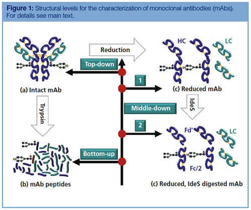
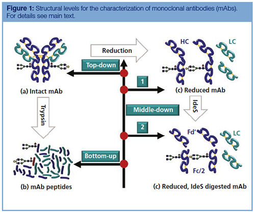
Structural Levels of Protein Characterization: Fundamental Considerations: Using an antibody molecule as an example, Figure 1 illustrates the structural levels that may be relevant for protein characterization in a biopharmaceutical context. Traditionally, the analysis of protein sequence and PTMs is performed after digestion with a protease, yielding a specific set of peptides (Figure 1[b]) that is analyzed by HPLC and MS. This approach is termed bottom-up analysis, offering the advantage that even small changes in a peptide, for example, an additional oxygen atom, entail a large relative change in the physicochemical properties of the peptide, which may be readily detectable by HPLC and MS.
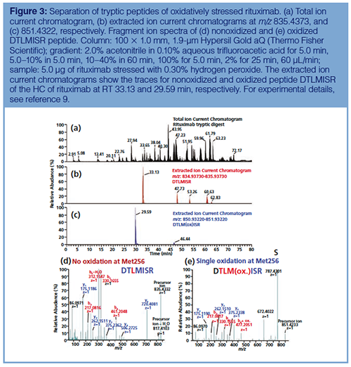
For example, oxidation of DTLMISR, a methionine-containing peptide of the therapeutic antibody rituximab, results in a mass shift from 834.4269 to 850.4218 Da, representing a relative mass difference of 1.88% that can readily be distinguished by MS. Moreover, incorporation of an additional oxygen atom into the peptide will decrease its hydrophobicity, which may facilitate the separation of oxidized and nonoxidized peptide variants by reversed-phase HPLC. Finally, sequence information may be gained upon gas-phase fragmentation and detection of fragment ions by MS, enabling both the identification and localization of modifications of specific amino acids. Apart from being laborious, time-consuming, and prone to the introduction of artifacts during proteolytic digestion, the bottom-up approach is limited in that the context of modifications within the different intact molecular protein species is lost.
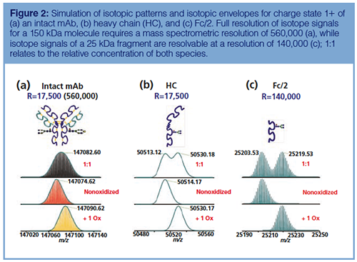
On the other hand, protein analysis may be attempted at the intact molecule level, as realized in so-called top-down approaches (Figure 1[a]). Numerous examples have successfully shown that therapeutic mAbs may efficiently be analyzed as intact proteins using HPLC and MS methods (15,16). Studies on the gasâphase fragmentation have also demonstrated that a considerable amount of sequence information may be collected from fragments generated from intact proteins by collision-induced dissociation (CID), electron capture dissociation (ECD), or electron-transfer dissociation (ETD) (17). Nevertheless, subtle changes in protein structure may be difficult to detect as a result of limitations in chromatographic or mass spectrometric capabilities to discern the change. To stay with the example given above, incorporation of one oxygen atom into an intact mAb molecule will result in a mass shift from 147074.62 to 147090.62 Da (+16 Da), representing a relative mass difference of 109 ppm, which is well within the typical mass accuracies of modern TOF and orbital trap mass spectrometers. Simulation of the isotopic patterns of two mAb species differing in a single oxygen atom, however, shows that the isotopic envelopes merge into a single broad distribution even at a mass spectrometric resolution sufficient to separate neighbouring isotope peaks. Thus, distinction of oxidized- and nonoxidized mAb species by MS at the intact protein level is not feasible (Figure 2[a]).
In order to obtain smaller protein subunits for investigation, tetrameric antibody molecules can be dissociated into two heavy and two light chains (HC and LC, respectively) by reduction of intermolecular disulfide bonds, yielding two 25 kDa LC and two 50 kDa HC subunits. While the isotopic patterns of the HC molecules are still not completely resolvable by MS (Figure 2[b]), the width of the isotopic pattern of a 25 kDa fragment is narrow enough to facilitate clear separation of oxidized- and nonoxidized species (demonstrated for a 25 kDa Fc/2 fragment in Figure 2[c]). Such fragments can be obtained upon proteolytic digestion with papain or IdeS, which specifically cleave within the so-called hinge region of the HC (Figure 1[d]). The two resulting fragments of almost identical size (Fd’ and Fc/2; approximately 25 kDa each) are ideally suited for the analysis of oxidation variants because they can readily be resolved by MS, especially as the main oxidation sites are located in the Fc domain (18).
Bottom-Up Determination of Antibody Oxidation at the Proteolytic Peptide Level: The classical approach to comprehensive protein characterization comprises chromatographic separation of proteolytic peptides by ion-pair (IP)-reversed-phase HPLC followed by sequence analysis of the separated peptides by tandem MS. Figure 3 illustrates that oxidation of methionine facilitates a clear chromatographic separation of the corresponding peptides. As expected, the incorporation of oxygen results in a decrease in hydrophobicity and hence a decrease in chromatographic retention of the oxidized variant of the peptide. Relative peak areas of the chromatographic peaks that are well separated may be used to relatively quantify the extent of oxidation at the peptide level. In addition, fragment spectra as shown in Figure 3(d) and 3(e) unambiguously identify the site of oxidation as methionine at position four of the peptide. Apart from oxidation monitoring, the high resolving power and mass accuracy of modern high-resolution mass spectrometers enables the detection of mass differences as little as 1 Da, which is characteristic for the deamidation of asparagine or glutamine residues, making peptide mapping one of the most powerful tools for the detection of protein modifications.
Investigation of Antibody Oxidation at the Intact Protein Level: As investigation of mAb oxidation at the intact protein level would eliminate the need for sample preparation by digestion and hence avoid oxidation artifacts, the separation of oxidized rituximab variants on a large-pore polymerâbased column developed for the separation of mAbs was attempted. Comparison of the chromatographic profiles of control and stressed sample in Figure 4(a) reveals a slight decrease in retention time (by approximately 5 s) and an increase in the peak width at half height of the eluting peak profile for the oxidized protein (from 11 to 15 s). These effects can be attributed to reduced hydrophobicity of the oxidized protein variants as well as increased complexity of the sample because oxidation may occur at different sites, resulting in a range of diverse protein species. Nevertheless, oxidized mAb variants cannot even partially be separated from nonoxidized species by means of IP-reversed-phase chromatography.
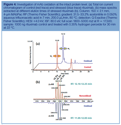
The corresponding mass spectra reveal a shift towards higher molecular mass of the oxidized protein, as shown in Figure 4(b) for the 44+ charge state of rituximab consisting of multiple signals that arise from the different glycoforms. When comparing corresponding glycoforms of oxidized and control samples, shifts in the range of m/z 0.32–0.37 translating into mass differences of 14–16 Da are detectable. Again, resolution of signals for different oxidized mAb species is impossible and the masses observed for the stressed sample represent averages of different singly- and multiply-oxidized protein variants, rendering the approach not viable for the exact determination of protein oxidation.
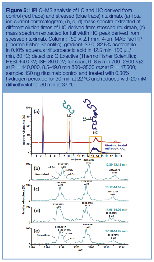
Investigation of Antibody Oxidation in the HC: Moving one step down from the intact protein level of an antibody, Figure 5 depicts the HPLC–MS analysis of LC and HC derived from a control and an oxidativelyâstressed rituximab sample. LC and HC were efficiently separated when eluted with a very shallow gradient of 32.0–32.5% acetonitrile in 12.5 min. When compared to the control sample, significant broadening of the chromatographic peak corresponding to oxidized HC was observed, indicating partial separation of different oxidized species (Figure 5[a]), although separation into discrete peaks was not attainable. Extraction of mass spectra from different time intervals within the chromatographic peak revealed that higher oxidized species, as evidenced by higher mass, eluted earlier compared to mono- or nonoxidized HC species (Figure 5[b–e]). Incorporation of up to three oxygen atoms, consistent with the occurrence of three methionines in the HC sequence, was observable in the mass spectra. Nevertheless, mass spectral resolution of the different oxidation variants allowing their unambiguous differentiation and quantification was still not feasible.
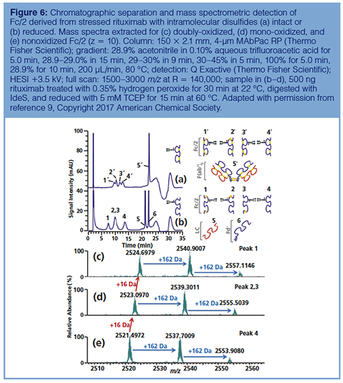
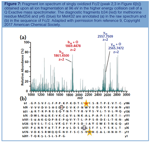
Investigation of Antibody Oxidation in the Fc/2 fragment: Finally, the analysis of methionine oxidation was performed in Fc/2 generated upon proteolysis with IdeS, an enzyme cleaving within the hinge region of the HC sequence. Interestingly, when intramolecular disulfide bridges in the HC domain were kept intact, a separation of four oxidized variants of Fc/2 (one nonoxidized, two singlyâoxidized, one doubly-oxidized species) was observable (Figure 6[a]). On the other hand, the two monoâoxidized Fc/2 species coeluted after the intermolecular disulfide bridges were cleaved upon reduction (Figure 6[b]). This observation suggests that changes in the threeâdimensional structure induced by methionine oxidation play an important role in the chromatographic separation of oxidized proteins or protein fragments (8–10). Moreover, the baseline separation of non-, mono- and doubly-oxidized species enabled the quantitative determination of oxidized protein variants both by UV-spectroscopic and mass spectrometric detection with relative process standard deviations between 7 and 14% (9). MS not only confirmed the +16 Da mass shifts as a result of the incorporation of oxygens (Figure 6[c–e]), but also allowed the localization of oxidation sites by middle-down fragmentation of Fc/2 (Figure 7).
Conclusions
The ability of analytical technologies to distinguish between oxidized and nonoxidized protein species is limited by several factors. Bottomâup analysis of tryptic peptides facilitates detailed analysis of even minor modifications and simultaneously provides sequence information deduced from tandem MS experiments. Nevertheless, the context of the different modifications in the whole protein is undetectable in this analytical approach. At the intact protein or HC level, efficient separation of oxidation variants of IgG1 type mAbs by reversed-phase HPLC or IP-reversed-phase-HPLC is generally impossible, mostly because of the minimal differences in hydrophobicity upon incorporation of additional oxygen atoms. In mass spectrometry, the natural width of the isotopic pattern limits the capability to distinguish oxidized proteins from their nonoxidized analogues to molecules smaller than approximately 25 kDa. Thus, oxidation monitoring in therapeutic mAbs is possible after decomposition into light and heavy chains, followed by proteolytic cleavage of the heavy chain into two fragments of 25 kDa. Moreover, at this molecular scale, non-, mono-, and doubly-oxidized variants can be separated by IP-reversedâphaseâHPLC.
Acknowledgement
The financial support by the Austrian Federal Ministry for Digital and Economic Affairs and by a startâup grant of the State of Salzburg is gratefully acknowledged.
Conflict of Interest Statement
Sandoz, a Novartis Division, and Thermo Fisher Scientific provide financial support for the Christian Doppler Laboratory for Innovative Tools for Biosimilar Characterization. The salary of Therese Wohlschlager is fully funded; Christian G. Huber’s salary is partly funded by the Christian Doppler Laboratory for Biosimilar Characterization. The authors declare no other competing financial interest.
References
- J. Njarðarson, http://njardarson.lab.arizona.edu/ Accessed 15 May 2018.
- M. Butler, Applied Microbiology and Biotechnology68, 283–291 (2005).
- A.J. Chirino and A. Mire-Sluis, Nature Biotechnology22, 1383–1391 (2004).
- X. Zhong and J.F. Wright, International Journal of Cell Biology2013, 19 (2013).
- N. Jenkins, L. Murphy, and R. Tyther, Molecular Biotechnology39, 113–118 (2008).
- H.S. Lu, P.R. Fausset, L.O. Narhi, T. Horan, K. Shinagawa, G. Shimamoto, and T.C. Boone, Archives of Biochemistry and Biophysics362, 1–11 (1999).
- I.C. Forstenlehner, J. Holzmann, K. Scheffler, W. Wieder, H. Toll, and C.G. Huber, Analytical Chemistry86, 826–834 (2014).
- I.C. Forstenlehner, J. Holzmann, H. Toll, and C.G. Huber, Analytical Chemistry web-published, (2015).
- C. Regl, T. Wohlschlager, J. Holzmann, and C.G. Huber, Analytical Chemistry89, 8391–8398 (2017).
- J. Holzmann, A. Hausberger, A. Rupprechter, and H. Toll, Analytical and Bioanalytical Chemistry405(21), 6667–6674 (2013).
- C. Horváth, B.A. Preiss, and S.R. Lipsky, Analytical Chemistry39, 1422–1428 (1967).
- F.E. Regnier and J. Kim, Analytical Chemistry90, 361–373 (2018).
- J.B. Fenn, M. Mann, C.K. Meng, S.F. Wong, and C.M. Whitehouse, Science246, 64–71 (1989).
- A. Makarov, Analytical Chemistry72, 1156–1162 (2000).
- J. Mohr, R. Swart, M. Samonig, G. Böhm, and C.G. Huber, Proteomics 10, 3598–3609 (2010).
- A. Beck, S. Sanglier-Cianferani, and A. Van Dorsselaer, Analytical Chemistry84, 4637–4646 (2012).
- N. Bandeira, V. Pham, P. Pevzner, D. Arnott, and J.R. Lill, Nature Biotechnology26, 1336–1338 (2008).
- C. Chumsae, G. Gaza-Bulseco, J. Sun, and H. Liu, J. Chromatogr. B Analyt. Technol. Biomed. Life Sci.850, 285–294 (2007).
Therese Wohlschlager is a postdoctoral researcher at the University of Salzburg, Austria. Within the Christian Doppler Laboratory for Biosimilar Characterization she focuses on the development of analytical tools for the characterization of glycosylated biopharmaceuticals.
Christof Regl is a PhD student at the University of Salzburg and part of the Christian Doppler Laboratory for Biosimilar Characterization. His research project deals with the analysis of therapeutic mAbs ranging from bottom-up to top-down strategies.
Christian Huber trained as an analytical chemist at the University of Innsbruck and is currently a professor of chemistry for biosciences at the University of Salzburg. He also leads the Christian Doppler Laboratory for Biosimilar Characterization, which cooperates with Novartis and Thermo Fisher Scientific. His research interests include proteome and metabolome analysis as well as in-depth protein characterization by means of HPLC and MS.
Koen Sandra is the editor of “Biopharmaceutical Perspectives”. He is the Scientific Director at the Research Institute for Chromatography (RIC, Kortrijk, Belgium) and R&D Director at anaRIC biologics (Ghent, Belgium). He is also a member of LCGC Europe’s editorial advisory board. Direct correspondence about this column to the editor-in-chief, Alasdair Matheson, at alasdair.matheson@ubm.com

Common Challenges in Nitrosamine Analysis: An LCGC International Peer Exchange
April 15th 2025A recent roundtable discussion featuring Aloka Srinivasan of Raaha, Mayank Bhanti of the United States Pharmacopeia (USP), and Amber Burch of Purisys discussed the challenges surrounding nitrosamine analysis in pharmaceuticals.

.png&w=3840&q=75)

.png&w=3840&q=75)


.png&w=3840&q=75)


.png&w=3840&q=75)


