LC–MS-MS Total Drug Analysis of Biological Samples Using a High-Throughput Protein Precipitation Method
LCGC North America
This article describes the use of a 96-well, solvent-resistant filtration plate for total drug analysis in blood plasma.
Sample preparation for total drug analysis in blood can be achieved by a variety of techniques, including centrifugal precipitation, liquid–liquid extraction and solid-phase extraction (SPE). Plasma proteins are precipitated using organic solvents (such as acetonitrile) and the precipitate is centrifuged into a pellet (1–3). For reproducible and accurate results, good protein removal by solvent precipitation and separation of the drug containing supernatant is critical. This separation typically is achieved with either long or multiple centrifugations. Protein precipitation ("crashing") is the preferred method for early animal studies. The increased number of samples created the need for higher throughput, multiwell techniques. In addition, the trend toward smaller plasma volumes combined with much lower centrifugal forces (~2000g for plates versus 15,000g for microfuge tubes) presents an obstacle to obtaining high quality, reproducible, liquid chromatography–mass spectrometry (LC–MS) ready samples
Efforts to improve sample preparation throughput have been made using 96-well filtration plates (4–7). Validation of the protein precipitation filtration method for the drug salbutamol using one of three different prototype filter plates gave acceptable results using vacuum filtration, but the method still involved multiple steps (8). This article describes the use of a 96-well, solvent-resistant filtration plate for total drug analysis in blood. Samples are prepared in the same manner as the centrifugal pelleting method but instead of pelleting and transferring the supernatant to obtain protein-free drug solution, the precipitated protein and the drug are separated by centrifugal-driven filtration. Pipetting the supernatant is eliminated and clean, protein-free drug solution is collected in a 96-well plate below the filter plate. This is particularly difficult for small plasma volumes. The polytetrafluorethylene (PTFE) membrane in the filter plate has high precipitated protein retention and is very clean with extremely low extractable content. The result is high sample recovery and no interfering contaminants. Together with detection by LC–MS-MS, this approach drives a high-throughput total drug analysis method that is faster and simpler than existing methods.
Comparison of the centrifugal pellet and centrifugal filter-plate methods was conducted using five discovery screening programs. Thirteen compounds (A, B, C, D, E, F, G, H, I, J, K, L, and M) were evaluated in one of five matrices: rat plasma, rat serum, dog plasma, mouse serum, and cyno monkey plasma. Validation of the bioanalytical methods, precision, and accuracy all were explored in the five programs.
Experimental Overview
In experiment I, bioanalytical assay validation in rat plasma with compounds A and B was compared using both the pellet and the filter-plate methods. In experiments II–IV, standard curves as well as quality control (QC) sample performance in running actual program samples were compared from both methods (side by side) for the drugs and animal models indicated. In experiment V, only the filter-plate method was run. The following is a summary of the five experiments that were performed:
Filtration versus Pellet Protein Precipitation
I. Validation in rat plasma.
Compounds A and B.
II. Standard curves in mouse serum.
Pharmacokinetic study compounds C, D, E, and F.
III. Standard curves in rat and mouse serum.
Biology study compounds G, H, I, J, and K.
IV. Standards and QC samples in dog plasma.
Compound L.
V. Injection reproducibility.
Comparison of (two) injections of same set of compound M in cyno monkey plasma calibration curve and QC samples at beginning and end of an assay (7-h run).
Materials and Methods
Serum and Plasma Samples: Plasma and serum samples were supplied by BMS Discovery Biology, Pharmacokinetics, and Toxicity departments.
Compounds were supplied by BMS Discovery Chemistry department.
HPLC-grade acetonitrile was obtained from Burdick & Jackson (Muskegon, Michigan). 98% GR formic acid was obtained from EMD (Germany). Deionized water (18.2 Ω·cm) was purified by a Milli-Q water purification system from Millipore Corporation (Billerica, Massachusetts).
Sample Preparation Procedures
A) Standard Protein Precipitation (Pellet) Method
Standard protein precipitation sample preparations were performed in a 96-well plate format with a Quadra-96 automated liquid handler from Tomtec (Hamden, Connecticut). Aliquots of 25 μL of the plasma/serum sample were treated with 50 μL of acidified (0.1% formic acid) acetonitrile containing 0.5 μg/mL of the internal standard followed by vortex mixing for 10 min. The supernatant was then separated from the precipitated proteins by a 30-min centrifugation (;1480g) and was transferred to a second 96-well plate. Typically, the volume of pellet and the level of solution in each vial were not consistent. Often, another 10-min centrifugation step was required to concentrate protein particles to avoid their transfer into the final aliquot for analysis. An aliquot of 10 μL of the clear supernatant was analyzed by LC–MS-MS.
B) Filter-Plate Protein Precipitation Method
Sample preparations were performed in a 96-well plate format with a Tomtec Quadra-96 automated liquid handler. First, 50 μL of acidified (0.1% formic acid) acetonitrile containing 0.5 μg/mL of the internal standard was added to a 96-well MultiScreen Solvinert plate from Millipore Corporation (Billerica, Massachusetts). 25 μL aliquots of serum–plasma samples from the animal studies were pipetted into the wells followed by vortex mixing for 3 min. The filter plate was stacked over a 0.5-mL, 96-well collection plate with a narrow-diameter 0.2-mL, 96-well plate insert (VWR) and centrifuged for 5 min at ;1480g. An aliquot of 10 μL was injected onto a column for analysis. The standard curves and QC samples were prepared in the same fashion as the samples.
LC–MS-MS Method: A high performance liquid chromatography (HPLC) system consisted of two LC-10ADvp pumps with a SCL-10Avp System Controller from Shimadzu (Columbia, Maryland) and a CTC Analytics HTS PAL autosampler from LEAP Technologies (Carrboro, North Carolina) equipped with a cooling stack autosampler tray maintained at 5 °C. The column used was a 50 mm × 2.1 mm, 5-μm Atlantis C18 column from Waters Corporation (Milford, Massachusetts) maintained at 40 °C. The mobile phase, consisting of 10 mM ammonium formate, 0.1% formic acid, and 0.2% acetonitrile in water (A) and acetonitrile with 0.1% formic acid (B), was run at a flow rate of 0.3 mL/min. The initial mobile phase composition was 95% A and 5% B. After sample injection, the mobile phase composition was held constant for 0.5 min and then changed linearly to 5% A and 95% B over 1.5 min and held at that composition for an additional 1.0 min. The mobile phase was then returned to initial conditions over 0.1 min, and the column was reequilibrated for 1.5 min. The total analysis time was 4.6 min.
The HPLC system was interfaced to a Quattro Premier LC–MS-MS tandem mass spectrometer from Waters/Micromass, (Manchester, UK) equipped with an electrospray interface operating in the positive ionization mode. Ultrahigh pure (UHP) nitrogen was used as the cone and desolvation gases at flow rates of 80 L/h for cone and 1000 L/h for desolvation. The desolvation temperature was 300 °C and the source temperature was 150 °C. Data acquisitions were via selected reaction monitoring (SRM). Ions representing the (M+H)+ species for each BMS compound. Only 12 compounds were mentioned in the introduction.] and the internal standard were selected in MS1 and collisionally dissociated with UHP argon at a pressure of 2 × 10⁻³ mbar to form specific product ions which were subsequently monitored by MS2.
LC–MS-MS SRM analyte peak area response to internal standard peak area response ratio was used for quantitation.
Results and Discussion
Most centrifugal precipitation methods use only one centrifugation step. However, it is important to recognize that a more rigorous, two-step method was used in all the comparisons reported in this study to achieve better results. The time of centrifugation in the pelleting method for a 0.5-mL, 96-well plate varied from 10 to 30 min. We performed a 30-min centrifugation to ensure the quality of protein precipitation and the bioanalytical assay in our routine.

Table I: Assay summary of experiment I
Results of the validation study (Experiment I) are shown in Table I. The standard curve consisted of four replicates at each of seven concentration levels ranging from 0.5 to 1000 ng/mL. QC samples were prepared in eight replicates at five concentration levels from 0.5 to 800 ng/mL. Sample preparation using the filter-plate method gave comparable results to the regular pellet method for compounds A and B in rat plasma. Both standard curves are linear over the 0.5–1000 ng/mL range and percent standard deviation (%SD) is acceptable for both methods, though somewhat lower for the filter plate. Using both methods, QC samples were run (n=8) at 0.5, 1, 8, 80, and 800 ng/mL for compound A. Results are summarized in Table II and are very comparable, indicating that precision and accuracy are similar for both methods. Internal standard responses also were consistent throughout the run for both pellet method and filter-plate method.
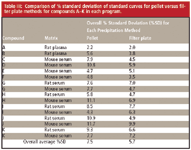
Standard curves at 10 concentration levels from 0.5 to 1000 ng/ml in duplicates using both methods were generated for compounds C, D, E, F, G, H, I, J, and K (Experiments II and III). Overall %SD data from standard curves for all compounds A–K are summarized in Table III. Except in the case of compound I in mouse serum, %SD is approximately the same or lower in the filter-plate method compared to the pellet method. The overall %SD is 7.5% for the pellet method and 5.7% for the filter-plate method. The filtration-plate method reduces bioanalytical assay variability at low end of standard curves.

Table II: Precision and accuracy of QC samples for Compound A in Experiment I
In Experiment IV, the pellet and filter-plate methods were compared in a study of compound L in dog plasma. These results demonstrated a 20.9% overall standard deviation compared with the 5.1% observed for the filter-plate method on the same samples. Although improved precision and accuracy were observed in the majority of filter-plate samples (Table III), the reasons for dramatically improved Experiment IV filter-plate performance is still under investigation.

Table III: Comparison of % standard deviation of standard curves for pellet versus filter plate methods for compounds AâK in each program.
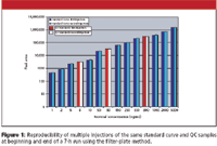
To determine the stability of samples over multiple injections using the filter-plate method, a reinjection study was performed using compound M in Experiment V. The standard curve and QC samples (8, 80, and 800 ng/mL) were run in duplicate at the beginning and end of the study. When using the pellet method, the second injection of a sample is often not reproducible following a long run. Comparison of peak areas for first injection and second injection of the same samples indicate excellent reproducibility using the filter-plate method (see Figure 1). This improved reproducibility is most likely due to the increased homogeneity of the filtered sample and the decrease in the amount of insoluble protein. Furthermore, the correlation coefficients of the standards and QC samples for the first and second injection of the filtered samples were calculated to be 0.9993 and 0.9997, respectively.

Figure 1
Conclusions
The filter-plate method is fast and simple. The total sample preparation time was reduced from 50 min for the pellet method to less than 10 min using the filter plate. Because a double centrifugation method was used for comparison, a significant time reduction was achieved. Due to the small sample volume for many animal study samples, longer centrifugation is needed to ensure a concentrated pellet that will not be disturbed during decanting. Therefore, a shorter centrifugation can impact sample homogeneity and specifically affect both reproducibility and column life. When compared with protocols with shorter centrifugation steps, even more significant improvement in data quality should be seen with the filter-plate method.
Preparing serum or plasma samples for total drug analysis using the 96-well, solvent-resistant filter plate offers comparable or better results to the traditional centrifugal pelleting method. Drug concentrations of samples analyzed in the filter plate method were consistent with the pellet method, however, the filter plate method improved precision, accuracy, and reproducibility of re-injection of samples. The cleaner and more homogenous extracts afford better quality data and longer column life.
References
(1) F. Neilsen-Kudsk, I. Magnussen, and P. Jakobsen, Acta Pharmacol, Toxicol. 42, 226 (1978).
(2) G.P. Butrimovitz and V.A. Raisys, Clin Chem. 25, 1461 (1979).
(3) R.L. Nation, G.W. Peng, V. Smith, and W.L. Chiou, J. Pharm. Sci. 67, 805 (1978).
(4) X. Ding, H. Luo, J. Bleecker, Castles, and P. Rudewicz, Proc. 52th ASMS Conf. Mass Spectrom. & Allied Top., MP219 (2004).
(5) M. Cleeve, R. Calverley, G. Davies, S. Merriman, A. Howells, J. Labadie, and C. Desbrow, Proc. 53th ASMS Conf. Mass Spectrom. & Allied Top., WP192 (2005).
(6) C. Lindermuth, J. Thomas, and D. Goykhman, Proc. 53th ASMS Conf. Mass Spectrom. & Allied Top., WP194 (2005).
(8) R.A. Biddlecombe and S. Pleasance, J. Chrom. B 734, 257–265 (1999).

Regulatory Deadlines and Supply Chain Challenges Take Center Stage in Nitrosamine Discussion
April 10th 2025During an LCGC International peer exchange, Aloka Srinivasan, Mayank Bhanti, and Amber Burch discussed the regulatory deadlines and supply chain challenges that come with nitrosamine analysis.


