Laser Desorption Postionization Mass Spectrometry
Photoionization has long been used to probe molecular structure on a fundamental level, but it has also been used in many mass spectrometry applications. This overview describes the mechanisms of and technical requirements for molecular photoionization based on single photon, multiphoton, and ultrashort pulse strategies under analyte conditions ranging from vacuum to atmosphere. One application of photoionization is the use of laser-based single photon ionization to enhance the signal while minimizing differential detection in analyses that probe solid samples by laser desorption or ablation. This laser desorption postionization method offers the possibility of foregoing matrix application to the sample while achieving higher lateral resolution. Results are presented for femtosecond (fs) laser desorption postionization mass spectrometry (LDPI-MS) using 7.9 eV single photon ionization for the detection of drug compounds that have previously been found to have low ionization efficiencies by secondary ion mass spectrometry. Geological applications of LDPI-MS are also discussed. Finally, a few commercial implementations are mentioned.
Short pulse lasers with a wide range of wavelengths have been popular in mass spectrometry for desorption/ablation of solid samples. Supplementing these methods with laser postionization increases ionization yields, improves lateral resolution in mass spectrometry imaging, and generally expands analytical opportunities.
Popular Methods in Mass Spectrometry (MS) Imaging
A major goal in mass spectrometry (MS) imaging is analyzing sub-micrometer features and integrated depth profiling, but these are limited by the trade-off in established methods between chemical information and lateral resolution (1). The most widely used MS imaging technique is matrix assisted laser desorption–ionization (MALDI) (2), which ionizes samples by mixing them with an organic matrix, then irradiating the resultant film with nanosecond (ns) laser pulses. Typical lateral resolution in MALDI is ~20 μm, with no capacity for in situ depth profiling, although a new implementation of atmospheric pressure MALDI has improved the resolution to a few micrometers (3). Although less popular than MALDI, secondary ion mass spectrometry (SIMS) was the first MS imaging method to demonstrate molecular imaging and permit depth profiling (4). Pulsed cluster ion beam sources have pushed the lateral resolution in SIMS of molecular species to slightly below one micrometer. Nano-SIMS uses atomic ion probes that enable much higher lateral resolution of ~50 nm, but is limited to the detection of only atomic ions, CN-, and other small fragment ions that nevertheless have been used for sophisticated molecular analyses (5). Desorption electrospray ionization (DESI) has been limited to ~10 μm lateral resolution without any capability for direct depth profiling (6). MALDI, SIMS, and DESI have all been shown to be quite powerful for MS imaging, but they also suffer from various shortcomings that can sometimes be addressed by the addition of laser postionization (4,7–9).
Volatilizing Analytes by Laser Desorption or Ablation
The molecular analysis of tissue, biofilms, and synthetic organic films can be achieved by laser desorption or ablation coupled with laser postionization to facilitate MS imaging with enhanced detection of neutrals (8,9). We begin by considering the initial step, where the term laser desorption (LD) generally refers to a lower energy emission of analyte into the gas phase whereas the term laser ablation (LA) refers to an explosive ejection. It should be noted that LD and LA are often used interchangeably by the MS community (for example, LD and LA are used interchangeably in MALDI, which proceeds by a more explosive mechanism).
The mechanism of analyte volatilization in LD, LA, MALDI, and laser desorption ionization (LDI) vary with wavelength, fluence, and pulse length, along with the optical properties of the sample (8,9). Pulse lengths can vary from ~10 ns to less than 100 fs; all can be used for ablation or desorption with varying results. Mechanisms depend in part on the optical properties of the target: ultraviolet nanosecond–laser desorption (UV ns-LD) of metal targets often proceeds by laser-induced thermal desorption whereas semiconductor targets can additionally undergo desorption induced by electronic transitions. MALDI most commonly employs UV ns-LD, but proceeds by a more complex desorption–ionization mechanism.
A more universal ablation occurs when utilizing ultrashort, <100 fs laser pulses that can ablate any material regardless of its optical properties while imparting little to no thermal damage to the underlying sample (8,10). These unique characteristics of femtosecond laser ablation (fs-LA) have led to several popular applications. Sub-100 fs lasers are used to perform fs-LA on biological cells and tissues in laser surgery with minimal damage to the adjacent tissue (10). Inductively coupled plasma–mass spectrometry (ICP-MS) also sometimes employs fs-LA to volatilize geological and biological samples for elemental analysis (11). Finally, fs-LA has been coupled to gas chromatography–mass spectrometry (GC–MS) for analyzing petroleum-bearing fluid inclusions (12). These prior applications of fs-LA have inspired its use in sampling for MS imaging, as described below.
Ion Formation and Introduction to Postionization
We have so far ignored the process of ion formation in LD, LA, MALDI, and LDI. Laser desorption, ablation, and ionization are inextricably linked in MALDI and LDI. MALDI and SIMS ion yields are typically <10-3 (4,8,9), leaving behind considerable sample undetected and limiting lateral resolution. MALDI also requires extensive and often complex sample preparation, including matrix application, that often varies with the sample type. Ion formation in other ns-LDI-MS strategies varies widely with the sample type and preparation (8). MS imaging with fs-LDI has also been demonstrated for lipids from pancreatic tissue (13), but limited molecular ion formation makes fs-LDI-MS most promising for elemental analysis (8,14).
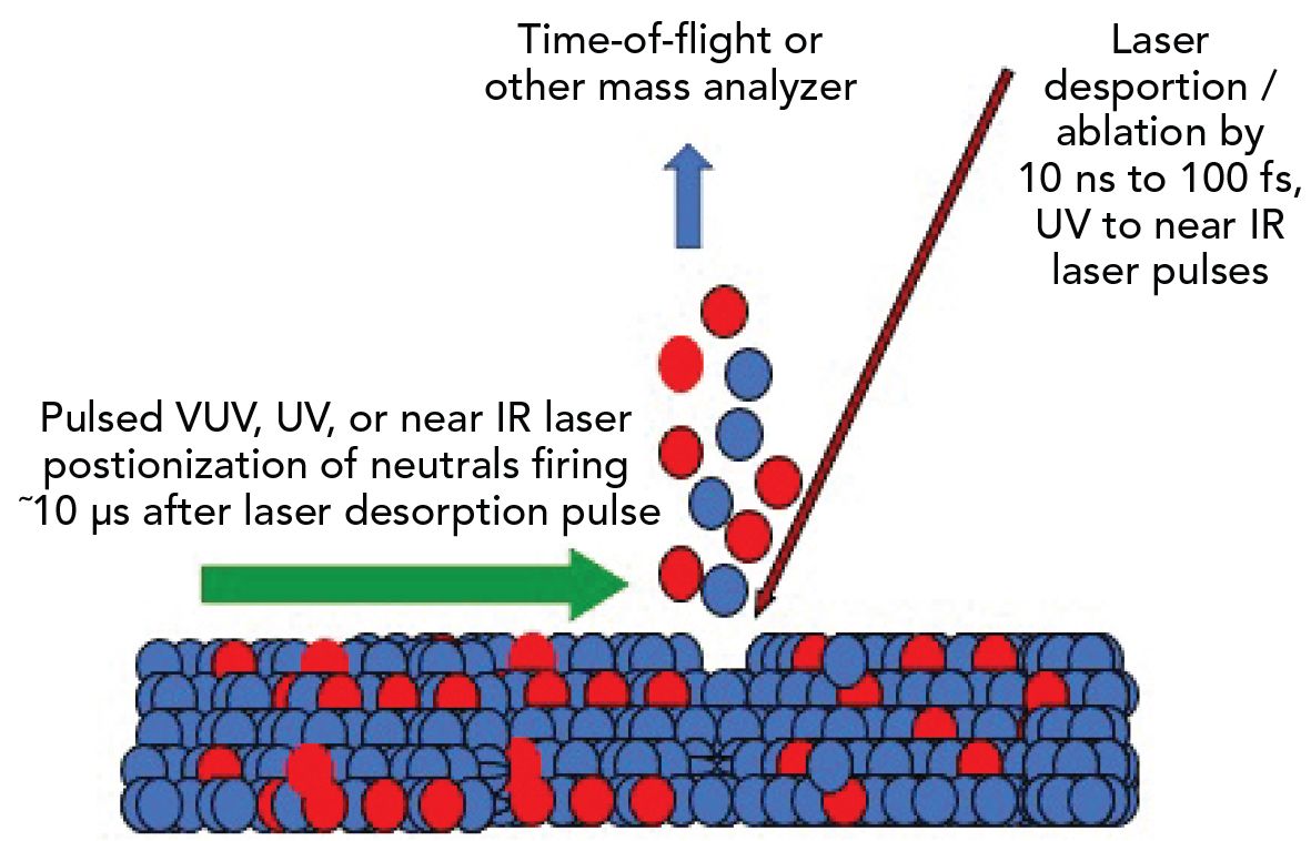
LDPI-MS, also known by the terms two laser MS, two-step laser MS, or laser ablation laser ionization MS (7,8,15), is an MS imaging technique that uses one laser pulse to desorb or ablate molecules from a solid sample held under vacuum and a second, temporally delayed laser pulse to photoionize the resultant plume of gaseous species, as shown in Figure 1. Non-optical methods of postionization, such as electrospray, chemical, and plasma-based ionization, have also been coupled with LD and LA to enhance ion yields beyond what is possible with MALDI or ns-LDI (16).
Figure 1: Schematic representation of laser desorption postionization mass spectrometry (LDPI-MS). The purple arrow follows the path of a pulsed laser beam that desorbs a solid sample to volatile neutrals that are postionized by a pulsed laser, as represented by the green arrow.
Laser Postionization via Single Photon Ionization
A common optical postionization strategy has been single photon ionization (SPI), which can occur when the energy of an incident photon exceeds the ionization energy of a gas phase molecule (8). Most organic compounds have ionization energies below ~10 eV and will undergo SPI by irradiation with vacuum ultraviolet (VUV) light of a similar or higher photon energy. One trend within organic homologous compounds is that ionization energies decrease as molecular weight increases (15). SPI has a simple mechanism and relatively low limits of detection that can reach down to the mid-low parts-per-billion range (15).
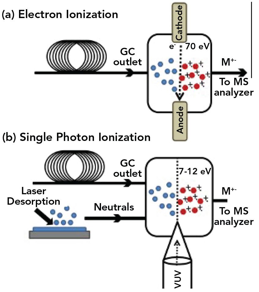
It is instructive to compare SPI with electron impact (EI), as shown in Figure 2 as coupled to gas chromatography and for SPI, laser sampling. Both methods initially generate radical cations, M+, which have long been considered to be less stable than the protonated molecules, MH+, typically generated by electrospray and chemical ionization. For decades, EI has been used for identifying molecular species by comparison of the mass spectral fragmentation patterns of separated compounds to those of known compounds in EI-MS libraries (17). However, SPI has higher ionization efficiency than EI as well as lower fragmentation when performed with photons no more than a few eV above the ionization energy (15). This may partially explain why there have been few reports of postionization of molecular species by electron impact of molecules volatilized by LD or LA.
Figure 2: This schematic shows formation of a radical cation precursor via (a) electron impact (EI) and (b) single photon ionization (SPI). The diagram represents either gas chromatography (GC) or in the case of SPI, laser desorption, to volatize neutrals for ionization. Figure used with permission from reference (7).
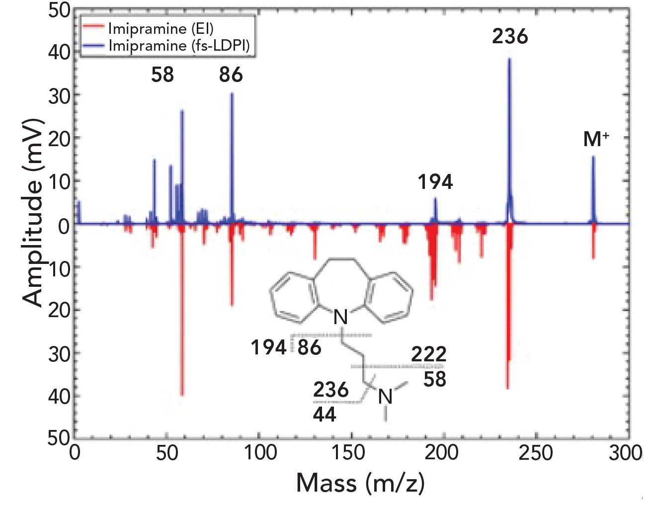
A recent study showed that three drug compounds that could not be easily detected by SIMS were observed using LDPI-MS, where ablation was performed with ~75 fs, 800 nm laser pulses, and SPI with ~7 ns, 7.9 eV laser pulses (see below) (18). The LDPI mass spectrum of imipramine displays higher intact precursor ion M+ compared with its corresponding EI mass spectrum in Figure 3. Furthermore, although the most intense peaks from EI-MS were replicated by LDPI-MS, the latter showed overall less fragmentation. The similarity between LDPI-MS and EI-MS was observed with the two other compounds successfully examined in this study (18). SPI-MS often displays major fragments as similar to those seen in EI-MS, but almost always displays the M+ that can be missing from EI-MS of labile compounds and additionally leads to a lower overall extent of fragmentation (19). This similarity implies that it may be possible to use EI-MS libraries to reliably interpret LDPI-MS performed with SPI.
Figure 3: Femtosecond-LDPI mass spectra of imipramine compared with EI mass spectra. Figure used with permission from reference (18).
The use of SPI for postionization in LDPI-MS is limited by the performance of sources of pulsed VUV radiation. The most convenient laboratory VUV source for LDPI-MS is the 157 nm fluorine excimer laser. Fluorine lasers are reasonably priced and reliable. Their intensities are in the range of ~1 mJ/pulse, allowing high efficiency detection via saturation of SPI. Furthermore, their pulse repetition rate is ~500 Hz, high enough for rapid imaging of relatively large sample areas. However, the fluorine laser photon energy is only 7.9 eV, generally limiting SPI to molecules such as polyaromatic hydrocarbons, tertiary amines, some fused ring drug compounds (15), and certain clustered ion species (20).
SPI of the wider array of organic compounds with higher ionization energies is possible with a VUV source that displays a correspondingly higher photon energy. Third harmonic generation of the 355 nm output from a standard ns pulsed Nd:YAG laser produces 118 nm wavelength (10.5 eV photon energy) laser pulses that serve as the more universal source for SPI alluded to above (15). However, this strategy produces <1 μJ/ pulse that cannot saturate SPI and leads to low overall ionization efficiency. The low repetition rates of the high power Nd:YAG lasers needed for VUV generation also limit imaging speed. Finally, the low intensity VUV beam is difficult to manipulate for inexperienced personnel and requires considerable engineered improvements before it can be readily used for general analytical applications.
Overall, expanding the use of SPI in LDPI-MS requires a more robust laboratory source of pulsed VUV radiation with ~10 eV photon energy, ~1000 pulse rate, and at least 100 μJ/pulse. High harmonic generation from ultrashort pulse lasers might one day fulfill these requirements if considerably higher intensity VUV pulses can be generated (21).
Postionization Using Multiphoton to Strong-Field Ionization
An alternative postionization to SPI is to instead utilize multiphoton strategies. SPI yields are linearly dependent on the number of photons, but resonance-enhanced multiphoton ionization (REMPI) becomes possible with exponentially higher laser intensities, in the range of 107–1011 W/cm2 (9,22). Furthermore, the excitation outlet pathway in REMPI proceeds through at least one intermediate excited state.
Figure 4a depicts the simplest form of REMPI: Two photon resonant ionization where one photon excites to an intermediate state via a strong optical absorption (which can be observed in UV-vis absorption spectroscopy), then a second photon excites out of the intermediate state to form a radical cation. An example of a resonant two photon ionization would be using 266 nm, ns laser pulses to excite a π-π* transition in a polyaromatic hydrocarbon like anthracene, then excite out of the π* state to form an ion. The two photons collectively impart 9.3 eV of energy to the polyaromatic hydrocarbon, well in excess of its ionization energy. The specific molecular electronic structure determines the types of REMPI pathways possible, the requisite laser wavelengths, and the extent of dissociation of the precursor.
Figure 4: Schematic of the two multiphoton ionization mechanisms: (a) resonance-enhanced multiphoton ionization (REMPI), (b) nonresonant multiphoton ionization (NRMPI) and (c) strong-field ionization, which can occur via ion tunneling.
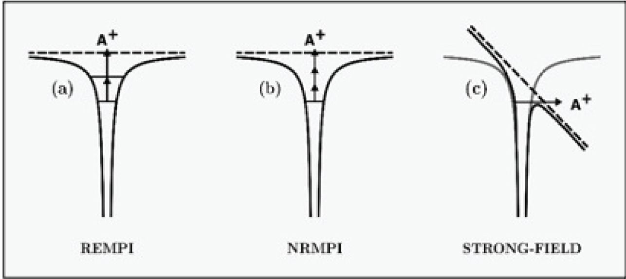
REMPI is traditionally performed with ns-laser pulses, but transitioning to shorter laser pulses can reduce fragmentation that often occurs through other pathways out of the intermediate states (9,22). Femtosecond-laser MS is performed with pulses shorter than a picosecond that lead to less dissociation in REMPI and can open up new ionization pathways less dependent on specific molecular structure (23). Femtosecond laser MS can also proceed by non-resonant multiphoton ionization (NRMPI) when yet higher laser intensities ~1013 W/cm2 are used and has the added advantage that it does not require the laser wavelength to be resonant with an excited state (Figure 4b) (9).
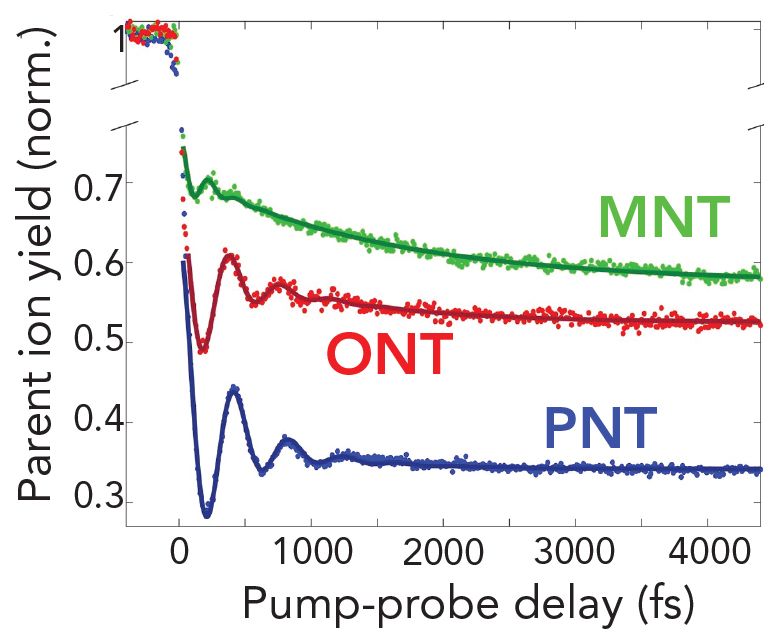
The higher laser intensities that allow NRMPI, when accessed via sub-100 fs IR laser pulses, can also lead to strong-field ionization (9,24). Strong-field ionization can occur by tunneling through the barrier to ionization via high intensity electric field-induced distortion of molecular electronic states induced by ultrashort laser pulses (9). Pulse duration, laser intensity, wavelength, and molecular electronic structure control how a specific molecule will undergo strong-field ionization. FIGURE 5 shows how structural isomers can be quantified in mixtures using a dual pulse fs IR laser technique in which a pump fs laser pulse ionizes the mixture and creates a radical cation in a ground state, then a time-delayed probe laser pulse (from the same laser) induces dissociation (24). The nitrotoluene ion signal varies with pump-probe delay time for its structural isomers o-, m-, and p-nitrotoluene (ONT, MNT, and PNT, respectively). Preliminary results found that the relative concentration of each isomer in binary and ternary mixtures could be determined to better than 10% accuracy without any preliminary use of chromatographic separation. Such a strategy would be very powerful when applied to LDPI-MS imaging, which like most MS imaging methods lacks a chromatographic front end. Femtosecond-laser MS also potentially allows coupling with sophisticated computational modeling of the ionization event that might find predictive utility in analysis (24).
Figure 5: Time dependent pump-probe experiments used for quantification of structural isomers in a mixture. Nitrotoluene ion signal varies with pump-probe delay time for its structural isomers o-, m-, and p-nitrotoluene (ONT, MNT, and PNT, respectively).
Applications of LDPI-MS
Nanosecond-LDPI-MS has found applications in analysis of biofilms, tissue samples, organic electronics, and geological samples (7,8,15,16). For example, LDPI-MS was able quantify antibiotics in biofilms and differentiate biofilms of different strains of E. coli (7,8).
A version of LDPI-MS is under consideration for incorporation into space flight instruments being developed for analysis of icy moons, comets, and carbonaceous asteroids (25). The molecular analyzer for complex refractory organic-rich surfaces (MACROS) is an instrument package utilizing ns-pulse lasers that has been developed at the NASA Goddard Space Flight Center. One of MACROS’ four basic operation modes for in situ sample analysis uses ns pulsed 266 nm IR laser for desorption and a ns pulsed laser for postionization via REMPI. L2MS is considered a “detailed” mode in MACROS because it does not require sample preparation, has a lower limit of detection, and is highly sensitive to detection of aromatic organic molecules of great interest to the mission.
The application of LDPI-MS using fs-laser ablation is at an earlier stage (8), but has already been shown to allow imaging of an organic electronic compound at a lateral resolution of 2 μm by taking advantage of the nonlinearity of fs-LA with laser intensity (8). Alternatively, the application of experimentally robust fs laser postionization to LDPI-MS will allow faster imaging and analysis of a wide range of compounds. Femtosecond laser MS has been coupled with gas chromatography for the ultrasensitive analysis of explosives (23,24) and pesticides (26), indicating the promise of its use in postionization for LDPI-MS.
Postionization via Photoionization at Elevated Pressures
We have focused on LDPI-MS performed with the sample, desorption, ablation, and postionization all occurring under high vacuum. However, there are several advantages to performing the analogous analysis at atmospheric pressure (8). The sample does not dehydrate and does not need to undergo the slow evacuation process required in traditional mass spectrometers. The plume of species desorbed or ablated from a sample immediately collides with air molecules, which enhances dissociation of a larger species, slowing down molecular velocities and possibly leading to oxidation (this final event also possibly being a disadvantage). Atmospheric pressure photoionization is most commonly initiated with SPI by VUV radiation and subsequently leads to chemical–ionization-like events that can result in the formation of protonated molecules instead of radical cations (27). Such protonation processes show great potential for expanding the breadth of LDPI-MS applications and may have driven the application of non-laser based postionization (16). Finally, most commercial mass spectrometers currently have atmospheric pressure interfaces, so coupling a LDPI front end to them avails one of their exquisite performance in terms of mass resolution and accuracy as well as tandem capabilities.
Commercial Instruments
Utilizing any analytical method relies on its commercial availability, but such options for LDPI-MS remain limited as of this writing. MALDI-2 is a method that uses a ns-pulse UV postionization laser to induce secondary ionization in the gas phase of plumes ablated by MALDI (28). Postionization in MALDI-2 occurs at intermediate pressures within an ion mobility device, leading to the declustering of higher mass species formed by MALDI. The pressure-dependent mechanism of MALDI-2 is probably more akin to that of atmospheric pressure photoionization than direct laser photoionization of a molecule isolated in vacuum. Perhaps foremost amongst the impressive advantages of UV laser postionization in MALDI-2 is that it enhances signal intensities of membrane lipids and other species detected in a tissue sample by up to two orders of magnitude over those observed by traditional MALDI. This higher signal also allows increased lateral resolution for MS imaging and Bruker is now selling an instrument with a MALDI-2 option.
Another two-laser commercial instrument currently in development by Exum Instruments is laser ablation laser ionization time-of-flight mass spectrometry (LALI-TOF-MS) (29). LALI-TOF-MS utilizes a 213 or 266 nm ns-pulsed laser to ablate into a pressure gradient and a second 266 nm, ns-pulsed laser for multiphoton ionization. The LALI-TOF-MS instrument is initially being developed for elemental analysis but will permit identification of a range of molecular species that can be expanded with the modification of the laser postionization strategy.
The development of LDPI-MS and related methods will continue to be driven by the rapid improvement and lowered costs of sub-ns-pulsed lasers, but requires coupling to higher performance mass analyzers to increase user acceptance. Readers interested in a more rigorous discus- sion of laser postionization are referred to a recent book on the fundamentals and the entire range of applications of photo-ionization in mass spectrometry (9).
Acknowledgments
Luke Hanley and Teodora Zagorac are supported by grant CHE-1904145 from the U.S. National Science Foundation (LH ORCID: 0000-0001-7856-1869. TZ ORCID: 0000-0001-7467-8736).
References
(1) R. Trouillon, M.K. Passarelli, J. Wang, M.E. Kurczy, and A.G. Ewing, Anal. Chem. 85, 522–542 (2012).
(2) A. Palmer, D. Trede, and T. Alexandrov, Metabolomics 12, 107 (2016).
(3) S. Heiles, M. Kompauer, M.A. Müller, and B. Spengler, J. Amer. Soc. Mass Spectrom. 31, 326–335 (2020).
(4) I.S. Gilmore, S. Heiles, and C.L. Pieterse, Ann. Rev. Anal. Chem. 12, 201–224 (2019).
(5) J. Nuñez, R. Renslow, J.B. Cliff III, and C.R. Anderton, Biointerph. 13, 03B301 (2018).
(6) R. Yin, J. Kyle, K. Burnum-Johnson, K.J. Bloodsworth, L. Sussel, C. Ansong, and J. Laskin, Anal. Chem. 90, 6548–6555 (2018).
(7) C. Bhardwaj and L. Hanley, Nat. Prod. Rep. 31, 756–767 (2014).
(8) L. Hanley, R. Wickramasinghe, and Y.P. Yung, Ann. Rev. Anal. Chem. 12, 225–245 (2019).
(9) L. Hanley and R. Zimmerman, eds., Photoionization and Photo-Induced Processes in Mass Spectrometry: Fundamentals and Applications (Wiley-VCH, Berlin, Germany, 2021).
(10) A. Vogel and V. Venugopalan, Chem. Rev. 103, 577–644 (2003).
(11) F. Poitrasson and F.-X. d’Abzac, J. Anal. At. Spectrom. 32, 1075–1091 (2017).
(12) H. Volk and S.C. George, Organic Geochemistry 129, 99–123 (2019).
(13) A.V. Walker, L.D. Gelb, G.E. Barry, P. Subanajouy, A. Poudel, M. Hara, I.V. Veryovkin, G.I. Bell, and L. Hanley, Biointerph. 13, 03B416 (2018).
(14) A. Cedeño López, V. Grimaudo, A. Riedo, M. Tulej, R. Wiesendanger, R. Lukmanov, P. Moreno-Garcia, E. Lortscher, P. Wurz, and P. Broekmann, Anal. Chem. 92, 1355–1362 (2020).
(15) L. Hanley and R. Zimmermann, Anal. Chem. 81, 4174–4182 (2009).
(16) X. Ding, K. Liu, and Z. Shi, Mass Spectrom. Rev., in press (2020) https://doi.org/10.1002/mas.21649
(17) S. Stein, Anal. Chem. 84, 7274–7282 (2012).
(18) C.L. Pieterse, I. Rungger, I.S. Gilmore, R.C. Wickramasinghe, and L. Hanley, J. Phys. Chem. Lett. 11, 8616–8622 (2020).
(19) G. Isaacman, K.R. Wilson, A.W.H. Chan, D.R. Worton, J.R. Kimmel, T. Nah, T. Hohaus, M. Gonin, J.H. Kroll, D.R. Worsnop, and A.H. Goldstein, Anal. Chem. 84, 2335–2342 (2012).
(20) M.T. Blaze, L.K. Takahashi, J. Zhou, M. Ahmed, G.L. Gasper, F.D. Pleticha, and L. Hanley, Anal. Chem. 83, 4962–4969 (2011).
(21) T. Popmintchev, M.-C. Chen, D. Popmintchev, P. Arpin, S. Brown, S. Ališauskas, G. Andriukaitis, T. Balciunas, O.D. Mucke, A. Pugzlys, A. Baltuska, B. Shim, S.E. Schrauth, A. Gaeta, C. Hernandez-Garcia, L. Plaja, A. Becker, A. Jaron-Becker, M.M. Murnane, and H.C. Kapteyn, Science 336, 1287–1291 (2012).
(22) U. Boesl, J. Mass Spectrom. 35, 289–304 (2000).
(23) K.W.D. Ledingham and R.P. Singhal, Int. J. Mass Spectrom. Ion Process. 163, 149–168 (1997).
(24) K. Moore Tibbetts, Chem. European J. 25, 8431–8439 (2019).
(25) S.A. Getty, J. Elsila, M. Balvin, W.B. Brinck- erhoff, T. Cornish, X. Li, J. Ferrance, A. Grubisic, and A. Southard, Molecular Analyzer for Complex Refractory Organic-Rich Surfaces (MACROS). in 2017 IEEE Aerospace Conference. 2017. https://doi.org/10.1109/ AERO.2017.7943706
(26) X. Yang, T. Imasaka, and T. Imasaka, Anal. Chem. 90, 4886–4893 (2018).
(27) T.J. Kauppila, J.A. Syage, and T. Benter, Mass Spectrom. Rev. 36, 423–449 (2017).
(28) J. Soltwisch, H. Kettling, S. Vens-Cappell, M. Wiegelmann, J. Müthing, and K. Dreisewerd, Science 348, 211–215 (2015).
(29) J. Williams and J. Putman, Spectroscopy 35(5), 9–16 (2020).
Teodora Zagorac and Luke Hanley are with the Department of Chemistry at University of Illinois at Chicago, in Chicago, Illinois. Direct correspondence to: LHanley@uic.edu

New Method Explored for the Detection of CECs in Crops Irrigated with Contaminated Water
April 30th 2025This new study presents a validated QuEChERS–LC-MS/MS method for detecting eight persistent, mobile, and toxic substances in escarole, tomatoes, and tomato leaves irrigated with contaminated water.

.png&w=3840&q=75)

.png&w=3840&q=75)


.png&w=3840&q=75)


.png&w=3840&q=75)


