High-Speed Process Aggregate Monitoring with UHP-SEC-MALS
The Application Notebook
At-line process monitoring (ALPM) of protein aggregation during processing and purification is made feasible by the 5-min runs typical of UHP-SEC. ALPM is particularly valuable in production because it can minimize the losses encountered when a process begins to fail. The addition of multi-angle light scattering (MALS) analysis to UHP-SEC deepens the quality and reliability of information obtained in near-real-time.
We demonstrate rapid UHP-SEC-MALS analysis under conditions mimicking different manufacturing and purification processing steps as well as stresses applied to test stability. MALS, RI, and dynamic light scattering (DLS) data provided robust, reproducible, and reliable quantification of molar mass, size, and amount of each species in a given sample.
Materials and methods
UHP-SEC was performed with an Acquity UPLC pump and autosampler (Waters Corporation) and BEH SEC column (200 à , 1.7 µm, 4.6 mm à 150 mm; Waters). The effluent of the SEC column flowed through an inline UVâvis detector (Waters), µDAWN multi-angle light scattering detector with internal WyattQELS dynamic light scattering detector, and Optilab UT-rEX dRI detector (all three from Wyatt Technology). For each chromatogram, 2 µL to 5 µL sample was injected with an overall run time of 5 min per sample. Data collection and analysis were performed with ASTRA software (Wyatt). Monoclonal antibody 1 (mAb1) represents various phases of the purification process, ending with purified sample. The suffixes -C1, -C2, and -C3 represent different fractions collected during the purification process.
A different monoclonal antibody, mAb2, was subjected to stresses representing stability conditions. In addition to MALS analysis similar to mAb1, DLS measurements were performed to assess any conformational changes.
Results
Light scattering chromatograms for two replicates of each of the three conditions are overlaid in Figure 1. The starting material (mAb1-C1) contains approximately 70% monomer, with the rest of the mass represented by aggregate and fragment species. Sample mAb1-C2 represents an intermediate purification process, resulting in a solution of 85% monomer. The final purified antibody (mAb1-C3) is >95% monomer by mass. UHP-SEC-MALS provided rapid quantitation of each mAb sample to verify the identity of SEC peaks, making this technique perfect for ALPM. In addition, the replicates overlay perfectly as do the molar masses determined from each replicate, indicating the robustness of this method.
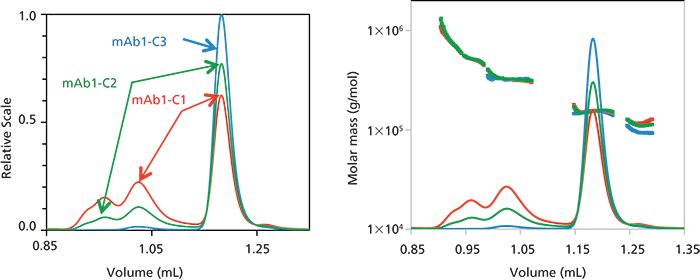
Figure 1: Left: Overlay of light scattering chromatograms for mAb1 undergoing different stages of purification for condition 1 (red), condition 2 (green), and condition 3 (blue). Right: Overlay of refractive index concentration signals and molar masses calculated from light scattering.
Five different species were defined based on the mAb1-C1 chromatogram. The peak definitions for these species are shown in Figure 2. The table in Figure 2 shows how effectively the different aggregate fractions are removed, while the monomer is enriched, as the purification process progresses. The molar mass of each species also appears consistent for collected fraction.
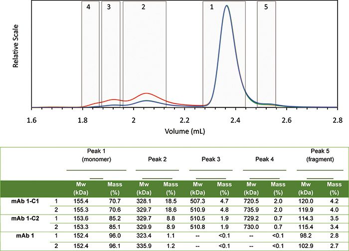
Figure 2: Peak definitions (top) and resulting molar mass and mass fraction (bottom) of each species. Plots correspond to light scattering (red), RI (blue), and UV (green) signals. RI and UV signals are barely distinguishable from each other for monomer and aggregates, indicating identical extinction coefficients, but differ for the fragment.
The molar mass of eluting species measured by the µDAWN and UT-rEX is extremely robust and reliable, despite the low concentration of certain peaks. For example, in the case of the purified mAb1, even though the maximum concentration of aggregate is only 0.8 µg/mL as it elutes from the column, the measured molar mass is within 2% of the molar mass measured for conditions 1 and 2, where the eluting concentration is significantly higher (Peak 2 Mw, Figure 2).
In another test, mAb2 was subjected to three different stress conditions and characterized by UHP-SEC-MALS-DLS, with results shown in Figure 3. Both conditions 1 and 2 caused mAb2 dimer formation, where condition 2 was shown to produce about 20% more dimer than condition 1. On the other hand, mAb2 subjected to condition 3 underwent quite different changes as a result of the applied stress: little dimer formation, but a shift in the molar mass of the fragment, and a shift in elution time of the monomer. DLS data indicate no conformational change of the monomer, suggesting that the later elution time is the result of a chemical modification leading to column interaction.
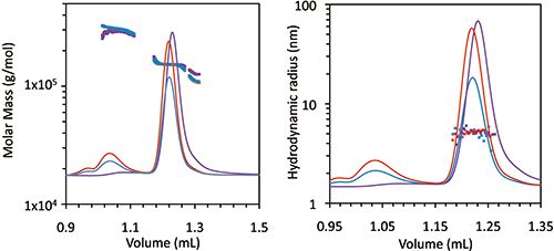
Figure 3: Left: Refractive index chromatograms for mAb2 under stability conditions 1 (blue), 2 (red), and 3 (purple). Molar mass values from MALS of the monomer, dimer, and fragment peaks have been overlaid. Right: Light scattering chromatograms for mAb2, plus hydrodynamic radii values of the monomer measured by online dynamic light scattering.
Conclusions
In summary, we demonstrated the use of high-speed UHP-SEC combined with MALS and DLS for process monitoring of antibody samples. With this technique, measurements may be performed in as little as 5 min, enabling monitoring of purification and other processing steps, as well as high-throughput stability screening. The µDAWN and UT-rEX provided essential and reliable quantification of molar mass and hydrodynamic radius of aggregates and fragments that would not be possible with analytical UHPLC alone. These data provide important information about the purification process, such as whether the aggregates are sheared during the process or whether new aggregate species are formed. The addition of MALS, DLS, and RI to UHP-SEC is critical to ascertain conclusively species identification and conformation.

Wyatt Technology
6300 Hollister Avenue, Santa Barbara, CA 93117
tel. +1 (805) 681-9009, fax +1 (805) 681-0123
Website: www.wyatt.com

SEC-MALS of Antibody Therapeutics—A Robust Method for In-Depth Sample Characterization
June 1st 2022Monoclonal antibodies (mAbs) are effective therapeutics for cancers, auto-immune diseases, viral infections, and other diseases. Recent developments in antibody therapeutics aim to add more specific binding regions (bi- and multi-specificity) to increase their effectiveness and/or to downsize the molecule to the specific binding regions (for example, scFv or Fab fragment) to achieve better penetration of the tissue. As the molecule gets more complex, the possible high and low molecular weight (H/LMW) impurities become more complex, too. In order to accurately analyze the various species, more advanced detection than ultraviolet (UV) is required to characterize a mAb sample.


