High Resolution LC–MS Analysis of Reduced IgG1 Monoclonal Antibody Fragments Using HALO Protein C4
A new fused-core particle designed specifically for the separation of proteins and monoclonal antibodies with 3.4 ?m particle size and a thin 0.2 ?m porous shell demonstrates high resolution of the heavy and light chain fragments of IgG1.
A new fused-core particle designed specifically for the separation of proteins and monoclonal antibodies with 3.4 µm particle size and a thin 0.2 µm porous shell demonstrates high resolution of the heavy and light chain fragments of IgG1.
The focus of many pharmaceutical companies has shifted to larger biotherapeutic molecules as potential treatments for disease. These biomolecules present a new set of challenges compared to their small molecule predecessors when complete characterization is considered. Variants can be found through different glycans, deamidations, enzymatic clipped polypeptides, and so on, not to mention the potential for aggregate formation. Rapid reversed-phase LC analysis is convenient for its ability to be interfaced to online mass spectrometry. The HALO Protein C4 bonded phase is ideal for this application with its optimized design for high recovery and stability at the high temperatures and low pH required by the analysis.
Experimental Conditions
Column: 100 mm × 2.1 mm Halo Protein C4; gradient: 29–32% B in 20 min; mobile phase A: 0.5% (v/v) formic acid with 20 mM ammonium formate; mobile phase B: 45% acetonitrile, 45% isopropanol/0.5% (v/v) formic acid with 20 mM ammonium formate; temperature: 80 °C; flow rate: 0.4 mL/min; instrument: Shimadzu LCMS 2020; detection: 280 nm; MS-2020 single quadrupole MS instrument using ESI at +4.5 kV capillary voltage, 2 pps scan rate from 500 to 2000 amu m/z. Component masses for these measurements were determined by deconvolution using MagTran software, v. 1.02, based on the ZScore algorithm developed by Zhang and Marshall (1).
Results
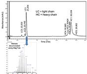
The addition of a small amount of ammonium formate to formic acid in the mobile phase improves the peak shapes. An optimized, shallow gradient allows resolution of the variants of both the heavy and light chain fragments of IgG1 as shown in Figure 1. The MS spectra can then be deconvoluted to determine the masses associated with each peak.

Figure 1: LCâMS separation of reduced IgG1. Column: 2.1 mm i.d à 100 mm HALO Protein C4; Flow rate: 0.4 mL/min. Mobile phase A: 0.5 % formic acid with 20 mM ammonium formate; mobile phase B: 45% AcN/45% IPA/ 0.5 % formic acid with 20 mM ammonium formate; gradient: 29â32% B in 20 min.; temperature: 80 °C; detection: 280 nm; MS conditions: Shimadzu LCMS-2020, ESI +4.5 kV, 2 pps, 500â2000 m/z.
Conclusions
HALO Protein C4 demonstrates the low pH and high temperature stability that is required to analyze reduced and alkylated IgG1 using mass-spec friendly mobile phase. The use of 80 °C enables improved peak shape while the high resolution allows complete analysis of the IgG1 fragments that are present.
References
(1) Z. Zhang and A.G. Marshall, J. Am. Soc. Mass Spectrom.9, 225 (1998).

MAC-MOD Analytical, Inc.
103 Commons Court, Chadds Ford, PA 19317
tel. (800) 441-7508; fax (610) 358-5993
Website: www.mac-mod.com

SEC-MALS of Antibody Therapeutics—A Robust Method for In-Depth Sample Characterization
June 1st 2022Monoclonal antibodies (mAbs) are effective therapeutics for cancers, auto-immune diseases, viral infections, and other diseases. Recent developments in antibody therapeutics aim to add more specific binding regions (bi- and multi-specificity) to increase their effectiveness and/or to downsize the molecule to the specific binding regions (for example, scFv or Fab fragment) to achieve better penetration of the tissue. As the molecule gets more complex, the possible high and low molecular weight (H/LMW) impurities become more complex, too. In order to accurately analyze the various species, more advanced detection than ultraviolet (UV) is required to characterize a mAb sample.


