Dried Blood Spots in Mass Spectrometry-based Protein Analysis. Recent Developments including Sampling of other Biological Matrices and Novel Sampling Technologies
The dried blood spot (DBS) sampling technique has been around for decades, predominantly for small molecules and mainly in newborn screening. Although determination of proteins after DBS sampling is usually performed with immunometric assays, the combination with mass spectrometry (MS) is gaining interest. This article provides an overview of DBS sampling for mass spectrometry-based protein analysis. The first part will focus on clinical applications for DBSs and on sampling other biological matrices apart from whole blood, including dried matrix spots (DMSs). The second part will explore the new frontiers of the DBS sampling technology, including novel sampling materials/devices, and novel combinations with mass spectrometry. Examples of use in both qualitative and quantitative protein analysis are highlighted as well as examples using both bottom-up and top-down proteomics approaches.

The dried blood spot (DBS) sampling technique has been around for decades, predominantly for small molecules and mainly in newborn screening. Although determination of proteins after DBS sampling is usually performed with immunometric assays, the combination with mass spectrometry (MS) is gaining interest. This article provides an overview of DBS sampling for mass spectrometry-based protein analysis. The first part will focus on clinical applications for DBSs and on sampling other biological matrices apart from whole blood, including dried matrix spots (DMSs). The second part will explore the new frontiers of the DBS sampling technology, including novel sampling materials/devices, and novel combinations with mass spectrometry. Examples of use in both qualitative and quantitative protein analysis are highlighted as well as examples using both bottom-up and top-down proteomics approaches.
Dried blood spot (DBS) sampling was introduced in 1913 when Ivar Bang demonstrated this less-invasive sampling strategy in glucose monitoring (1). In 1963 Susi and Guthrie applied DBS sampling for phenylketonuria screening in newborns (2) and the technique gained widespread acceptance. In the beginning, the strategy was mainly used in newborn screening of phenylketonuria and other small molecule metabolic markers. However, as a result of the advantages of DBS sampling, including small sampling volumes, improved stability of analytes, less biohazard risk and easy transport, the technique was also soon evaluated for other analytes. The first reports of DBSs for sampling and storage of proteins are from the early 1970s (3,4). In one work, Thielmann and Aquino (3) demonstrated that blood could be sampled and stored on filter paper prior to electrophoretic separation of hemoglobin S from hemoglobin A. In another work, Laurell (4) described a test for α1-antitrypsin deficiency using DBSs and electrophoretic separation. Since then, the technique has also had widespread use for the analysis of proteins in a wide range of applications (5), the majority of the applications described use immunoassays to determine the proteins.
Although immunoassays are highly sensitive and widely used, mass spectrometry (MS) has certain advantages with respect to specificity and differentiation power. MS is therefore a powerful tool in quantitative protein determination and in the study of the proteome. In the 1980s, the first reports of MS determination of proteins in DBSs can be found. Wada et al. (6) used molecular secondary ion mass spectrometry (SIMS) in a study of DBSs from 80 000 Japanese neonates: A double-focused mass spectrometer with magnetic scanning was used for structural analysis of the detected hemoglobin variants after purification using chromatography and subsequent tryptic digestion of the isolated variants. As the blood samples were obtained from newborns, the use of DBS sampling with its low invasiveness and low sample volumes were advantageous. The large sample set (80 000) combined with the differentiation power of the MS made it possible to easily detect and confirm abnormal globins of low prevalence. Following this, very few reports of mass spectrometric determination of proteins from DBSs can be found throughout the 1990s where the main use of MS for protein analysis of DBSs was to characterize hemoglobin variants.
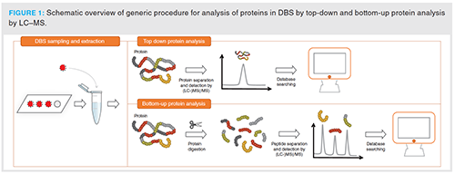
Overall, the majority of reports on DBS sampling in protein analysis by MS, especially from the earlier days, are on the determination of hemoglobin variants, mainly for use in newborn screening (7–10). Hemoglobin is a high abundance protein and determination of this protein by MS is – even from very small sample volumes such as DBSs – relatively straightforward. The protein can be determined either directly (top-down) or after a simple digestion step (bottom-up) without extensive clean up. This is demonstrated in reference 11. A schematic overview of the top-down and bottom-up procedure after DBS sampling is illustrated in Figure 1.
For many other proteins of interest, such as biomarkers, doping agents or drugs, the situation is more complex. This is because the amount present in the blood sample is extremely low compared to the highly abundant proteins (such as hemoglobin, albumin and immunoglobulins) in blood. For these analytes the applicability of DBS sampling has been dependent on the development of more sensitive analytical instrumentation as well as on the development of highly selective sample preparation methods.
After the turn of the century, reports on MS-based protein analysis from DBSs have increased. A variety of applications are described from newborn screening (8–10, 12–14), to biomarker analysis (15–21) and discovery (22–26), doping analysis (27–33), and bioanalysis of therapeutic proteins (drug discovery and therapeutic drug monitoring) (34–36). Different sampling materials and devices such as volumetric absorptive microsampling (VAMS) (17,37) and water-soluble sampling material (38,39) have been introduced as well as materials with incorporated functionality for protein pretreatment and enrichment (40,41).
The combination of DBS sampling and MS in the analysis of proteins has been reviewed twice previously, in 2014 and 2017 (42,43). These reviews focus on clinical perspectives in DBS protein quantification using MS, and challenges and opportunities in MS analysis of proteins from DBSs. The present review will give a comprehensive overview of the progress in DBS sampling for MS-based protein analysis with emphasis on the last five years. The first part will focus on clinical applications for DBS sampling (biomarker determination, doping analysis and the analysis of therapeutic proteins) and on the sampling of other biological matrices apart from blood (dried matrix spots [DMSs]). The second part will explore the new frontiers in DBS sampling technology such as novel sampling material/devices, and novel combinations with MS. Examples of use in both qualitative and quantitative protein analysis will be given, as well as examples using both bottom-up and top-down proteomics approaches.
Clinical applications
Biomarker determination and discovery
DBS sampling started out as a sampling technique for biomarker determination in newborn screening of inborn errors of metabolism, and is still the method of choice. One important reason for increased and continued use of DBSs in these programmes is the increase in MS sensitivity. In addition, DBS sampling has a large potential for other biomarker applications because blood sampling for screening of diseases or treatment follow-up could easily be performed by the patient, without going to the phlebotomist/hospital/doctor’s office. This is especially beneficial when sampling in remote areas or if the goal is to monitor otherwise healthy individuals such as athletes.
Combining DBS sampling with MS is also advantageous for protein biomarker analysis because MS is the analysis method of choice for many of the analytes in the newborn screening programme. In addition, the multiplexing and differentiating capability of the MS will be an extra benefit in protein biomarker analysis.
A majority of the publications applying DBS sampling and MS is within biomarker analysis, either targeted biomarker determination where validated biomarkers are determined or in proteomics studies for biomarker discovery. Most of the biomarker studies published use a bottom-up approach. Top-down studies are also available, but mainly in evaluation of new concepts (discussed in more detail in a separate section). To our knowledge, there are currently still no protein biomarkers routinely analyzed from DBSs by LC–MS. As a consequence of this, limited information about the reproducibility of the published LC–MS methods for analysis of protein biomarkers from DBSs is available. There are a couple of factors that suggests that high reproducibility of DBS LC–MS methods might be a bigger challenge than for other methods, especially for low-abundance biomarkers: In addition to variations arising from DBS sampling itself such as low sample volume, varying hematocrit and non-complete elution from the cards, LC–MS- based protein analysis by the bottom-up approach consists of several steps contributing to the variability such as denaturation, alkylation, proteolysis, immunocapture (especially for low abundance proteins), solid-phase extraction etc. The majority of the published papers circumvent the challenge of varying hematocrit by spotting a fixed volume and using the whole (16,30) or the majority of the spot (19), or by using a normalization step when sampling only a portion of the spotted blood (20). Improving and simplifying both the DBS workflow and the MS-based protein analysis will increase the likelihood for these methods to be used by the routine laboratory in the future.
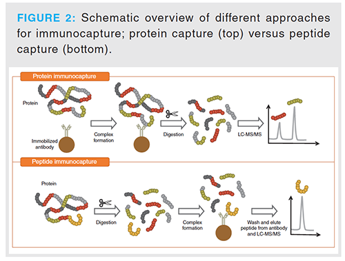
Targeted biomarker determination: Examples of methods describing biomarker quantification using DBS sampling are the determination of ATP7B protein as a potential screen for Wilson’s disease (19) and the determination of Yersinia pestis markers (15). In both examples the proteins are determined from DBSs using the bottom-up approach, the most commonly applied approach in biomarker quantification by MS. As the abundance of both ATP7B protein and the Yersinina pestis markers in blood is low, a selective sample clean-up step, immunocapture, has been included in the analytical procedure. Immunocapture is a common clean-up and enrichment procedure for low-abundant proteins in biological matrices prior to MS (44), and can be performed both before and after the digestion step (Figure 2). Both approaches are described in combination with DBSs for biomarker analysis. For example, the Yersinia pestis markers (15) and human chorionic gonadotropin (hCG; a biomarker for pregnancy, ovarian and testicular cancer as well as doping agent for male athletes) (16) were determined using immunocapture prior to digestion. ATP7B protein (19), and a range of clinically-relevant plasma proteins such as albumin, apolipoproteins and c-reactive protein (20) on the other hand, were determined using immunocapture after digestion. Further sensitivity can be achieved by using nano liquid chromatography (nano LC–) MS/MS for the subsequent analysis (15,16,19): For hCG > 10 times improvement in detection and quantification limits was achieved when transferring a method for the determination of hCG in DBSs from a micro LC–MS/MS system to a nano LC–MS/MS system (16).
Biomarker discovery: Biomarker discovery studies using DBS sampling are also described, both by targeted and non-targeted approaches. A targeted approach with multiple reaction monitoring (MRM) was done for several of the studies: Examples are assessment of schizophrenia risk (23) and screening for primary immunodeficiency disorder (25). In addition, two separate publications demonstrate that a targeted approach can be used for reproducible MS determination of a high number of proteins 97 (22), and 82 (24) from DBSs.
Non-targeted biomarker discovery is described for identification of biomarkers for athletes in different training states (using data independent acquisition). The goal was to identify two different clusters of proteins to define acute versus chronic physiological changes related to functional overreaching (26).
Determination of protein modifications: Another potential application of DBS sampling in protein analysis by MS is the determination of protein modifications. Both post-translational modifications (PTMs) such as site-specific N-glycopeptides of serological proteins (45) and environmentally-induced modifications such as the oxidation of cysteine 34 of human serum albumin (46), age-associated methylation of hemoglobin (47), reactive nitrogen and oxygen species-induced modifications in hemoglobin (48,49) are reported. For these analytes it is especially important to evaluate the stability of the modifications and/or if the modifications studied can be artificially induced during the storage of the DBSs in air.
The main challenge for widespread use of DBSs in MS-based biomarker determination are the quantification of low-abundant proteins from the complex matrix and the low sample volumes used in DBS sampling. This may be resolved using affinity clean-up (for targeted applications) and nano-LC–MS as described for ATP7B protein (19). An additional challenge for use in remote area sampling is that you will need to send the sample to a laboratory with advanced equipment and that the response time will most likely be increased (15).
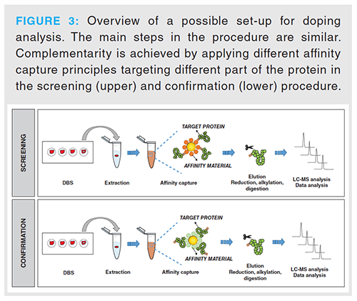
Doping analysis: DBS sampling has emerged as an interesting sampling alternative in doping analysis (30), and also for protein and peptide doping agents. This is highlighted by the attention the World Anti-Doping Agency (WADA) has focused on alternative sampling matrices such as DBSs. The advantages are linked to the ease of sampling which makes it both possible to directly determine the level of circulating substances immediately prior to (or after) the competition, and the low invasiveness for the athlete. The latter is especially advantageous for “out-of-competition” testing because no trained phlebotomist is necessary for drawing the blood sample. Furthermore, since doping samples sometimes have to be stored for several years (up to 10 years) it is a great advantage that storage of DBS samples is less demanding (both related to temperature and space requirements) than conventional urine and blood samples. Recent examples of protein-doping agents that can be determined by MS from DBS are insulin (33), the synthetic human adrenocorticotropic hormone tretracosactide hexaacetate (29) and the erythropoietin-stimulating protein sotatercept (30). The latter uses a bottom-up approach and high-resolution mass spectrometry (HRMS) determination for both screening and confirmation. Sufficient sensitivity is achieved by affinity clean-up using protein G in the screening procedure and activin A in the confirmation procedure. The screening method can also detect other IgG-based doping agents such as luspatercept and bimagrumab. In addition, as high resolution full-scan MS data is collected retrospective analysis can be performed to detect further IgG-based drugs with performance-enhancing properties. For confirmation a selective extraction of sotatercept was enabled using activin A (30). Figure 3 illustrates a set-up for screening and confirmation in doping analysis using two different affinity approaches for sample clean-up.
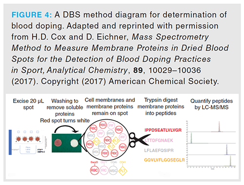
Another recent progression in doping analysis where proteins are monitored from DBSs is to determine blood doping by autologous blood or recombinant human erythropoietin (31,32). In the described method (31,32) the membrane proteins are isolated and subsequently protein digested before LC–MS/MS determination of two reticulocyte membrane proteins (Figure 4); one of the two membrane proteins is only present in immature reticulocytes while the other is present on both red blood cells and reticulocytes. The ratio of these two proteins is more sensitive to changes in erythropoiesis than the conventional way of only monitoring percentage reticulocytes (31,32). Hence the method has a potential in blood doping analysis.
The major challenge for increased use of DBS sampling in doping analysis of proteins is most likely the current need of specific (affinity based) sample enrichment procedures which makes targeted screening of multiple analytes more difficult than for small molecule doping agents.
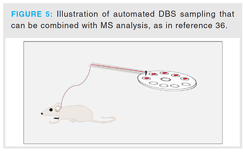
Bioanalysis of therapeutic proteins: In drug discovery, there has been an increasing focus on using microsampling techniques to reduce the volume of blood and hence the number of animals as test subjects in drug development (50). This trend is also valid in the development of biopharmaceuticals, such as for therapeutic proteins/antibodies, which is a fast-growing class of drugs on the market. The use of both DBS sampling and MS for analysis of therapeutic proteins during drug discovery has been described, although not extensively. A recent example is an automated system based on DBS sampling using serial microsampling (36). The system was used for studying the pharmacokinetics (PK) of a therapeutic protein, and only one mouse was necessary to collect a full PK curve as opposed to one mouse per data point using conventional sampling methods (blood serum). An illustration of an automated DBS sampling system is shown in Figure 5. The major difference compared to traditional PK studies will be that the curves are obtained for whole blood and not serum or plasma which commonly is the matrix of choice to establish dose response curves. After elution from the DBSs the target protein was enriched using protein A and digested prior to analysis.
Other sampling matrices/dried matrix spots
Most bioanalytical methods are developed and validated using serum or plasma as the biological matrix, and for biomarkers reference levels are established using the same matrices. A challenge with DBS sampling for use in biomarker analysis is therefore a lack of reference levels using capillary blood as sample matrix. As a result of this, the advantages of small sample volume and increased stability of dried analytes makes it worthwhile in some occasions to also use the dried sampling format for other biological matrices than whole blood. In addition, for analytes that are mainly distributed in plasma (such as plasma proteins), the sensitivity might be higher when using serum or plasma compared to whole blood. As increased stability is often seen for dried matrices, another advantage is that demanding and costly sample freezing necessary for storage and shipping of liquid samples can be avoided (51). Synovial fluid, cerebrospinal fluid, saliva, tears and urine, as well as the processed blood matrices serum and plasma are all biological matrices that can be used for dried matrix spotting. In analysis of proteins by MS, the use of other matrices is scarce, but has been described for diagnostic proteins such like hCG (16) as well as for two biomarkers for Yersinia pestis (F1 antigen and low calcium response V antigen) (15), site-specific N-glycopeptides of serological proteins (45), and serum proteins in a study on patients with intrauterine growth restriction (a pathological pregnancy condition) (51).
DMS sampling for protein analysis by MS is described mainly for serum (16,41,45,51,52). Other matrices used are plasma (both human [16] and mouse [15]), urine (16) and (mouse) spleen (15).
The analyte determined from most different matrices is hCG. It has been determined from dried plasma, serum and urine spots in addition to from DBSs (16). In addition, it has been determined from dried serum spots using a smart sampling device combining the sampling step with immunoaffinity capture (41,52). Figure 6 shows MS chromatograms at the limit of detection (LOD) of hCG from four different matrices spotted on conventional DBS cards.
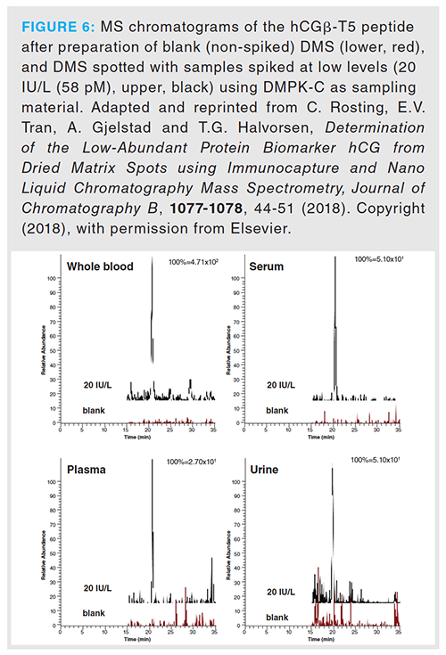
LODs in the lower IU/L-level (corresponding to 14.5–30.5 pM) were observed for hCG from all matrices (Table 1). The repeatability at this low level was satisfactory (relative standard deviation [RSD] < 25%) for all matrices except plasma (Table 1). The reason for the high RSD observed from plasma was speculated to be due to the anticoagulant because a similar effect was observed for whole blood drawn using different anticoagulants previously (38). This suggests that urine and serum might be more suitable alternative matrices for spotting, and that attention has to be paid to the anticoagulant when using plasma as the sample matrix and during method development for DBSs when whole blood is collected in tubes containing anticoagulant.
Exploring New Frontiers in DBS Sampling
New sampling materials and devices: Recently there has been a focus on new sampling materials and devices that can overcome some of the challenges often associated with DBS sampling. These new materials and devices may improve the repeatability and reproducibility of LC–MS methods for proteins from DBSs, and hence increase the likelihood for these methods to be used in a routine laboratory. Traditionally, pure cellulose cards (most commonly non-impregnated to avoid denaturation) are used for DBS sampling of proteins. The major challenges are poor extraction recoveries from blood spots and the effect of hematocrit on quantification. The issue of poor extraction recoveries has been approached by introducing water-soluble sampling materials: In 2015 water-soluble carboxymethyl cellulose was introduced as a potential sampling material in protein analysis from blood spots (38). Rosting et al. evaluated the material for the analysis of hCG from DBSs. The method was later refined and evaluated for other sample matrices (plasma, serum, urine) and it was demonstrated that lower LODs were obtainable for all matrices (except whole blood where comparable LODs were seen) using the water-soluble material compared to conventional sampling cards (16).
Other materials such as volumetric absorptive microsampling (VAMS) focus on the possibility to sample an accurate volume to avoid the potential issue of varying blood hematocrit. Blood hematocrit will have an impact on quantification when only a portion of the blood spot is used for further analysis, hence sampling using VAMS eliminates the volume variation and is expected to improve the reproducibility of the analysis. VAMS has been evaluated for proteins by MS in both a generic set-up using six non-human model proteins (37), and in quantification of circulating cardiovascular risk associated apolipoproteins (17). Despite VAMS being described to be less affected by blood hematocrit because of the sampling of accurate volume, an effect of the hematocrit on the recovery was seen for two out of six model proteins at low hematocrit (20%) in the work using non-human model proteins and a quite wide hematocrit range (20-60%) (37). The reasons for this are not clear but it is described that although the VAMS collect a constant volume independent of hematocrit, extreme hematocrit levels might influence extraction recoveries from VAMS (53,54).
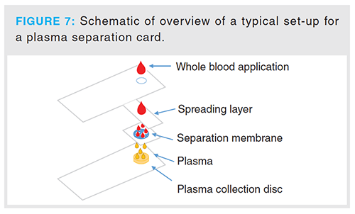
Another way of ensuring accurate volume is by using plasma separation cards. These cards separate the blood cells from the blood plasma and sample an accurate volume of the blood plasma. Although applied in protein analysis from dried serum spots (51) after directly spotting serum on the base sheet of the plasma separation card, these cards have not yet been used for spotting whole blood in protein analysis by MS. A schematic overview of a typical set-up for a plasma separation card can be found in Figure 7.
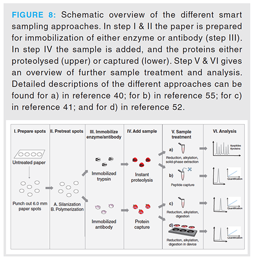
So-called smart sampling materials have recently been introduced. These sampling materials integrate protein relevant sample preparation to the sampling step and hence reduce the sample preparation time consumption significantly (40,52,55). The main advantage of this concept is to utilize the sample drying time to perform part of the sample preparation. This will also reduce the number of manual steps in the procedure and could have a positive effect on the reproducibility of the analyses. Both proteolytic enzymes (trypsin) and antibodies have been covalently immobilized to paper polymerized with 2-hydroxyethyl methacrylate-co-2-vinyl-4,4-dimethyl azlactone (pHEMA-VDM), and good performance has been described in quantitative proof-of-concept studies (52,55). A schematic overview of the different applications employing smart materials is shown in Figure 8.
Novel combinations with mass spectrometry: Most described methods for MS-based protein analysis of DBSs uses liquid chromatography coupled to ESI-MS. Novel approaches of protein analysis, DBS sampling and MS are the use of paper spray (56,57), and the use of liquid extraction surface analysis (LESA) in combination with direct electrospray MS (58,59).
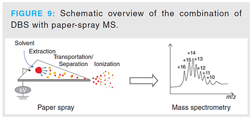
The combination of DBS sampling with paper-spray MS is convenient because in DBS sampling the sample is already spotted on paper. Nevertheless, only a couple of top-down examples of this combination are described, in these applications the paper is modified prior to use to reduce the hydrophilicity of the paper and hence reduce the interaction between the analytes and the paper (56,57). A schematic overview of the process is shown in Figure 9.
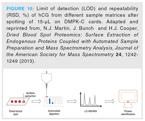
Applying LESA to DBSs allows for direct electrospray analysis from the dried sample after the addition of a small volume of extraction solvent on top of the DBSs. The technique has been combined with MS both with and without high field asymmetric waveform ion mobility spectrometry (FAIMS). The top-down approach is especially advantageous in combination with LESA because the extraction drop can then be directly injected to the MS (59–61). Additionally, non-targeted bottom-up proteomics examples are also described (58). Possibilities for automating the digestion process after LESA are also available (62). A schematic overview of an automated process for digestion of DBSs after LESA can be found in Figure 10. Automated workflows are advantageous to ensure robust sample handling and increased sample throughput.
DBS as a sampling format is also well suited for combinations with other direct ionization techniques such as desorption electrospray ionization (DESI). However, for full utilization of this concept to occur the potential issue of matrix effects, that is, ion suppression, which is also seen with paper spray, must be resolved.
Conclusion
Recently, there has been an extensive increase in the use of DBS as a sampling method prior to protein analysis by mass spectrometry. The sampling technique has been applied to a wide range of analytes in a variety of applications followed by LC–MS detection. The use, however, of the sampling technique in MS analysis of proteins is not widespread. The main reasons for this are most likely the quantification challenges observed as a result of varying hematocrit as well as correlation with already established methods using serum or plasma as the sample matrix. In addition, the small sample volumes used in DBS sampling can be a challenge for low-abundant proteins, where pre-concentration of a larger sample volume is often necessary to obtain sufficient sensitivity. The first issue can be circumvented using novel sampling materials
/formats that ensures sampling of a fixed volume while still being able to use blood from a finger or heel prick. The second is also solvable, but will need reference levels for the biomarkers/doping agents/drugs to be established in DBSs and correlated to existing levels and matrices. The latter can be solved by selective sample preparation techniques and sensitive instrumentation. Development of new so-called smart sampling materials for protein analytes will potentially increase the advantages of DBS sampling to other sampling formats because part of the sample preparation can be performed simultaneously with sampling. However, the time-reducing advantages of the smart sampling approaches will first be fully utilized when combined with direct MS techniques, which is something that will require improvements in these techniques to reduce matrix effects and improve the sensitivity of the analysis.
References
- I. Bang, Fresenius, Zeitschrift für analytische Chemie52, 521–523 (1913).
- R. Guthrie and A. Susi, Pediatrics32, 338–343 (1963).
- K. Thielmann and A.M. Aquino, Clin. Chim. Acta35, 237–238 (1971).
- C.B. Laurell, Scand. J. Clin. Lab. Invest.29, 247–248 (1972).
- J.D. Freeman, L.M. Rosman, J.D. Ratcliff, P.T. Strickland, D.R. Graham, and E.K. Silbergeld, Clin. Chem.64, 656–679 (2018).
- Y. Wada, T. Fujita, K. Kidoguchi, and A. Hayashi, Human Genetics72, 196–202 (1986).
- F. Boemer, Y. Cornet, C. Libioulle, K. Segers, V. Bours, and R. Schoos, Clin. Chim. Acta412, 1476–1479 (2011).
- J. Hachani, S. Duban-Deweer, G. Pottiez, G. Renom, C. Flahaut, and J.M. Périni, Proteom. Clin. Appl.5, 405–414 (2011).
- S.J. Moat, D. Rees, L. King, A. Ifederu, K. Harvey, K. Hall, G. Lloyd, C. Morell, and S. Hillier, Clin. Chem.60, 373–380 (2014).
- B.J. Wild, B.N. Green, and A.D. Stephens, Blood Cells Molecules and Diseases 33, 308–317 (2004).
- M.E. McComb, R.D. Oleschuk, A. Chow, W. Ens, K.G. Standing, H. Perreault, and M. Smith, Anal. Chem.70, 5142–5149 (1998).
- A. deWilde, K. Sadilkova, M. Sadilek, V. Vasta, and S.H. Hahn, Clin. Chem. 54, 1961–1968 (2008).
- S. Henning, M. Mormann, J. Peter-KataliniÄ, and G. Pohlentz, Amino Acids41, 343–350 (2011).
- F. Boemer, O. Ketelslegers, J.M. Minon, V. Bours, and R. Schoos, Clin. Chem., 54, 2036–2041 (2008).
- A. Rifflet, S. Filali, J. Chenau, S. Simon, F. Fenaille, C. Junot, E. Carniel, and F. Becher, Eur. J. Mass Spectrom. 25, 268–277 (2019).
- C. Rosting, E.V. Tran, A. Gjelstad, and T.G. Halvorsen, J. Chromatogr. B1077–1078, 44–51 (2018).
- I. van den Broek, Q. Fu, S. Kushon, M.P. Kowalski, K. Millis, A. Percy, R.J. Holewinski, V. Venkatraman, and J.E. Van Eyk, Clin. Mass Spectrom.4, 25–33 (2017).
- C.M. Henderson, J.G. Bollinger, J.O. Becker, J.M. Wallace, T.J. Laha, M.J. MacCoss, and A.N. Hoofnagle, Proteom. Clin. Appl.11, Article Number: 1600103 (2017).
- S. Jung, J.R. Whiteaker, L. Zhao, H.W. Yoo, A.G. Paulovich, and S.H. Hahn, J. Proteome Res.16, 862–871 (2017).
- M. Razavi, N.L. Anderson, R. Yip, M.E. Pope, and T.W. Pearson, Bioanalysis8, 1597–1609 (2016).
- M. Razavi, V. Farrokhi, R. Yip, N.L. Anderson, T.W. Pearson, and H. Neubert, Clin. Chem.65, 492–494 (2019).
- A.G. Chambers, A.J. Percy, J. Yang, and C.H. Borchers, Mo. Cel. Proteomics 14, 3094–3104 (2015).
- J.D. Cooper, S. Ozcan, R.M. Gardner, N. Rustogi, S. Wicks, G.F. van Rees, F.M. Leweke, C. Dalman, H. Karlsson, and S. Bahn, Transl. Psychiatry7, Article Number: 1290 (2017).
- S. Ozcan, J.D. Cooper, S.G. Lago, D. Kenny, N. Rustogi, P. Stocki, and S. Bahn, Sci. Reports 7, Article Number: 45178 (2017).
- C.J. Collins, I.J. Chang, S. Jung, R. Dayuha, J.R. Whiteaker, G.R.S. Segundo, T.R. Torgerson, H.D. Ochs, A.G. Paulovich, and S.H. Hahn, Front. Immunol.9 Article Number: 2756 (2018).
- D. Nieman, A. Groen, A. Pugachev, and G. Vacca, Proteomes6, Article Number: 33 (2018).
- H.D. Cox, J. Rampton, and D. Eichner, Anal. Bioanal. Chem.405, 1949–1958 (2013).
- I. Möller, A. Thomas, H. Geyer, W. Schänzer, and M. Thevis, Anal. Bioanal. Chem.403, 2715–2724 (2012).
- L. Tretzel, A. Thomas, H. Geyer, P. Delahaut, W. Schänzer, and M. Thevis, Anal. Bioanal. Chem.407, 4709–4720 (2015).
- T. Lange, K. Walpurgis, A. Thomas, H. Geyer, and M. Thevis, Bioanalysis11, 923–940 (2019).
- H.D. Cox and D. Eichner, Anal. Chem.89, 10029–10036 (2017).
- H.D. Cox, G.D. Miller, A. Lai, D. Cushman, and D. Eichner, Drug Testing and Analysis9, 1713–1720 (2017).
- A. Thomas and M. Thevis, Drug Testing and Analysis10, 1761–1768 (2018).
- J. Kehler, N. Akella, D. Citerone, and M. Szapacs, Bioanalysis3, 2283–2290 (2011).
- B.G. Sleczka, C.J. D’Arienzo, A.A. Tymiak, and T.V. Olah, Bioanalysis4, 29–40 (2012).
- Q. Zhang, D. Tomazela, L.A. Vasicek, D.S. Spellman, M. Beaumont, B. Shyong, J. Kenny, S. Fauty, K. Fillgrove, J. Harrelson, and K.P. Bateman, Bioanalysis8, 649–659 (2016).
- I.K.L. Andersen, C. Rosting, A. Gjelstad, and T.G. Halvorsen, J. Pharm. Biomed. Anal.156, 239–246 (2018).
- C. Rosting, A. Gjelstad, and T.G. Halvorsen, Anal. Chem.87, 7918–7924 (2015).
- C. Rosting, C.Ø. Sæ, A. Gjelstad, and T.G. Halvorsen, Bioanalysis8, 1051–1065 (2016).
- Ø. Skjærvø, T.G. Halvorsen, and L. Reubsaet, Analyst143, 3184–3190 (2018).
- Ø. Skjærvø, E.J. Solbakk, T.G. Halvorsen, and L. Reubsaet, Talanta195, 764–770 (2019).
- S. Lehmann, A. Picas, L. Tiers, J. Vialaret, and C. Hirtz, Crit. Rev. Clin. Lab. Sci.54, 173–184 (2017).
- N.J. Martin and H.J. Cooper, Expert Rev. Proteomic.11, 685–695 (2014).
- T.G. Halvorsen and L. Reubsaet, Trend. Anal. Chem95, 132–139 (2017).
- N.Y. Choi, H. Hwang, E.S. Ji, G.W. Park, J.Y. Lee, H.K. Lee, J.Y. Kim, and J.S. Yoo, Anal. Bioanal. Chem.409, 4971–4981 (2017).
- Y. Yano, H. Grigoryan, C. Schiffman, W. Edmonds, L. Petrick, K. Hall, T. Whitehead, C. Metayer, S. Dudoit, and S. Rappaport, Anal. Bioanal. Chem.411, 2351–2362 (2019).
- H-J.C. Chen and S.W. Ip, Chem. Res.Toxicol.31, 1240–1247 (2018).
- H-J.C. Chen, C.H. Fan, and Y.F. Yang, Chem. Res.Toxicol.29, 2157–2163 (2016).
- H-J.C. Chen and Y.-C. Teng, J. Food Drug Anal.27, 526–530 (2019).
- P.J. Prince, K.C. Matsuda, M.W. Retter, and G. Scott, Bioanalysis2, 1449–1460 (2010).
- M. Wölter, M. Russ, C.A. Okai, W. Rath, U. Pecks, and M.O. Glocker, Eur. J. Mass Spectrom.25, 381–390 (2019).
- Ø. Skjærvø, T.G. Halvorsen, and L. Reubsaet, Anal. Chim. Acta1089, 56–65 (2019).
- Y. Mano, K. Kita, and K. Kusano, Bioanalysis7, 1821–1829 (2015).
- Z. Ye and H. Gao, Bioanalysis9, 349–357 (2017).
- Ø. Skjærvø, T.G. Halvorsen, and L. Reubsaet, Anal. Meth.12, 97–103 (2020).
- J. Li, Y. Zheng, W. Mi, T. Muyizere, and Z. Zhang, Anal. Meth.10, 2803–2811 (2018).
- Y. Ren, H. Wang, J. Liu, Z. Zhang, M.N. McLuckey, and Z. Ouyang, Chromatographia76, 1339–1346 (2013).
- C. Rosting, J. Yu, and H.J. Cooper, J. Proteome Res.17, 1997–2004 (2018).
- N.J. Martin, R.L. Griffiths, R.L. Edwards, and H.J. Cooper, J. Am. Soc. Mass Spectrom.26, 1320–1327 (2015).
- R.L. Griffiths, A. Dexter, A.J. Creese, and H.J. Cooper, Analyst140, 6879–6885 (2015).
- V.A. Mikhailov, R.L. Griffiths, and H.J. Cooper, Int. J. Mass Spectrom.420, 43–50 (2017).
- N.J. Martin, J. Bunch, and H.J. Cooper, J. Am. Soc. Mass Spectrom.24, 1242–1249 (2013).
Trine Grønhaug Halvorsen is professor in pharmaceutical analysis at the Department of Pharmacy, University of Oslo, Norway. She works with new strategies for analysis of protein biomarkers in biological samples by LC–MS/MS. Her main focus is on novel sampling materials and sample preparation of low-abundance markers.
Cecilie Rosting is a post doctor at the National Institute of Occupational Health in Norway and her research interest is the analysis of biological samples by LC–MS/MS. She has experience in the analysis of protein biomarkers and small molecular metabolites, microsampling techniques and biomonitoring in occupational health.
Øystein Skjærvø received his Ph.D. at the Department of Pharmacy, University of Oslo, Norway. He works with innovative sampling materials for combined sampling and sample pretreatment for LC–MS analysis. The sampling materials are fabricated towards low-volume sampling of biological matrices containing protein biomarkers.
Leon Reubsaet is professor in pharmaceutical analysis at the Department of Pharmacy, University of Oslo, Norway. He works with targeted protein determination in biological samples using mass spectrometry. His main focus is on development of paper-based smart samplers and affinity sample preparation.
Accelerating Monoclonal Antibody Quality Control: The Role of LC–MS in Upstream Bioprocessing
This study highlights the promising potential of LC–MS as a powerful tool for mAb quality control within the context of upstream processing.

.png&w=3840&q=75)

.png&w=3840&q=75)


.png&w=3840&q=75)


.png&w=3840&q=75)


