On-Column Sample Degradation
This month's instalment of "LC Troubleshooting" presents two examples of sample degradation inside the liquid chromatography (LC) column. Depending upon the type of samples you analyse, sample degradation might or might not be a problem that you encounter regularly. However, most of us run a sufficiently wide variety of sample types over our careers that we will probably run into some samples that do not behave as expected.
This month's instalment of "LC Troubleshooting" presents two examples of sample degradation inside the liquid chromatography (LC) column. Depending upon the type of samples you analyse, sample degradation might or might not be a problem that you encounter regularly. However, most of us run a sufficiently wide variety of sample types over our careers that we will probably run into some samples that do not behave as expected.
The examples presented here are related pyridazine-containing and related aniline-containing chemical compounds with molecular weights in the 500 Da range, in the context of the drug discovery process. Each assay is for product purity, so a major peak with > 95% of the peak area is expected. When additional peaks appear in the chromatogram, they signal potential contamination or degradation of the sample. These two case studies illustrate some of the techniques that can be used to help determine if the problem is contamination or degradation and that can help to isolate the source of degradation. The analytical conditions are not disclosed fully because of the proprietary nature of the methods.
Epimer Ratio Changes
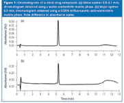
The first example is a method used to determine chemical purity together with epimeric purity of a chiral compound. The method used a reversed-phase column, had been confirmed suitable for epimeric ratio determination, and was in routine use. The typical chromatogram normally appeared as shown in Figure 1(a), where the b-epimer accounted for almost all of the peak area, with a very small a-epimer peak just in front of it; the a-peak was not present in most samples. One day, however, when the method was set up, the chromatogram of Figure 1(b) was obtained, exhibiting approximately 5% a-epimer. As can be imagined, a sudden change in the epimeric purity of a chiral drug substance can create panic in a development drug candidate identification process. Fortunately, there was a nuclear magnetic resonance (NMR) method that was available for structural analysis in addition to the purity determination LC method. When the same samples were analysed by NMR, they showed the expected high b-epimer ratio (a-epimer < 0.5%).

Figure 1
One of the techniques used to isolate any analytical problem is to systematically change one variable at a time until the problem source is discovered. It is most convenient to make easy changes first, and work towards method changes that require more work. The first step was to make a blank injection to ensure no spurious peaks were present. Next, a new preparation of the analytical sample was run to be sure that it was not contaminated. The same results were obtained. Next, a fresh batch of mobile phase was prepared and the problem disappeared. Now, the question was what aspect of the mobile phase was responsible for the problem. Examination of the mobile phase preparation records and an interview of the technician identified the problem source. There were two methods in the laboratory that used the same column type for different products. One method used a trifluoroacetic acid–acetonitrile mobile phase and the other used water–acetonitrile. The method of Figure 1(a) called for the water–acetonitrile mobile phase.
When trifluoroacetic acid was added intentionally to this mobile phase, results similar to Figure 1(b) were found. However, from a chemical point of view, it was hard to imagine how trifluoroacetic acid caused epimerization from the b- to a-epimer. At that point, we looked at peak areas and it was apparent that the area of the b-peak was decreased dramatically in the presence of trifluoroacetic acid [note the difference in absorbance scales in Figures 1(a) and 1(b)]. In addition, a very noisy baseline was observed early in the chromatogram [Figure 1(b)], indicating the presence of degradants, from void volume to elution volume of the b-peak. As a result, relative ratio a/b was increased drastically. So, by means of peak area examination, it was clear that the trifluoroacetic acid in the mobile phase selectively degraded the b-enantiomer, leading to an "apparent" unexpected a/b ratio. It appeared that the technician inadvertently pulled the wrong recipe from the file. To prevent this problem from happening in the future, specific containers properly labelled for trifluoroacetic acid–containing mobile phases were added to ensure that the proper mobile phase was used.
Unexpected Impurities
The second example involved an analyte with an aniline functional group. A method was being developed to determine the purity of the drug product. A water–acetonitrile gradient was used with a 5 μm particle diameter Type-B C18 column operated at 45 °C and 1 mL/min. A study was made to identify the column that gave the best peak shape for the analyte of interest. After several attempts over several days, a column was found that gave good peak shape for the standard, but to the chemist's alarm, several degradant peaks appeared as shown in the chromatogram of Figure 2(a). The overall sample purity was 62% by area; one of the degradant peaks accounted for approximately 16% of the total area. Structural analysis of the same sample by NMR indicated that the sample was > 95% pure. When the major degradant peak was interpreted in LC–mass spectrometry (MS) analysis, the structure was consistent with degradation of the active ingredient.

Figure 2
At first, it was speculated that the sample had degraded upon storage. NMR was run again, with the same > 95% purity results. Small changes of the sample preparation conditions did not correlate with the chromatographic observations. Because of the experience with the reversed-phase method discussed previously, the mobile phase was reformulated, but no change was observed.
Modify Column Conditions
The next suspected cause of degradation was interaction between the sample and the column. To test this, it was hypothesized that if the sample were exposed to the column for a shorter time, it would degrade less. A series of gradient modifications were made, so that the gradient started at 5% acetonitrile [normal conditions, as in Figure 2(a)], 15% acetonitrile and 30% acetonitrile; the gradient slope (per cent change per minute) was kept constant to keep selectivity constant. The results are shown in Figure 3. As is expected, a higher organic starting condition will result in a shorter retention time for the main peak. Notice that the amount of degradation dropped as the initial percentage of acetonitrile increased. Two possible explanations for these observations are suggested. First, exposure to more water could cause sample instability. However, the earlier tests of sample stability in various solvents before injection did not show any instability in water over the time frame of the observations. This left the exposure time to the column as another explanation.

Figure 3
The problem column was what is sometimes called a "lightly loaded" or "high aqueous mobile phase" C18 column. The concentration of C18 groups on the column was < 2 μmol/m2 of column packing. A "fully bonded" phase generally will have > 3 μmol/m2 of coverage. The low coverage of bonded phase also means that the exposed silanol (-Si-OH) content on the silica particle surface will be increased. Interaction with silanol groups can be one source of strong sample–column interactions, especially for compounds with basic functional groups such as the aniline group found in the present instance. To further explore exposed silanols as the problem source, the sample mobile phase and sample were used with a high-coverage (>3 μmol/m2 ) Type-B C18 column from the same manufacturer. A different column from the same manufacturer was chosen in an attempt to minimize column-to-column differences because of other reasons than the bonded phase coverage. When the sample was run on the high-coverage column, no degradation was observed [Figure 2(b)]. To check that the degradants could be seen on the second column if they were present, force-degraded sample was injected on the high-coverage column. Although the elution pattern was somewhat different between columns, as expected, the degradants' peaks did show up in the chromatogram from the high-coverage column.
One more experiment was tried to help understand the degradation process. It was known that under forced degradation conditions (acid, base, heat, light, oxidation), the drug substance was quite stable in acid media. The mobile phase was modified to include 0.1% acetic acid in the aqueous phase. With the acidic mobile phase, no degradation was observed on the lightly loaded C18 column. It is not determined if the acid stabilized the compound or somehow reduced the reactivity of the column.
Practical Implications
When we think of sample degradation inside the chromatographic column, most often we think of biological molecules, such as proteins, peptides and enzymes that can change conformation or lose biological activity through the chromatographic process. Degradation of small molecules (< 1000 Da) is much less common and rarely seen or reported by most workers. When extra peaks appear in stability indicating or sample-purity methods, it can be a red flag, indicating a problem in the production process. As is the situation in the first example discussed here, it is wise to check carefully all the method steps to make sure that no mistakes were made. A reanalysis of the suspect samples might be worthwhile, especially when one considers the cost and effort required to isolate and identify a production problem. Sometimes, a careful examination of the peak areas can be a good starting point for understanding the source of unexpected results.
During method development, unexpected results are more likely than for validated methods. When such problems are found and you identify conditions that appear to eliminate the problems, be sure to perform a sufficient number of experiments to convince yourself that your conclusions are indeed correct. In the second example, rather than stopping as soon as the high-coverage C18 column was found to give good results, the workers went further to confirm the results. To be sure that they were not being misled by the nonelution of degradants' peaks, a sample known to contain degradants was injected to confirm that the peaks could be seen under the selected conditions. Be sure to take into account your understanding of the chemistry of the subject compound, such as its stability under different pH conditions, excessive heat or light exposure. And finally, bear in mind that symptoms of degradation can include noisy baselines, extra peaks, missing peaks, misshapen peaks and peak-area-ratio changes.
Frederic Bell is a senior scientist in the Drug Discovery Department of Fournier Pharma, France a member of the Solvay Group. With his group, he uses a wide variety of techniques, including LC, NMR and MS to support organic synthesis and design of new drug substances. He also serves as a teacher of chromatographic analysis within the University of Burgundy, Dijon, France.
"LC Troubleshooting" editor John W. Dolan is vice-president of LC Resources, Walnut Creek, California, USA; and a member of the Editorial Advisory Board of LCGC Europe. Direct correspondence about this column to "LC Troubleshooting", LCGC Europe, Advanstar House, Park West, Sealand Road, Chester CH1 4RN, UK.
Readers can also direct questions to the Chromatography Forum at http://www.chromforum.com

.png&w=3840&q=75)

.png&w=3840&q=75)


.png&w=3840&q=75)


.png&w=3840&q=75)


