Chromatography Applications in Drug Discovery of Therapeutic Proteins
LCGC North America
In this month's installment of "Directions in Discovery," the authors discuss how, with the arrival of combinatorial libraries and high-throughput screening, pharmaceutical firms can develop new models of drug discovery that not only lessen the initial capital outlay involved in drug discovery, but also refine the discovery process.
Bringing a new protein drug from inception to market typically takes more than 10 years and costs over $800 million. The primary stage of this process — drug discovery — represents a very significant element in the entire cycle. With the arrival of combinatorial libraries and high-throughput screening, pharmaceutical firms can develop new models of drug discovery that not only lessen the initial capital outlay but also refine the discovery process. These models will be based upon identification of the genetic sequences that code for a particular protein involved in the disease process. Once the protein molecule with a target therapeutic value is identified, cell culture using established cell line can be used to generate the material. Such a task can be accomplished in a relatively short period of time if the expression system using standardized fermentation-cell culture platform technology is in place. For preliminary investigation of bioactivity, typical amounts required at this stage are in the microgram to milligram range. Additionally, purity generally does not need to exceed 95%. To facilitate purification, the protein molecule with an appropriate tag (or double tag in some cases) could be expressed using transient transfection in E. coli or other heterologous expression systems. Operating the tag as a purification handle, the protein can be adsorbed selectively from the feedstock. Examples of customary and less conventional protein tag systems (1-5) are listed in Table I. The corresponding one-step process for preferential and speedy purification of the tagged protein also is shown in the table.

Tim Wehr
After chromatography and buffer exchange into formulation media such as phosphate-buffered saline, the quality of the compound should be assessed before biological characterization. Traditional tests include electrophoresis, high performance liquid chromatography (HPLC), Western blot, endotoxin determination by limulus amoebocyte lysate assay, crystallization, and X-ray crystallographic study. Results of these tests can give an indication of the suitability of the material. If the test results are satisfactory, the material can be submitted for the cell-based assay to gain a more realistic sense of how it will perform in a cellular system. What was once a difficult and tedious procedure to establish potency has become much more rapid and consistent. Companies have developed highly automated systems for cell-based assays. These instruments allow scientists to culture living cells very closely related to the cell types found in specific organs. This gives researchers better insight into how cell type can affect the activity of the compound being evaluated. The cell-based assay also can provide an indication of cytotoxicity. This permits companies to cut their losses on drug candidates at an early stage of development, well before they reach clinical trials. To complement the cell-based assay, it is useful to determine the affinity of the protein for the receptor-antigen using kinetic measurement that utilizes surface plasmon resonance technology as the basis for detecting and monitoring protein interactions.
For proteins other than monoclonal antibodies, one consideration is detagging the protein after purification. While small tags such as poly-His normally do not need to be removed, others could be removed by proteolytic cleavage using commercially available systems. A selected protease can identify a recognition site at which it cuts preferentially. In any event, after using the protein tag and purification system to obtain micro quantities of proteins to demonstrate bioactivities successfully, the untagged protein usually is preferred in the next phase of purification for drug discovery. The protein obtained will allow further in-depth study or a full spectrum of toxicology investigation. At this point, the protein can be produced by semistable, then transient, then stable transfection in an established cell line. A semistable cell line generally produces low-titer material that, typically in large volume, would be sufficient for cursory chromatography method development. To facilitate purification development, parallel study can be started to develop an enzyme-linked immunosorbent assay (ELISA) or a Western blot assay. Such analytical tools are useful in guiding the evolution of a purification step. Once the later fermentation methods generate sufficient amounts of proteins, one can proceed to develop a purification train with a goal of generating protein meeting purity requirement (generally > 95%) and endotoxin concentration (generally < 0.3 EU/mg of protein). This process might not be optimal but it should be sufficient for initiating preclinical investigation such as pharmacokinetic study, toxicology, and early characterization work. To arrive at a final product, a typical purification train during protein drug development can involve three consecutive sequences consisting of capture, intermediate, and polishing steps. The steps should be orthogonal for optimal purification. An overview of the steps is discussed as follows.

Figure 1: Murine IgG1 purification using ion-exchange chromatography and ceramic hydroxyapatite chromatography. Ion-exchange column: UNOsphere S (Bio-Rad Laboratories, Hercules, California). Ceramic hydroxyapatite column: CHT (Bio-Rad Laboratories).
Capture step: This could be accomplished by a variety of precipitation procedures such as the use of nonionic water-soluble polymers, ammonium sulfate, ethanol, or divalent metal ions. The procedure causes the protein to leave solution and form insoluble particles that then are collected by centrifugation-filtration. However, it could present certain shortcomings such as product insolubility, difficulty in eliminating the reagent, temperature control, generation of aerosol during continuous centrifugation, and other undesirable mechanical complexity. Additionally, reversal of protein solubility might be difficult, especially with sulfhydryl derivatives. Therefore, many of the efforts on the capture step have been directed toward adsorptive-based chromatography, involving, for example, ion exchange, affinity adsorption, and hydrophobic interaction. Selection of the chromatography media for the capture step normally complies with the following attributes:
- Clearance of protein components in the medium, host cell contaminants, and DNA is expected to be at least one order of magnitude.
- Because the titer normally is low at this point, the harvest volumes generally are high. The capture step is expected to dewater the load material substantially, thus reducing the volume by at least one order of magnitude.
- Eluate from the capture step is conditioned so that it will be ready for the intermediate step.
- Good protein recovery (generally 85% or better).
One technology on the horizon is the use of membrane chromatography. This involves disposable capsules or cartridges that allow flow rates significantly faster than traditional column chromatography methods. It can serve as a viable step for rapidly capturing the protein of interest.
Intermediate step: The bulk of the protein contaminants and the volume should be reduced sufficiently at this stage. For convenience, selection of the intermediate step is undertaken so that unnecessary dilution or buffer exchange of the feedstock can be avoided. As examples, hydrophobic interaction chromatography or immobilized metal affinity chromatography could follow an ion-exchange step because both modes of operation can accept high conductivity in the load if the previous step entails elution of the product in high salt. Ideally, if favorable chromatography conditions for selective adsorption and elution can be found, one might even consider the intermediate step as being the polishing step. However, in situations where the eluate from the intermediate step still does not match the purification criteria required of the protein for in vitro and in vivo characterization, then one could consider a polishing step.

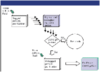
Figure 2: An example of protein drug discovery leading to pre-Phase I investigation.
Polishing step: After the intermediate step, the protein solution is already substantially pure, except for a limited amount of trace contaminants such as aggregates, truncated species, or host cell proteins. The step should then be one of high selectivity or high efficiency. In some situations, size-exclusion chromatography can be employed. It is a traditionally low productivity step, but it can present purification potential, such as aggregate removal, that other approaches do not offer. One might also consider the elution buffer of this step so that the eluate containing the product is already in the formulation of choice. Figure 1 shows an example of a purification train (6) consisting of a capture step and an intermediate step. The process produces purified murine IgG1 from tissue culture fluid. A polishing step can be considered for further development of the process.
Taken together, the discovery process of a protein drug candidate can be summarized in Figure 2. It illustrates the initial stage of screening proteins using a tagged protein purification chromatography system. Based upon the physical and biological characterization of the protein, a decision can be made to determine the viability of the project. In cases where untagged protein is preferred for further in-depth study, it then would trigger the development of a purification chromatography train to provide sufficient quantities of proteins. The protein obtained will be used for more extensive preclinical investigation before initiation of a Phase I clinical study.

Table I: Small-scale purification using tag systems
References
(1) P.C. Simons and D.L. Vanderjagt,
Anal. Biochem.
82
, 334-341 (1977).
(2) J. Porath, J. Carlsson, I. Olsson, and G. Belfrage, Nature 258(5536), 598-599 (1975).
(3) R.A. Kagel, G.W. Kagel, and V. Garg, Biochromatography 4(5), 246-252 (1989).
(4) L.F. Hare and E. Lee, Biochemistry 28(2), 574-580 (1989).
(5) S.R. Narayanan, J. Chromatogr. 658, 237-258 (1994).
(6) H. Lai and S. Franklin, Bio-Rad Bull. 2780 (2002).
Paul K. Ng was with the Pharmaceutical Division, Bayer HealthCare, Berkeley, California. He is currently with the Protein Separations Division, Life Sciences Group, Bio-Rad Laboratories, Hercules, California, and can be contacted at e-mail: paul_ng@bio-rad.com.
Michael H. Coan is currently with the Pharmaceutical Division, Bayer HealthCare, Berkeley, California.
Tim Wehr "Directions in Discovery" editor, is staff scientist at Bio-Rad Laboratories, Hercules, California. Direct correspondence about this column to Direct correspondence about this column to "Directions in Discovery," LCGC, Woodbridge Corporate Plaza, 485 Route 1 South, Building F, First Floor, Iselin, NJ 08830, e-mail lcgcedit@lcgcmag.com.

Common Challenges in Nitrosamine Analysis: An LCGC International Peer Exchange
April 15th 2025A recent roundtable discussion featuring Aloka Srinivasan of Raaha, Mayank Bhanti of the United States Pharmacopeia (USP), and Amber Burch of Purisys discussed the challenges surrounding nitrosamine analysis in pharmaceuticals.
Extracting Estrogenic Hormones Using Rotating Disk and Modified Clays
April 14th 2025University of Caldas and University of Chile researchers extracted estrogenic hormones from wastewater samples using rotating disk sorption extraction. After extraction, the concentrated analytes were measured using liquid chromatography coupled with photodiode array detection (HPLC-PDA).


