Characterization of mAb Glycoforms on FcγRIIIA-Ligand by Analytical and Semi-Preparative Chromatography
Binding between monoclonal antibodies (mAbs) and effector cells directly impacts the accompanying immune response or antibody-dependent cell cytotoxicity (ADCC). N-glycans at Asn297 of the Fc region influence this binding, as they affect the specificity of IgG to the FcγRIIIA receptor of effector cells. To monitor FcR receptor binding, a recombinant FcγRIIIa-ligand has been commercialized into TSKgel FcR-IIIA high-performance affinity chromatography (AFC) columns that separate antibodies based on affinity differences to FcγRIIIa.
TSKgel FcR-IIIA-NPR has a linear loading range of 5–50 μg mAb, which is useful for analytical applications, but is highly limiting if collection of elution products is necessary for further purification or analyses. This is overcome by the semi-preparative TSKgel FcR-IIIA-5PW column, allowing collection of sufficient material for improved analytical workflows in a short time.
Recombinant FcγRIIIa-ligand, with enhanced physiochemical stability, is immobilized onto a non-porous particle for analytical applications, and onto a larger porous particle for semi-preparative applications.
Both columns share a comparable resolution while increased loading capacity and elution volumes are achieved on the TSKgel FcR-IIIA-5PW.
Here, intact mAb and papain-digested mAb were used to compare the scalability of semi-preparative TSKgel FcR-IIIA-5PW and to provide a methodology to broaden from analytical to semi-preparative mode while maintaining similar resolution between the two different capacity columns.
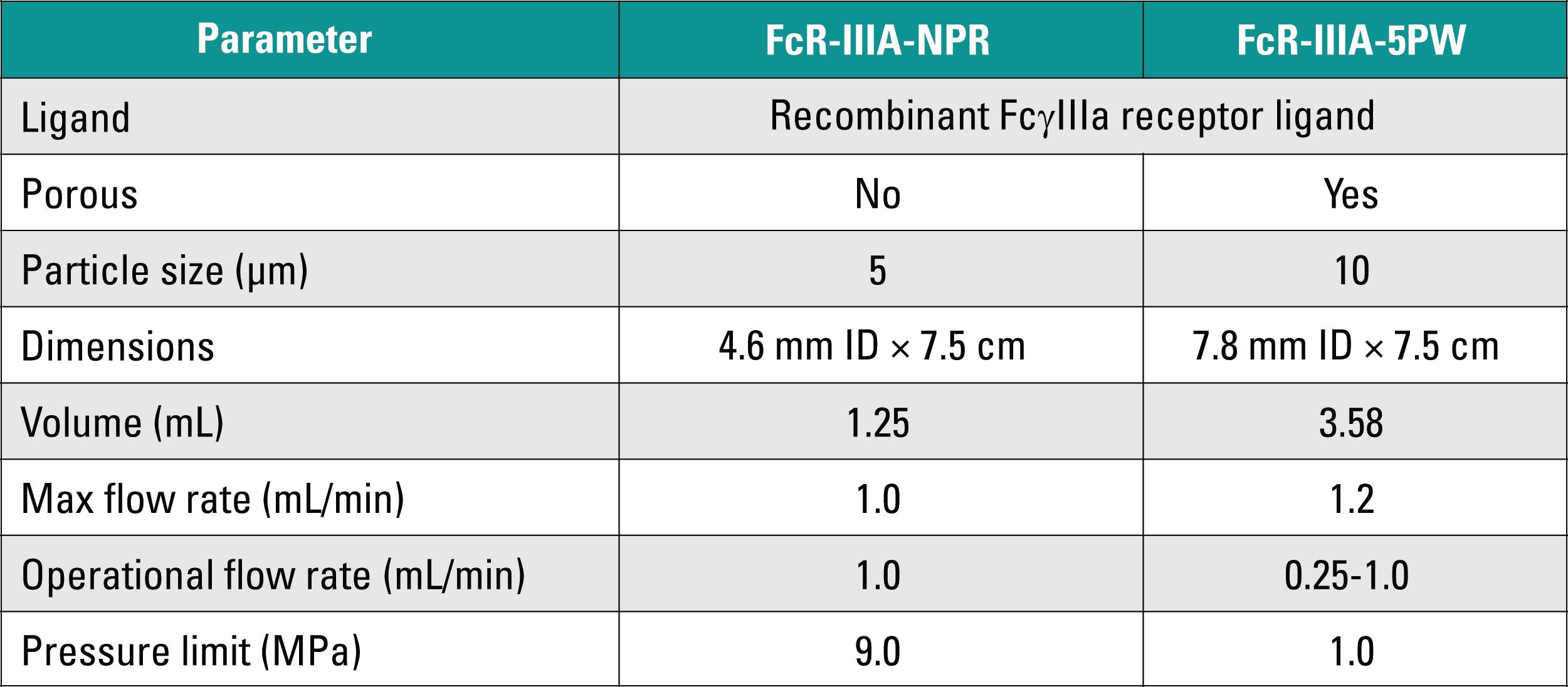
Table I indicates the specifications and operating conditions of analytical and semi-preparative TSKgel FcR-IIIA columns.
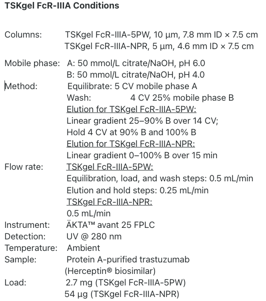
Materials and Methods
Separation of trastuzumab on the analytical and semi-preparative columns is seen in Figure 1, illustrating comparable peak profiles and ease of scaling using the same mobile phase and flow rate.
Figure 1: Separation of trastuzumab based on its glycoform pattern on analytical (A) and semi-preparative (B) TSKgel FcR-IIIA columns
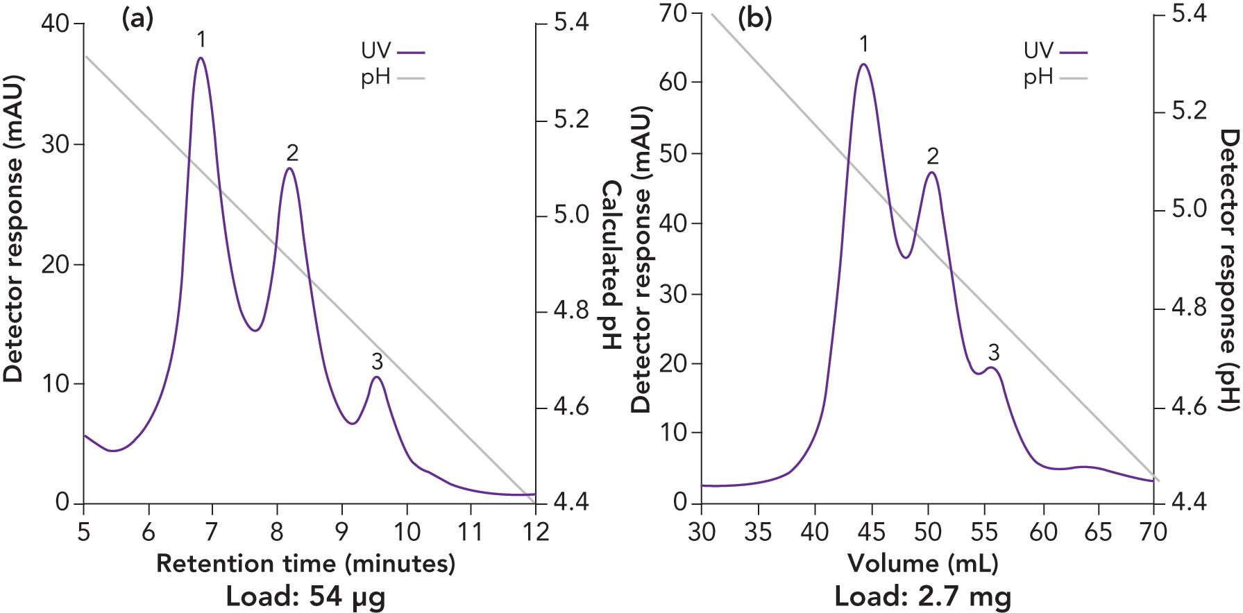
Figure 2 (panel A) illustrates fraction collection from the semi-preparative TSKgel FcR-IIIA-5PW column run and purity analysis for each peak. For semi-preparative peaks from 5 mg load (left panel), the fractions in the boxed area were pooled, buffer-exchanged, concentrated in spin concentrators, and subjected to the analytical TSKgel FcR-IIIA-NPR column for purity assessment. The analytical FcR runs (panels B-D) show that each pooled fraction contained predominantly homogeneous peak material and only minor amounts of other peaks were present.
Figure 2: Fraction collection from the semi-preparative column run and purity analysis for each peak
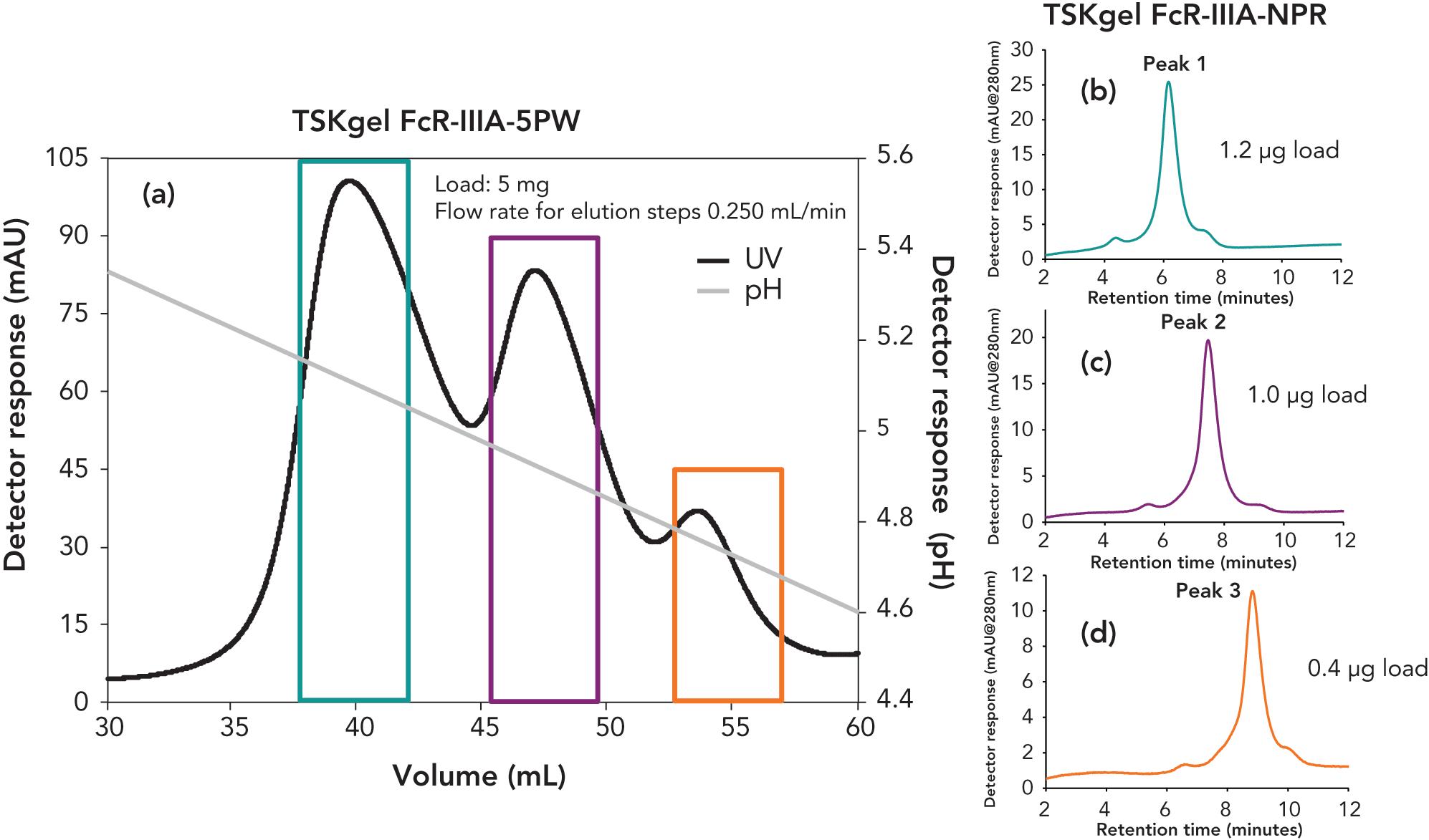
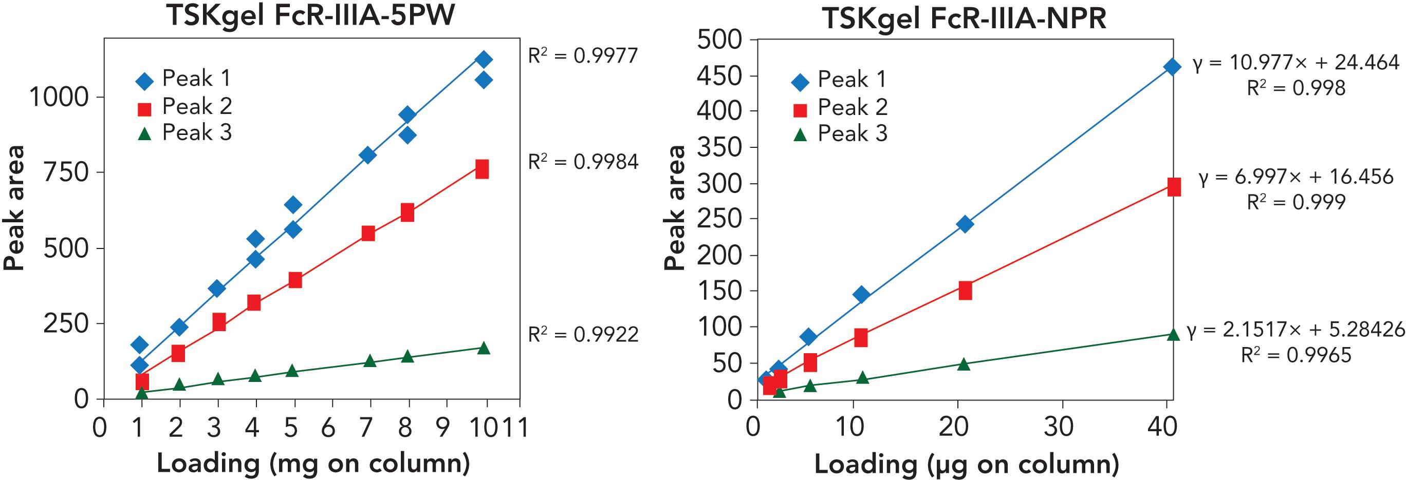
Figure 3 shows the linearity of peaks 1, 2, and 3 for different loads on the semi-preparative TSKgel FcR-IIIA-5PW (left figure) and TSKgel FcR-IIIA-NPR (right figure) columns. A wide range of loading capacity was confirmed: 2–50 μg on the NPR and 1–5 mg on the 5PW column. In larger loads up to 10 mg, peaks started to blend together (not shown). Thus, for the semi-preparative column, the recommended load range was determined as 0.5 mg to 5 mg of mAb.
Figure 3: Linearity with total peak area
To demonstrate binding specificity of the TSKgel FcR-IIIA columns to the Fc region of antibodies, papain (Figure 4; papain cleavage site after His-228 in trastuzumab) was used to digest intact IgG to its Fab and Fc fractions and the reaction mix was loaded onto the semi-preparative and analytical TSKgel FcR-IIIA columns (Figure 5). Intact mAb and the Fc fragment both bound onto the FcR-IIIA columns and eluted as typical three peaks.
Figure 4: Schematic illustration of mAb fractionation by papain digestion

Figure 5: Elution profiles of TSKgel FcR-IIIA columns for intact mAb and the papain-released Fc-fraction
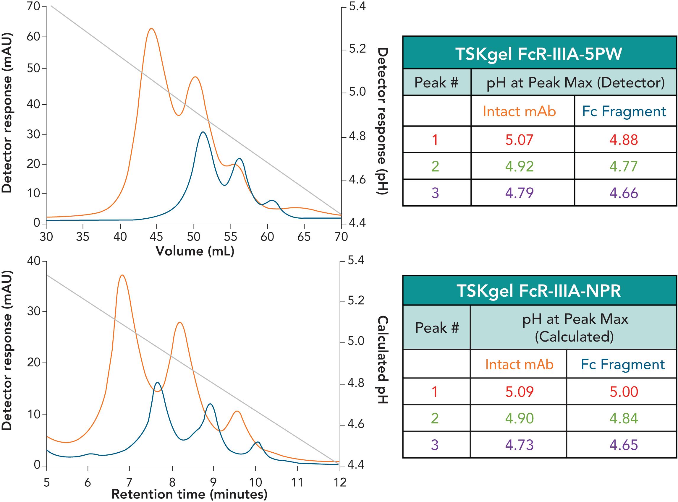
Intact mAb and the Fc fragment have comparable peak profiles between the analytical and semi-preparative formats, as demonstrated in Figure 5. Tighter binding of the Fc fragment relative to the intact mAb was observed on both columns, potentially due to the removal of steric hindrance of the Fab portions. Approximately 50× more material was loaded (2.7 mg vs 54 μg) on the semi-preparative TSKgel FcR-IIIA-5PW, allowing greater elution volumes and fraction collection. The purity of the three peaks eluted from the TSKgel FcR-IIIA-5PW columns are confirmed by the peak profile and different retention times when applied to the analytical TSKgel FcR-IIIA-NPR column. Each pooled fraction analyzed on the analytical column are predominantly homogeneous.
As reported, the fractions are linked to different glycan patterns, which can be confirmed by subjecting them to more detailed glycan analysis.
Conclusion
In summary, selectivity and resolution of intact mAb and papain-digested mAb can be maintained, even at high load, with use of the semi-preparative TSKgel FcR-IIIA-5PW column. The ability to collect more material per injection allows for more detailed ADCC activity estimation of specific mAb glycoforms or mAb modalities related to its Fc domain. These modalities can be separately studied through other orthogonal methods.

TSKgel and Tosoh Bioscience are registered trademarks of Tosoh Corporation.
ÄKTA is a trademark of Cytiva.
Herceptin is a registered trademark of Genentech, Inc.

Follow the Data to Grow: Why a Scientific Data Platform is Essential
October 28th 2024Innovation is the engine that powers a company’s growth and product development, and for enterprises with R&D laboratories, those lab environments are the greatest source of this innovation. In this white paper, learn how a platform approach to scientific data management, including semantic search, advanced analytics, and lab automation, leads to better enterprise decisions at the executive level, optimized lab performance, more discoveries, and stronger product pipelines.

