Bioanalytical Tools for the Characterization of Biologics and Biosimilars
LCGC North America
LC–MS and CE–MS are the go-to techniques for characterizing biologics, but often help is needed from a range of other methods.
Bioanalytical Tools for the Characterization of Biologics and Biosimilars
In spite of the recent slowdown in the approval rate of new biopharmaceutical entities, dozens of new biologicals were introduced in the United States and the European Union during the past 5 years, adding up to more than 200 products already on the market (1). One of the fastest growing segments in this market is biosimilars (also referred to as follow-on biologics or subsequent entry biologics). This emerging biotech sector requires novel bioanalytical tools to address the challenging regulatory aspects (2), which entail comprehensive characterization by sensitive and high resolution bioanalytical methods covering all four structural levels of biotherapeutic proteins (that is, from primary to quaternary). During the development phase of both the novel biologics and biosimilars, full sequence coverage, purity assessment, quantification, and product identification is necessary. In addition, quite a few other critical features should be analyzed, such as glycosylation (including their microheterogeneity and site specificity), mutation, phosphorylation, sulfation, disulfide linkages, oxidation, deamidation, glycation, proteolytic clipping, and several others. Analysis of isomerization, especially isoaspartic acid formation (isoAsp and isoD), is another important requirement for their analysis because it might reveal immunogenic structural changes (3). Identification of host-cell impurities, aggregation, and determination of higher order structures and isoforms are also highly important (4).
The analytical needs depend on the actual application in hand; for example, high throughput is a prerequisite during clone selection and high sensitivity is important during product release. To attain such levels of characterization, orthogonal bioanalytical methods should be applied including liquid chromatography (LC), capillary electrophoresis (CE), mass spectrometry (MS), and their combinations: LC–MS and CE–MS. These methods enable thorough analysis of intact proteins (top down), peptide digests (bottom up), and even in between (middle down or up) (5). The most frequently used proteolytic enzyme for these applications is trypsin that produces the majority of fragments below 3 kDa, a size range that is readily compatible with most present day MS instruments. Higher resolution mass spectrometers such as hybrid MS units enable routine analysis of larger fragments with higher charge states (6).
The Bioanalytical Toolset
Mass spectrometers are the most important workhorses in the characterization of biologics and biosimilars (7,8). Important features include mass resolution and accuracy. In this respect, Table I delineates the capabilities of the top-of-the-line MS systems these days.

Table I: MS capabilities
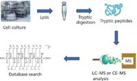
However, even for the best mass spectrometers on the market, the use of separation methods (LC or CE) before the mass analysis is highly recommended to decrease the complexity of the sample entering the device (9), as shown in Figure 1. Pre-MS separation also increases the dynamic range and decreases ion suppression in mass spectrometry. One of the preferred separation modes is LC, typically using narrow-bore or capillary columns with packing particle sizes of 3–5 µm or lower (1.7 µm). The porosity of the stationary phase should match the size of the solute molecules; for example, for peptides it should be 20 nm and for proteins 30 nm. The pressure drop on such systems ranges from 6000 to 15,000 psi depending on the particle size and the column length. Ultrahigh-pressure liquid chromatography (UHPLC) provides higher resolution and sensitivity for peptide mapping than regular high performance liquid chromatography (HPLC), enabling better sequence coverage and variant characterization (10). Other separation parameters, such as column temperature, are also important for special applications. Because of its higher speed of analysis, UHPLC is a good choice for high-throughput peptide mapping, providing fine details about differences between a biosimilar and its innovator product counterpart. State-of-the-art software tools help with automated searches of LC–MS datasets to assign peptides and annotate peptide variants in minutes even at low levels, a process that manually takes several days per data file. Differential plotting options enable quantitative comparison between different peptide maps of, for example, a biosimilar to an innovator product.

Figure 1
Capillary electrophoresis is another high efficiency separation method frequently used in bioanalysis providing fast separation (minutes to seconds) with predictable selectivity, requiring only small sample volumes (1–10 µL) (11). Features of CE also include quantification in a reasonable dynamic range. The option to use multicapillary devices can significantly increase the throughput of the analysis (for example, DNA sequencing). Coupling CE to a mass spectrometer is an emerging field of research. From the various modes of CE, capillary zone electrophoresis (CZE), capillary gel electrophoresis (CGE), and capillary isoelectric focusing (cIEF) are most frequently used for the analysis of biologics and biosimilars.
In the analysis of biotherapeutics, with the goal of good recovery, the hydrophobicity and size of the sample molecules should be considered as well as their aggregation tendency. Samples should be treated accordingly with detergents or denaturants. In peptide analysis by LC–MS, the size and hydrophobic or hydrophilic characteristics of the peptides should be considered. If the peptides are too small or hydrophilic then they are not retained. If the peptides are too big or hydrophobic then they are not recovered properly. To avoid these size-related issues, one option is to implement a multienzyme approach that provides suitable sized peptides for the analysis, keeping in mind that peptide sizes between 1000 and 3000 Da work well for most LC–MS systems. On-line LC–MS, for the analysis of larger peptides, may need a combination of collision-induced dissociation (CID) and electron-transfer dissociation (ETD). CID of an isolated charge-reduced species derived from ETD is an effective approach to determine phosphorylation sites and glycosylation modifications as shown in Figure 2 (12). To further analyze glycosylation modifications, hydrophilic interaction liquid chromatography (HILIC) stationary phases have been introduced using amide, diol, amine, aminopropyl, and zwitterionic (ZIC-HILIC) phases (13). However, normal-phase silica-based columns are still being used for glycopeptide or glycan analysis. Capillary electrophoresis (as well as CE–MS) is another good option to analyze glycosylated proteins or peptides (14). Rapid analysis of glycosylation, especially for immunoglobulins, is also possible by a novel microchip-based LC–MS approach that utilizes an integrated microfluidics-based sample preparation system before MS analysis (15). The built-in PNGase F enzyme reactor cuts off the IgG glycans, and the resulting glycosylamines are separated on a porous graphitized carbon column followed by detection in the connected MS unit.

Figure 2
Sample Preparation Issues
Effective sample preparation for the analysis and characterization of biologics and biosimilars is a key issue (16). This includes the collection and handling of the samples as well as the reproducibility of process protocols. Introduction of quality control (QC) points is highly important to monitor the process. Also, reagent components along with their potential effects on the sample during downstream processes (for example, detergents, denaturants, extreme pHs, and protein concentration) should be considered. System suitability tests are necessary to ensure the performance of the analytical approach. Thus, it is important that the sample preparation process is reproducible, including solubilization of all proteins of interest. Protein aggregation should be avoided during sample preparation and the concomitant analytical processes. Also, chemical modifications of the proteins or peptides should be prevented and interfering molecules should be removed. This is important during clone selection and stability studies, in which detection of undesired protein forms are crucial. Obtaining proteins of interest at a detectable level may involve the removal of abundant or nonrelevant classes of proteins. If digestion is among the sample preparation steps, its reproducibility and efficiency is also very important.
Analysis of Critical Features
Among the important critical features, analysis of post translational modifications, especially glycosylation, is becoming more and more important during the characterization of biotherapeutics. The typical goal is to decipher the heterogeneity of the attached glycans and recognize the multiple glycosylation sites per protein. As a matter of fact, each site can hold multiple glycan structures resulting in hundreds of glycoforms, which might be responsible for various biological activities (17). Many factors contribute to alterations in glycan processing during the production of therapeutic glycoproteins, thus their analysis during the process is crucial. With complicated samples such as complex glycan mixtures, the use of orthogonal separation methods is a key to provide full characterization. CE and CE–MS analysis of intact glycoproteins (18) or the released glycans (19) can achieve such a goal. Apparently, reversed-phase LC–based protein separation has limited resolving power for glycoforms. CE, however, in which separation is primarily based on charge and size, provides high resolving power for glycoforms and is helpful in the characterization of multiple glycosylation sites (14).
The comprehensive analysis of intact glycoproteins can be done by first separating them using chromatography or electric field–mediated techniques. Ion-exchange chromatography has limited resolving power, but isoelectric focusing provides high resolution in charge-based separation. Before the separation and analysis, appropriately gentle sample preparation steps such as size-exclusion chromatography or molecular weight cut-off filtration are necessary. Among the MS-friendly separation tools, reversed-phase HPLC has only limited resolution for glycoforms. CE separates analyte molecules based on their charge, mass, and molecular shape; however, it requires MS-compatible buffer systems and minimal dead volume at the electrospray ionization (ESI) interface. Typical approaches include using sheath flow, sheathless, or liquid junction methods (20). The orthogonality of the above listed separation methods should be fully utilized in the analysis of biotherapeutics, both for innovative biologics and biosimilars. It is especially important during the development phase as glycosylation processing (microheterogeneity and site specificity) is very host-cell dependent and sensitive to cell culture conditions.
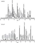
During MS based assignment of glycopeptides, the peptide backbone sequence is obtained by ETD after selecting the best candidates from the CID fragmentation. Confirmation can be accomplished by accurate mass determination or with orthogonal analytical methods such as CE, high-pH anion-exchange chromatography, or reversed-phase LC. Analysis of the released glycans is also possible by MS. However, simple MS can provide information only on glycan composition with respect to the number of hexoses (for example, galactose and mannose), N-acetyl-hexosamines (GlcNAc and GalNAc) as well as sialic acids (Neu5Ac and Neu5Gc), and provides no information on their linkages or positions (21). By using MS-MS with appropriate fragmentation modes (CID, post-source decay [PSD], ETD, or photodissociation) some linkage information can be obtained. This, again, emphasizes the necessity of a separation dimension before MS detection and analysis. With the use of MS-friendly high-resolution separation techniques, positional and linkage isomers can be separated and consequently analyzed. The HPLC separation of native glycans can be accomplished by porous graphitized carbon stationary phases, and derivatized glycans (2AB or 2AA) are better separated using HILIC separation mode. The charge-based separation by high performance anion exchange chromatography (HPAEC) is a good tool for determining characteristics such as the number of sialic acids. Another important separation tool before MS analysis is CE of highly charged fluorophore (aminopyrene trisulfonate [APTS] or 8-aminonaphthalene-trisulfonate [ANTS]) labeled glycans. Figure 3 compares the separation of IgG N-glycans by HILIC and CE–laser-induced fluorescence (LIF) methods. An important issue is the localization of the actual glycosylation sites, and because peptides with different glycoforms are eluted closely, MS-based verification is used to obtain such information.

Figure 3
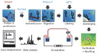
One of the most frequently used applications of CE in the analysis of biotherapeutics and biosimilars is the characterization of protein glycosylation, which requires preseparation derivatization with a charged fluorophore after the release of glycans (both N- and O-linked). This reductive amination-based, rapid, single-step labeling reaction (usually with APTS) features great efficiency (>90%) and provides a triple-charged fluorescent tag supporting excitation at 488 nm with 520-nm emission. Quantification is easy because only one fluorophore is conjugated to one sugar entity (23). Figure 4 delineates a CE-based glycan analysis workflow for therapeutic antibodies, starting with sample purification with protein A cartridges, through APTS labeling, sample cleanup, CE analysis, and database query. Such characterization of monoclonal antibody (mAb) N-linked glycans readily gives comprehensive information on their structural diversities, such as the presence of core fucosylation or immunogenic sugar residues, such as alpha 1→3 galactosylation or N-glycolylneuraminic acid. This assessment of glycosylation microheterogeneity is very important during clone selection, process control, and lot release for both the therapeutic recombinant mAbs and their biosimilar counterparts.

Figure 4
Disulfide mapping is another important characterization method, which requires treatment with multiple enzymes and multimode MS to reveal disulfide-linked sites and the sequence information of the linked peptides (24). ETD cleaves the disulfide bonds preferentially over the peptide backbone and CID cleaves the peptide backbone while leaving disulfide bonds intact. The specificity of enzymes used for disulfide mapping should be high enough to avoid miscleavages when multiple disulfide links are in close proximity. Another issue to consider is that enzyme digestion often fails to cleave a desired amino acid right next to a disulfide bond (25). Glycosylation in disulfide-linked peptides further complicates their analysis as the efficiency of peptide N glycanase F (PNGase F) can be poor or the peptide may not be fully recovered after deglycosylation, both leading to decreased sensitivity. Please note that when using ETD fragmentation, the disulfide bonds are preferentially cleaved over glycosidic bonds. Disulfide assignment by ETD or CID uses precursor masses to obtain the most likely disulfide-linked peptides. ETD obtains disulfide-dissociated or partially dissociated peptides, which can be confirmed by further fragmentation with CID.
Other Methods
Even though most of the analyses of biologics and biosimilars focus on the primary structure, the higher order structures of these molecules is also highly relevant, and they are invisible to the above described techniques. Several classical biophysical tools can be used for global structural analysis of biologics and biosimilars, such as analytical ultracentrifugation, size-exclusion chromatography, fluorescence, circular dichroism, isothermal titration calorimetry, and differential scanning calorimetry. These techniques can reveal information about secondary, tertiary, and quaternary structures, but currently there is still a lack of full understanding about the conformations of most therapeutic proteins.
Hydrogen or deuterium exchange using mass spectrometry (HX-MS) is another valuable method for protein conformation analysis because the kinetics of H–D exchange is highly dependent on the protein conformation (26). Advantages of HX-MS include relatively good resolution, picomole-range sample requirement, no restriction in protein size, and a relatively high tolerance of sample impurities. The approach provides data on conformational changes during function and helps identify interactions or surfaces with the aid of known structures. Alternatively, it also provides some useful information when the structure is unknown. Besides being a valuable tool for studying protein function, HX-MS is emerging as a method of quality control in biopharmaceutical development and a diagnostic tool for proper folding.
Closing Remarks
Biotherapeutics are highly complex molecules with complicated higher order structures that are subject to a plethora of post-translational modifications. The biologic substance (the molecule itself) and biologic product (the pharmaceutically formulated final product) also contain process- and product-related impurities such as aggregates, heterogeneous structures, and fragments (27). These factors underline the importance of the subject matter discussed in this column, helping to understand the analytical science accompanying the development of these complex therapeutic molecules and keeping pace with the rapidly developing regulatory framework for these products.
References
(1) G. Walsh, Nat. Biotechnol. 28, 917–924 (2010).
(2) http://www.fda.gov/downloads/Drugs/GuidanceComplianceRegulatoryInformation/Guidances/ucm122858.pdf, 2010.
(3) H.A. Doyle, R.J. Gee, and M.J. Mamula, Autoimmun. 40, 131–137 (2007).
(4) J. Carpenter, B. Cherney, A. Lubinecki, S. Ma, E. Marszal, A. Mire-Sluis, T. Nikolai, J. Novak, J. Ragheb, and J. Simak, Biologicals 38, 602–611 (2010).
(5) Z. Zhang, H. Pan, and X. Chen, Mass Spectrom. Rev. 28, 147–176 (2009).
(6) X. Han, A. Aslanian, and J.R. Yates, 3rd, Curr. Opin. Chem. Biol. 12, 483–490 (2008).
(7) C.A. Srebalus Barnes and A. Lim, Mass Spectrom. Rev. 26, 370–388 (2007).
(8) A.M. Altelaar and A.J. Heck, Curr. Opin. Chem. Biol. 16, 206–213 (2012).
(9) M. Ewles and L. Goodwin, Bioanal. 3, 1379–1397 (2011).
(10) U.D. Neue, M. Kele, B. Bunner, A. Kromidas, T. Dourdeville, J.R. Mazzeo, E.S. Grumbach, S. Serpa, T.E. Wheat, P. Hong, and M. Gilar, Adv. Chromatogr. 48, 99–143 (2010).
(11) B.L. Karger, A.S. Cohen, and A.J. Guttman, Chromatogr. 492, 585–614 (1989).
(12) J.J. Coon, Anal. Chem. 81, 3208–3215 (2009).
(13) G. Zauner, A.M. Deelder, and M. Wuhrer, Electrophor. 32, 3456–3466 (2011).
(14) D. Thakur, T. Rejtar, B.L. Karger, N.J. Washburn, C.J. Bosques, N.S. Gunay, Z. Shriver, and G. Venkataraman, Anal. Chem. 81, 8900–8907 (2009).
(15) N. Tang and K. Waddell, J. Biomol. Tech. 22, S70 (2011).
(16) Y. Jmeian and Z. El Rassi, Electrophor. 30, 249–261 (2009).
(17) A. Varki, R. Cummings, J. Esko, H. Freeze, P. Stanley, C.R. Bertozzi, G.W. Hart, and M.E. Etzler, Essentials of Glycobiology, Second Edition, (Cold Spring Harbor Laboratory Press, Cold Spring Harbor, New York, 2009).
(18) R. Haselberg, G.J. de Jong, and G.W. Somsen, Electrophor. 32, 66–82 (2011).
(19) A. Guttman, Nature (London) 380, 461–462 (1996).
(20) B.R. Fonslow and J.R. Yates, 3rd, J. Sep. Sci. 32, 1175–1188 (2009).
(21) Y. Mechref and M.V. Novotny, Mass Spectrom. Rev. 28, 207–222 (2009).
(22) S. Mittermayr, J. Bones, M. Doherty, A. Guttman, and P.M. Rudd, J. Proteome Res. 10, 3820–3829 (2011).
(23) Z. Szabo, A. Guttman, T. Rejtar, and B.L. Karger, Electrophor. 31, 1389–1395 (2010).
(24) Y. Wang, Q. Lu, S.L. Wu, B.L. Karger, and W.S. Hancock, Anal. Chem. 83, 3133–3140 (2011).
(25) G. Chen and B.N. Pramanik, Expert Rev. Proteomics 5, 435–444 (2008).
(26) S.R. Marcsisin and J.R. Engen, Anal. Bioanal. Chem. 397, 967–972 (2010).
(27) A.S. Rathore, Trends in Biotech. 27, 698–705 (2009).
András Guttman is a bioanalytical consultant, heading the Horváth Laboratory of Bioseparation Sciences, at the University of Debrecen in Hungary. He is also an adjunct professor in the Barnett Institute at Northeastern University in Boston, Massachusetts.

András Guttman
Anurag S. Rathore is a biotech CMC consultant and an associate professor with the Department of Chemical Engineering at the Indian Institute of Delhi, India.

Anurag S. Rathore
Ira S. Krull is Professor Emeritus of Chemistry and Chemical Biology at Northeastern University, Boston, Massachusetts, and a member of LCGC's editorial advisory board.

Ira S. Krull


.png&w=3840&q=75)

.png&w=3840&q=75)


.png&w=3840&q=75)


.png&w=3840&q=75)


