Analyzing Phosphorylated N-Glycans with Recovery on Bio-Inert LC Systems and PEEK-Lined HILIC Columns
LCGC Europe
Glycosylation is a critical quality attribute (CQA) that can impact on product safety and efficacy of protein biopharmaceuticals. Characterization of N-glycans is therefore of paramount importance for the pharmaceutical industry. Hydrophilic interaction liquid chromatography (HILIC) combined with fluorescence detection (FLD) and 2-aminobenzamide (2-AB) labelling is the golden standard for the analysis of N-glycans enzymatically liberated from biopharmaceuticals. However, for phosphorylated N-glycans, that is, those attached on lysosomal enzymes, irreproducible data and recovery issues are observed on conventional liquid chromatography (LC) instrumentation and columns, which can be attributed to the interaction of the phosphate moieties with stainless steel components in the flow path. This article demonstrates the analysis of phosphorylated glycans with full recovery on a bio-inert LC system and PEEK-lined HILIC column.
Glycosylation is a critical quality attribute (CQA) that can impact on product safety and efficacy of protein biopharmaceuticals. Characterization of N-glycans is therefore of paramount importance for the pharmaceutical industry. Hydrophilic interaction liquid chromatography (HILIC) combined with fluorescence detection (FLD) and 2-aminobenzamide (2-AB) labelling is the golden standard for the analysis of N-glycans enzymatically liberated from biopharmaceuticals. However, for phosphorylated N-glycans, that is, those attached on lysosomal enzymes, irreproducible data and recovery issues are observed on conventional liquid chromatography (LC) instrumentation and columns, which can be attributed to the interaction of the phosphate moieties with stainless steel components in the flow path. This article demonstrates the analysis of phosphorylated glycans with full recovery on a bio-inert LC system and PEEK-lined HILIC column.
Protein biopharmaceuticals are a rapidly growing class of therapeutics that are widely used for the treatment of various lifeâthreatening diseases (1,2). Around 40% of protein biopharmaceuticals are glycosylated and total glycan mass can range from 2–3% for monoclonal antibodies (mAbs) and up to 50% for erythropoietin (EPO). Glycosylation can impact on product safety and efficacy and is therefore considered a critical quality attribute (CQA). For example, terminal sialic acid residues on complex N-glycans regulate the half-life of EPO in the blood stream, core fucosylation influences the effector function of mAbs, and mannose-6-phosphate moieties on therapeutic enzymes are essential for trafficking to the lysosomes where the enzymes need to be catalytically active (1,2). Characterizing these glycan structures is therefore an essential requirement. Glycosylation analysis of protein biopharmaceuticals can be performed at different levels, that is, at protein, peptide, and glycan levels (3).
Hydrophilic interaction liquid chromatography (HILIC) combined with fluorescence detection (FLD) and 2-aminobenzamide (2-AB) labelling is the gold standard for analysis of N-glycans enzymatically liberated from biopharmaceuticals (1,2,3). While this method has a proven track record on neutral and sialylated N-glycans, irreproducible data with poor recovery is often encountered upon analyzing phosphorylated N-glycans originating from therapeutic lysosomal enzymes. This is attributed to the interaction of the phosphate groups with stainless steel components in the flow path. This phenomenon has also been observed for nucleotides and phosphopeptides (4–9). For N-glycans analysis, this issue is often alleviated by using mobile phases containing an ion-pairing reagent such as triethylamine (10,11) or by performing separations at high pH (pH > 12) using high-performance anion exchange chromatography with pulsed amperometric detection (HPAEC-PAD) (12,13).
This article demonstrates that phosphorylated glycans can be successfully analyzed by HILIC with the standard 2-AB-labelling method using the commonly applied mobile phase composition, that is, 100-mM NH4-formate pH 4.5 and acetonitrile, when the entire sample flow path is metal-free. To achieve this, the use of a bio-inert liquid chromatography (LC) system as well as a PEEK-lined HILIC column is mandatory.
ExperimentalMaterials: Acetonitrile and water were obtained from Biosolve. Ammonium formate was purchased from Sigma Aldrich. 2-AB-labelled RNase B and recombinant human acid alpha-glycosidase N-glycans (uncapped and capped) were provided by a local biotechnology company.
Methods: HILIC–FLD measurements were performed on an Agilent 1260 Infinity II Bio-Inert LC system equipped with a quaternary pump (G5654A), a multisampler (G5668A), a multicolumn thermostat (G7116A) with heat exchanger, and fluorescence detector (G7121B) with bio-inert flow cell. For LC–mass spectrometry (MS) experiments the above LC configuration was hyphenated to an Agilent QTOF LC–MS system (G6545A) equipped with JetStream electrospray ionization (ESI) source. LC and LC–MS data were acquired and analyzed using OpenLAB CDS version 2.1, MassHunter for instrument control (B06.01) and MassHunter for data analysis (B07.00) (Agilent Technologies), respectively.
Two columns were used in this study: a 2.1 mm × 100 mm, 2.7âµm AdvanceBio Glycan Mapping column (superficially porous particles-amide) (Agilent) in regular stainless steel housing and an equally dimensioned PEEKâlined column custom packed with identical superficially porous particles (Agilent). Elution was carried out with a linear gradient of (A) 100âmM NH4-formate pH 4.5 and (B) acetonitrile from 80% to 60%B in 38 min. The flow was set at 0.4 mL/min, the column temperature at 40 °C, and the injection volume was 1 µL. Excitation and emission wavelengths of the FLD were 260 nm and 430 nm, respectively. The operational parameters for the QTOF source were drying gas temperature: 300°C, drying gas flow: 8 L/min, nebulizer pressure: 35 psig, sheath gas temperature: 350 °C, sheath gas flow: 8 L/min, nozzle voltage: 1000 V, capillary voltage: 3500 V, and fragmentor voltage: 150 V. QTOF data were collected in centroid mode at a rate of 1 spectrum per second and acquisition range was 500–3200 m/z. The system was operated in the extended dynamic range mode (2 GHz).
Results and Discussion
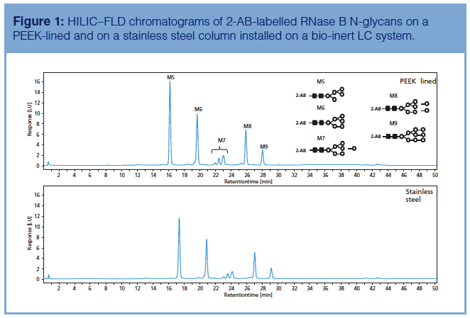
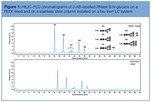
HILIC with FLD and 2-aminobenzamide (2-AB) labelling is currently the method of choice for the analysis of neutral and sialylated N-glycans liberated from biopharmaceuticals (1,2,3). Figure 1 shows the analysis of 2-AB-labelled neutral high mannose RNase B N-glycans on a stainless steel and PEEK-lined amide HILIC column installed on a bio-inert LC system. Very similar chromatograms are obtained on both columns for these neutral glycans. 2-AB-labelled glycans elute based on their polymerization degree, that is, the higher the number of glycosidic bonds, the more retention. Moreover, sufficient selectivity differences exist to separate compounds with the same polymerization, that is, M7 isomers which differ in the positioning of the mannose residue on the glycan tree. Both profiles were obtained on the bio-inert LC system, but similar results could be produced on a conventional stainless steel HPLC system.
The HILIC–FLD and HILIC–ESIâMS analysis of 2-AB-neutral and phosphorylated high mannose N-glycans originating from human acid alpha-glucosidase recombinantly expressed in yeast is shown in Figures 2 and 3, respectively. Human acid alpha-glucosidase catalyzes the hydrolysis of glycogen to glucose in the lysosomal compartment of the cell. Around 50,000 people worldwide have a deficiency of this enzyme, which leads to glycogen accumulation in the lysosomes causing muscle damage. These patients typically receive an enzyme replacement therapy with recombinant human acid alpha glucosidase (14). The presence of mannose-6-phosphate containing glycans is a CQA since these functionalities are responsible for targeting the enzyme to the lysosomes where it needs to break down glycogen (15).
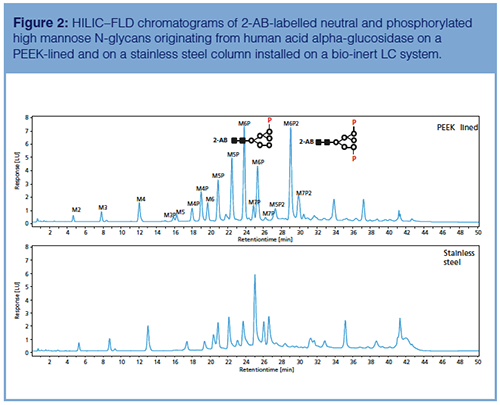
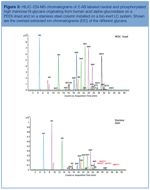
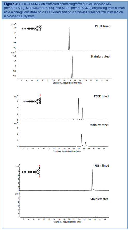
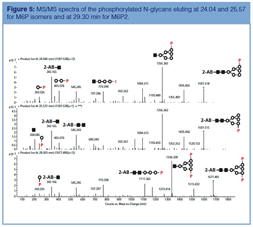
In contrast to the observations on the neutral RNase B N-glycans, an enormous discrepancy is noticed when analyzing the 2-AB-labelled glycans on a stainless steel or on a PEEK-lined column. The neutral glycans M2-8 behave equally well on both columns. The monophosphorylated glycans M2-8P show tailing peaks on the stainless steel column while biphosphorylated glycans M5-8P2 are not recovered at all from the latter column as a result of interaction between metal ions and phosphate moieties. The same recovery principle is known to apply for nucleotides, that is, AMP > ADP > ATP. On the PEEK-lined column, perfect Gaussian shaped peaks are observed for both mono- (M6P, for example) and biphosphorylated (M6P2, for example) structures showing the importance of removing all metal parts from the flow path (instrument, column including frits). Isomeric monophosphorylated glycans are observed as well, which differ in the positioning of the phosphate group on the α1-3 or α1-6 branch of the glycan tree. This is further supported by the extracted ion chromatograms of M6, M6P, and M6P2 presented in Figure 4. Some relevant MS/MS spectra are shown in Figure 5.
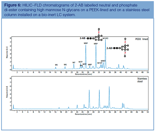
It is important to note that the above mentioned phenomena related to adsorption of phosphorylated species on metal ions of the LC system and column only occurs for the phosphate mono-ester containing N-glycans (PO4-M)2- and not for the phosphate di-ester containing N-glycans (M-PO4-M)1-. For the latter glycans, the profiles obtained on the stainless steel and PEEKâlined columns are exactly the same (Figure 6). Interestingly, these phosphate di-ester-carrying glycans are present on human acid alphaâglucosidase recombinantly produced in Yarrowia lipolytica, but the outer mannose residues are enzymatically removed during downstream processing to generate the active form of the enzyme, that is, carrying mannose-6-phosphate residues (15).
Conclusion
The HILIC analysis of phosphorylated N-glycans released from therapeutic enzymes using a bio-inert LC and PEEK-lined column has been described. It was demonstrated that these challenging structures can successfully be analyzed when the entire flow path is devoid of metal parts, that is, instrument and column inertness.
Acknowledgement
The authors acknowledge Sonja Schneider, Sonja Krieger, Linda Lloyd, and Udo Huber (Agilent Technologies).
References
- K. Sandra, I. Vandenheede, and P. Sandra, J. Chromatogr. A 1335, 81–103 (2014).
- S. Fekete, D. Guillarme, P. Sandra, and K. Sandra, Anal. Chem.88, 480–507 (2016).
- V. D’Atri, E. Dumont, I. Vandenheede, D. Guillarme, P. Sandra, and K. Sandra, LCGC Europe30(8), 424–434 (2017).
- A. Wakamatsu, K. Morimoto, M. Shimizu, and S. Kudoh J. Sep. Sci.28, 1823–1830 (2005).
- S. Liu, C. Zhang, J.L. Campbell, H. Zhang, K.K.-C. Yeung, V.K.M. Han, and G.A. Lajoie, Rapid Commun. Mass Spectrom.19, 2747–2756 (2005).
- R. Tuytten, F. Lemiere, E. Witters, W. Van Dongen, H. Slegers, R.P. Newton, H. Van Onckelen, and E.L. Esmans, J. Chromatogr. A1104, 209–221 (2006).
- Y. Asakawa, N. Tokida, C. Ozawa, M. Osamu, M. Ishiba, O. Tagaya, and N.Asakawa, J. Chromatogr. A1198–1199, 80–86 (2008).
- H. Sakamaki, T. Uchida, L.W. Lim, and T. Takeuchi, Anal. Sci.31, 91–97 (2015).
- H. Sakamaki, T. Uchida, L.W. Lim, and T. Takeuchi, J. Chromatogr. A 1381, 125–131 (2015).
- J.Y. Kang, O. Kwon, J.Y. Gil, and D.B. Oh, Anal. Biochem.501, 1–3 (2016).
- J.Y. Kang, O. Kwon, J.Y. Gil, and D.B. Oh, Data Brief7, 1531–1537 (2016).
- M.R. Hardy and J.S. Rohrer, in Comprehensive Glycoscience, J.P. Kamerling, Ed. (Elsevier, Amsterdam, The Netherlands, 2007), pp. 303–327.
- S. Kandzia, N. Grammel, E. Grabenhorst, and H.S. Conradt, in Cells and Culture, ESACT Proceedings, T. Noll, Ed. (Springer, Dordrecht, The Netherlands, 2010), pp. 867–873.
- A.T. van der Ploeg and A.J. Reuser, Lancet372, 1342–1353 (2008).
- P. Tiels, E. Baranova, K. Piens, C.D. Visscher, G. Pynaert, W. Nerinckx, J. Stout, F. Fudalej, P. Hulpiau, S. Tännler, S. Geusens, A. Van Hecke, A. Valevska, W. Vervecken, H. Remaut, and N. Callewaert, Nat. Biotechnol.30,1225–1231 (2012).
Koen Sandra is the editor of “Biopharmaceutical Perspectives”. He is the Scientific Director at the Research Institute for Chromatography (RIC, Kortrijk, Belgium) and R&D Director at anaRIC biologics (Ghent, Belgium). He is also a member of LCGC Europe’s editorial advisory board. Direct correspondence about this column to the editor-in-chief, Alasdair Matheson, at alasdair.matheson@ubm.com
Jonathan Vandenbussche is LC–MS Technician at RIC.
Pat Sandra is President of RIC and Emeritus Professor at Ghent University (Ghent, Belgium).

Accelerating Monoclonal Antibody Quality Control: The Role of LC–MS in Upstream Bioprocessing
This study highlights the promising potential of LC–MS as a powerful tool for mAb quality control within the context of upstream processing.
Common Challenges in Nitrosamine Analysis: An LCGC International Peer Exchange
April 15th 2025A recent roundtable discussion featuring Aloka Srinivasan of Raaha, Mayank Bhanti of the United States Pharmacopeia (USP), and Amber Burch of Purisys discussed the challenges surrounding nitrosamine analysis in pharmaceuticals.

.png&w=3840&q=75)

.png&w=3840&q=75)


.png&w=3840&q=75)


.png&w=3840&q=75)
