Analysis of Polymers and Protein Nanoparticles using Asymmetrical Flow Field-Flow Fractionation (AF4)
LCGC Europe
This article describes some of the latest developments for the analysis of polymers and nanoparticles.
Gelatin nanoparticles are used as a novel, biodegradable and well-tolerated drug carrier in tumour therapy and numerous other diseases. For a valid formulation approach in clinical studies an in-depth understanding of the molecular weight characteristics of this biopolymer is essential. Asymmetrical Flow Field Flow Fractionation (AF4) analysis was shown to be a fast and reliable analysis method for the pre-formulation screening of standard hydrophilic and hydrophobic prototype gelatins and also for mucoadhesive thiomers.
Polymer and protein-based drugs are becoming more common in the pharmaceutical industry and asymmetrical flow field-flow fractionation (AF4) offers a number of advantages over existing techniques, particularly for formulations containing polymers and nanoparticles. The interest in AF4 has grown since Giddings' break-through work in the nineties1 and the subsequent research by Fraunhofer2 has helped the technique gain wider acceptance and use. By combining this technique with multiangle light-scattering detectors (MALS), polymeric active ingredients can now be separated from polymeric excipients and particulate structures such as nanoparticles or virus-like particles (VLPs).3,4
Expert knowledge and practical expertise in this technique is, however, somewhat limited, but the advantages of AF4 are becoming more widely accepted. This article describes novel work in polymer and nanoparticle research and highlights the importance and potential of AF4 in this emerging field.
In practice established methods for protein and polymer analysis, such as reversed-phase high performance liquid chromatography (RP HPLC), size exclusion chromatography (SEC) and fluorimetry are often limited (e.g., shear force destruction of protein aggregates in stability studies due to the presence of the stationary phase or the need for fluorometric moieties in the sample). A typical packed column for these techniques requires high pressure — but this is not the case for AF4.
The separation principle of AF4, however, is based on a differential flow of a solvent in a channel and a respective perpendicular cross-flow to separate the analytes only by hydrodynamic properties in a fast, non-destructive, one-phase separation mechanism. One phase, non-destructive separation ensures very gentle separation conditions for large biomolecule analysis and aggregate detection, a rapid cost-effective analysis time and less unwanted interactions because of the small surface area of the channel compared with packed columns. Additionally, the smallest molecules can be analysed simultaneously with large proteins, aggregates or nanoparticles, which means AF4 offers a wider dynamic range than other chromatographic methods.
Figure 1 shows a schematic diagram of this separation channel. In theory macromolecules between 1 kDa and several GDa or particles between 2 nm and 100 μm can be separated by AF4.5
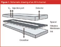
Figure 1: Schematic drawing of an AF4 channel.
Nanoparticles have the potential to offer numerous advantages for targeted, safe and effective drug delivery,6,7 especially when made of biocompatible and biodegradable polymers. Such nanoparticles could be useful for the targeted delivery of drugs to improve bioavailability, sustain drug effects at the target site, solubilize drugs for intravascular delivery and improve the stability of therapeutic agents against enzymatic degradation.8 Additionally, by modulating the polymer characteristics, the release of a therapeutic agent from nanoparticles can be controlled to achieve the desired therapeutic efficacy.9
Gelatin nanoparticles, for example, are a promising drug carrier system for future applications in various new approaches in gene therapy.10 Innovative gelatin prototypes with modifications of the backbone offer a further improvement regarding enhanced drug loading and modified targeting properties in vivo.
The aim of this investigation was to characterize the molar mass profile of two newly modified gelatin prototypes by AF4 and compare them with standard gelatin and fractionated gelatin, where low molecular weight (lmw) gelatin was removed from high molecular weight (hmw) gelatin. A thorough knowledge of the molar mass profiles in these highly lmw-reduced samples and in the prototype samples is mandatory for all nanoparticle formulation steps from gelatin as well as from other polymers such as chitosan. Hence, we also examined the molecular weight fractions of highly different chitosan–thiomer batches modified in two ways with sulphydryl groups and prompting different viscosities and chain lengths. In the course of method development we determined the optimum settings in terms of optical detector parameters as well as the correct type of membrane and elution profile for the separation of hydrophobic large proteins.
Key Points
With this data, an improved formulation process for nanoparticles was developed based on the traditional desolvation method developed by Coester.11 The physicochemical characterization of all nanoparticles was done by static light scattering (SLS) and compared with established gelatin nanoparticle formulations in terms of size, zeta potential and polydispersity index (PI).
Experimental
AF4 measurements of gelatins: As well as two hydrophobically modified gelatin prototypes (Gelita, Eberbach, Germany) called MS and MA, with succinate and dodecenylsuccinate residues, standard gelatin type A (Bloom ~175) from porcine skin (Sigma Aldrich, Munich, Germany) was analysed in this study. As control experiments in turn, measurements with two customized Gelita batches (VP306/VP413-2) that possessed less than 20% (w/w) peptides < 65 kDa were conducted. The AF4 analysis of these gelatins was performed on an AF1000-FOCUS system (Postnova, Landsberg am Lech, Germany) coupled with a UV detector (UV100 Thermo Scientific, Egelsbach, Germany), an RI detector (n-1000 WGE Dr. Bures, Germany) for concentration detection and a static light-scattering detector (miniDAWN Wyatt Technology Corporation, Dernbach, Germany) for molecular weight determination. The laser wavelength accounted for 690 nm, while slice collection was set to 1200. For molar mass determination the refractive index (RI) increment was set to 0.174 mL/g and the second virial coefficient was set to 0. The separation was achieved using a PBS buffer pH 6.0 as the mobile phase, a channel with 350 μm height and an ultrafiltration membrane consisting of regenerated cellulose with 5 kDa cut-off (Postnova). All proteins were dissolved in analysis buffer at a concentration of 2.5%. The channel flow-rate accounted for 1 mL/min, while the cross flow was adjusted to 0.05 mL/min over 10 min and then reduced to 0 mL/min, which resulted in a total measurement period of 20 min.
AF4 measurements of chitosans: The modification of the chitosan materials (Sigma) was conducted according to protocols described elsewhere.7,8 Chitosan low viscosity modified with N-acetylcysteine (Sigma) (lyophilized); chitosan low viscosity modified with thiobutylamidine (Sigma) (lyophilized); chitosan low molecular weight modified with N-acetylcysteine (lyophilized) and chitosan low molecular-weight modified with thiobutylamidine (lyophilized) were investigated with the same AF4 hardware set-up as for gelatin. The solvent and the running buffer for the chitosan samples were made of 0.3 M acetic acid, 0.2 M sodium acetate and sodium hydroxide/hydrochloric acid q.s. The pH was adjusted to 4 at a chitosan concentration of 0.1%. A membrane consisting of regenerated cellulose with a cut-off of 10 kDa was used in a 350 μm separation channel. The detector's dn/dc was set to 0.163 mL/g and the second virial coefficient to 0. For chitosan the cross flow was set to 1.0 mL/min at a channel flow of 1.0 mL/min while the focus time amounted to 350 s. The complete measurement period was 25 min.
Automatic microviscosimetry: Microviscosimetry experiments were performed using an AMVn microviscosimeter (Anton-Paar, Ostfildern, Germany). The viscosity of all investigated polymer solutions was determined by examining the rolling time of a steel sphere under the influence of gravity in an inclined cylindrical tube filled with the sample liquid. To ensure a constant temperature of 40 °C ± 0.01 °C, a built-in peltier was used. The viscosity was then calculated using the laws of Stoke:

with F being the frictional force, η the fluid viscosity and d the diameter of the spherical object. The molecular weight was approximated with the Mark-Houwink equation.14
Nanoparticle formulation: Nanoparticles from Sigma gelatin were prepared using the established two-step desolvation technique.15 In brief, a first desolvation step was performed with acetone (Sigma) as an antisolvent to remove the low molecular weight fractions of Sigma gelatin. After resolvation of the sediment with 40 °C warm highly purified water, nanoparticles were formed in a second desolvation step under constant stirring by addition of acetone at pH 2.5. The in situ nanoparticles were then stabilized by crosslinking with glutaraldehyde (Sigma). Before further analysis the nanoparticles were centrifuged and washed twice with highly purified water.
The MA and MS prototype gelatin nanoparticles were prepared from a 1% solution of gelatin by a novel desolvation method including several modifications to the previously mentioned two-step desolvation method: for succinylated gelatin the pH was set to 10, while for dodecenylsuccinylated gelatin the pH could be varied in a range from 2.4–10 to reach particles in the lower nanometer range. For a higher precision in the desolvation and to ensure a good size control, a peristaltic pump (Miniplus 3, Abimed Gilson, Langenfeld, Germany) was combined with an immersed needle for the first time where acetone is added via a 26 G steel needle (Sterican, Braun, Emmenbruecke, Germany) into the stirred solution. Crosslinking was achieved by the addition of glutaraldehyde, however, at a different concentration and pH.16
The chitosan nanoparticles were prepared by ionic gelation in 0.05% acetic acid solution at a polymer concentration of 2.5% (w/v) and a pH of 5.5. After complete dissolution of the polymer at 40 °C, a 0.2% (w/v) solution of sodium triphosphate pentabasic (Sigma) as the counter polyanion was added dropwisely (5 mL/min) under constant stirring until nanoparticles were formed. The final crosslinking was done by adding 10 μL of a 1 mM iodine (Sigma) solution. The anions and iodine were removed by dialysis against 0.1 M HCl over 12 h.
Nanoparticle characterization: The size distribution of all nanoparticles was analysed in aqueous dispersion by SLS (LA-950 laser difractometer, Retsch Technologies, Haan, Germany). Each size value and corresponding polydispersity index was the mean of 10 subruns. Static light scattering was used to screen larger agglomerates. All experiments were conducted with a refractive index of 1.59 (iabs = 0.01) and highly purified water as the dispersion medium. The zeta potential of the nanoparticles was measured with the Zetasizer Nano (Malvern) and flow through cells in highly purified water under controlled ionic strength conditions. All measurements were conducted in triplicate.
Results and Discussion
AF4 measurements of gelatins: At first, gelatin bulk material from Sigma-Aldrich was analysed to gain a benchmark for the following investigations of the modified samples. The molar mass was determined to comprise sizes ranging from 10 kDa to above 10000 kDa (Figure 2), which confirmed the data reported by Fraunhofer17 and exceeded the findings from size exclusion high performance liquid chromatography–MALS (SE-HPLC–MALS) analysis18 by more than one order of magnitude. The discrepancy between sizing data obtained from SE-HPLC and AF4 reflects the fundamental differences between these two separation techniques. While SE-HPLC separation takes place in a packed column, AF4 uses an open channel leading to lower hydrostatic pressure and therewith lower shear forces on the samples are encountered during analysis. High molecular weight specimens particularly suffer from degradation by increased shear forces and are, therefore, preserved and can be detected during AF4 analysis.19

Figure 2: Molecular weight distribution of standard Sigma gelatin. UV (curve) and MALS (dotted line) signal.
Figure 2 also shows how the high molecular weight fractions of gelatin almost elute over the whole experimental period. As a result of the broad variety of molecules possessing different molar masses in gelatin bulk material, a baseline separation of particular portions is excluded. Thus it was decided to only apply a weak separation force to expand the elution of the blend of molecules over a prolonged period to demonstrate the broad molecular variety within gelatin. A thorough understanding of the molecular weight distribution of natural polymers is essentially important for the formulation scientist. As will be discussed later, by delpeting certain fractions of gelatin, smaller and more homogenously distributed nanoparticles can be generated.
While very fast analysis is, of course, always possible with AF4 we demonstrate that the broad molecular weight distribution of the analysed gelatins has important implications in nanopartcile formulation. In this particular experiment, however, our goal was to highlight the broad molecular weight distribution of these gelatins, rather than a superfast analysis (which is, of course, possible).
As only the high molecular weight fraction of Sigma gelatin can be used for the preparation of homogenous nanoparticles it generally has to be processed by two-step desolvation. Manufacturing experiments in turn, conducted with two customized Gelita batches (VP306/VP413-2) that possessed less than 20% (w/w) peptides < 65 kDa resulted in successful one-step desolvation synthesis of gelatin nanoparticles exhibiting equivalent size and size distribution.15 These findings reveal the restriction that has to be especially made for the presence of low molecular weight portions in gelatin batches designated to one-step desolvation. The successful depletion of the low molecular weight fraction of gelatin is demonstrated in Figure 3.

Figure 3: UV signal (continuous line) and molecular weight (dots) calculated from respective UV and MALS data resulting from AF4 analysis of gelatin bulk material VP413-2 developed and provided from Gelita; the circle marks the low molecular-weight fraction.
In addition, gelatin sediment obtained from two-step desolvation after the first desolvation step — as the result from fractionation and used for the preparation of nanoparticles — also underwent AF4 analysis. Data from these experiments and from gelatin bulk material are displayed as function of their mean molecular weight in Figure 4.

Figure 4: Mean molecular weight fractions calculated from respective UV and MALS data resulting from AF4 analysis of (1) gelatin bulk material purchased from Sigma-Aldrich, (2) gelatin bulk material VP306, (3) VP413-2 developed and provided from Gelita, and of (4) gelatin sediment obtained after the first desolvation step from the manufacturing process of the gelatin nanoparticles.
Interestingly the clear shift of the mean molecular weight of the gelatin sediment (4) by more than one order of magnitude compared with the bulk material (1) is not a prerequisite for a successful one-step desolvation. A mean molecular weight between 400 and 500 kDa determined for the Gelita batches VP306 and VP413-2 was already sufficient to allow the new production process. Thus, derived from these findings and the specification of the applied gelatin batches a mean molecular weight of ~500 kDa and a threshold of a maximum of 20% (w/w) for the portion of low molecular weight fractions < 65 kDa could be defined as prerequisite for the successful manufacturing of gelatin nanoparticles by a one-step desolvation procedure.
The mean molecular weight of gelatin sediment ranges clearly above the one of the Gelita batches, which may not only be attributed to even more reduced amounts of peptides < 65 kDa far below 20% in the sediment. Thus, the fractionation of gelatin bulk material during two-step desolvation supposedly led to a depletion of molecular weight fractions bigger than 65 kDa.
In the process of method development for the analysis of MA and MS gelatins, several ultrafiltration membrane types were tested. After experiments with different materials and cut-off values regenerated cellulose with a cut-off of 10 kDa proved to be the most adequate. The results in terms of recovery rate, repeatability and signal quality are presented in Table 1. Regenerated cellulose is a very low protein binder and therefore ideally suited for analyses that require maximum sample recovery. In addition the membrane possesses a good solvent resistance with both aqueous and organic solvents, and is able to work over a wide pH range.

Table 1: Influence of the different membrane types on recovery rate and repeatability (defined as the intra day repeatability of a 100% value in per cent of six replicates).
The derivated hydrophobic prototype gelatins showed a molar mass distribution from 140–10000 kDa and 200–100000 kDa for MS and MA, respectively. While the average molecular weight of MS was 218 kDa, MA showed an average molecular weight of 395 kDa. The molar mass distribution of standard gelatin was found to be between 140–1000 kDa with an average molar mass of 158 kDa. (Figure 5). This is in accordance with the prior studies, where even fractions up to 10000 kDa were detected. The average recovery was about 97.4%.
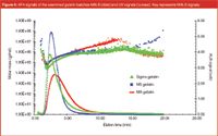
Figure 5: AF4 signals of the examined gelatin batches MALS (dots) and UV signals (curves). Key represents MALS signals.
AF4 Measurements of Chitosans
For the analysis of chitosan, the cumulative mass distribution was plotted for a better comparison with the gelatin samples. As seen in Figure 6 the distribution of molecular weight fractions in chitosan is much broader than for gelatin (Figure 7). Interestingly, chitosan lmw modified with thiobutylamidine as a potential nanoparticle crosslinker showed an increased amount of low molecular weight fractions compared with the unmodified chitosan lmw samples. A polymer crosslinking throughout the small molecular weight range could be the potential reason for this data. In contrast, the thiobutylamidine modification of low viscosity chitosan lead to higher molar mass profiles over the whole range, which might be based on the natural origin of chitosan and also on a partial depolymerization during the sulphhydryl modification process.
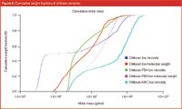
Figure 6: Cumulative weight fractions of chitosan samples.
Because the chitosans were modified with the potentially crosslinking sulphhydryl groups we compared the total amount of free sulphydryl groups and the existing disulphide bonds to the calculated molar masses. In Figure 8 these three parameters were correlated and it was shown, that a constant amount of disulphide bonds throughout the samples does not automatically account for higher molar masses of the polymers. The influence of these modifications is given exemplarily for the case of TBA-modified chitosan in Figure 9. For the N-acetylcysteine modification of chitosan lmw, the viscosity was so high, that a molar mass calculation could not be made. In all other cases the molar separation and molecular weight calculation was successful and reproducible. In conclusion, AF4 analysis was able to give a good idea of what the molecular weight distribution of the chitosan samples looks like and where modifications of the backbone lead to different retention behaviour.
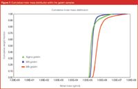
Figure 7: Cumulative molar mass distribution within the gelatin samples.
Automatic microviscosimetry: The molecular weight results from AF4 were in accordance with the calculated values from automatic microviscosimetry via the Mark-Houwink equation (Table 2). A correlation factor of 1.0 stands for an optimum compliance of the calculation with the measured values.
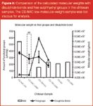
Figure 8: Comparison of the calculated molecular weights with disulphide bonds and free sulphhydryl groups in the chitosan samples. The CS-NAC low-molecular-weight-sample was too viscous for analysis.
Nanoparticle formulation and characterization: The prototype gelatin nanoparticles were prepared from a 1% solution of gelatin by a single desolvation method including several modifications to the above mentioned two-step desolvation method: the pH for negatively charged MS gelatin nanoparticles had to be adjusted to 10 for a homogeneous and small particle size, while MA gelatin with long hydrophobic side chains lead to nanoparticles in the whole pH range from 2.5–10. Paying tribute to the changed physicochemical properties of the protein, inert glass stirring beads had to be used for the preparation process to prevent aggregation during desolvation. Furthermore, the amount of acetone needed for desolvation and the amount of glutaraldehyde as a crosslinker had to be optimized because several changes in the functional groups of the protein had occurred during the prior modification.
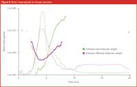
Figure 9: Molar mass signals of chitosan samples.
Resulting MA nanoparticles were 193 nm ± 16 nm (mean ± SD; n = 3) in size with a polydispersity index (PDI) of 0.107, while MS nanoparticles were larger, with 362 nm ± 22 nm (mean ± SD; n = 3) and a PDI of 0.053. Statistical analysis of the data was performed by a one way analysis of variance (Table 3).

Table 2: Molecular weight of Sigma gelatin and modified gelatin prototypes determined by automatic microviscosimetry compared with AF4 (mean value).
Chitosan nanoparticles prepared from the modified samples resulted in small and stable nanoparticles (Table 3). However, their polydispersity index was much higher than for the gelatin nanoparticles.

Table 3: Size and distribution of the nanoparticles as measured by SLS and zeta potential (n=3).
Conclusion
With the precise determination of the molecular weight distribution of modified gelatin and chitosan prototypes by AF4, an important basis for further studies concerning the development of small, uniform and stable gelatin and chitosan nanoparticles for drug delivery purposes was achieved.
The analysis of gelatin bulk material by the combination of asymmetrical flow field-flow fractionation and multiangle light scattering was accomplished as a continuation of earlier studies from Fraunhofer.2 At first, their basic results obtained for gelatin bulk material applied for the manufacturing of gelatin nanoparticles by two-step desolvation could be confirmed. Secondly, mean molecular weights of new customized gelatin quality characterized by the depletion of low molecular weight fractions during production could be successfully classified in between the mean molecular weight determined for gelatin bulk material from Sigma-Aldrich and gelatin sediment obtained from two-step desolvation.
These results demonstrated the impact of the low molecular weight fraction of gelatin for the manufacturing of gelatin nanoparticles and contributed to further understanding and description of gelatin nanoparticle synthesis by desolvation. This was at least feasible in a one-step attempt using particular batches of the customized Gelita material.15 The one-step desolvation is not only straightforward in terms of technological aspects because it simplifies the manufacturing procedure, but is also especially interesting for regulatory considerations. A simplified formulation process will increase the chance for regulatory approval and make post-approval changes easier and faster to realize due to the process being less complex. The simplicity of the whole gelatin nanoparticle formulation process makes them attractive for FDA approval.
One of the major drawbacks of the two-step desolvation is that gelatin nanoparticles are produced with bulk material obtained from the first desolvation step that is not exactly defined (i.e., varying laboratory equipment may lead to different fractionation outcomes in terms of molecular weight).
The first desolvation step within the two-step desolvation method requires a manual discarding of the supernatant, leaving the whole process with a formulator based variable.Just recently we standardized this process using a newly developed instrument that reproducibly fractionates the gelatins the same way each time. The successful application of the one-step desolvation for gelatin nanoparticle synthesis circumvents this problem.
It was further shown that hydrophobically modified large proteins can be sized by AF4. As with all quantitative analysis tools the application of the correct separation material, in this case the right membrane type and cut-off had to be taken into close consideration during the process of method development. We can state that the AF4 method described provides a fast and reliable tool for the analysis of proteins with a broad molecular weight distribution. This was confirmed not only with prototype gelatin samples, normal gelatin, but also with modified chitosan batches at different thiol-bond crosslinking rates. The adapted desolvation process proved to be a reliable way of producing mono-modal and small-sized nanoparticles confirmed by static light scattering. With the negative zetapotential of the prototype nanoparticles, these carriers can be used for electrostatic attachment of positive charged drug molecules, such as methotrexate. Additionally, the new prototype nanoparticles possess different surface properties that hold promise for a different body distribution pattern. Further in vivo studies are in progress to analyse the properties of those new gelatin nanoparticles.
In the future AF4 will definitely be used to a greater extent for the analysis of polymers, proteins and nanoparticles. New developments in the fields of liposomes, cells, bacteria and especially antibodies will raise the demand for a fast and reliable analysis method such as AF4, particularly when the standard separation methods for polymeric and colloidal analytes come to their limits, AF4 may be the new method of choice.
Stephan Schultes, Kathrin Mathis, Klaus Zwiorek and Jan Zillies are scientists in the group of Dr Coester and Professor Winter, all involved besides their individual projects in pharmaceutics in the development of AF4 applications.
Conrad Coester is principal investigator at the LMU, Department of Pharmacy. He is specializing on nanoparticulate drug delivery, focusing on numerous applications of gelatin nanoparticles.
Gerhard Winter is a full professor for Pharmaceutical Technology and Biopharmacy. After 12 years in the biotech industry he now focuses his research at the LMU since 1999 on parenteral dosage forms including depot systems, colloids and especially protein drugs.
References
1. J.C. Giddings, Science, 260(5113), 1456–1465 (1993).
2. W. Fraunhofer and G. Winter, European Journal of Pharmaceutics and Biopharmaceutics, 58(2), 369–383 (2004).
3. C. Augsten and K. Maeder, Light scattering for the Masses: Characterization of Poly(D,L-lactide-co-glycolide) Nanoparticles; Wyatt Technology Corporation Application Notes (2005).
4. R. Lang, and G. Winter, Light Scattering for the Masses: Characterization of Virus Like Particles by Asymmetrical Flow Field-Flow Fractionation; Wyatt Technology Corporation Application Notes (2006).
5. M. Schimpf, K.D. Caldwell and J.C. Giddings (eds.); Field-Flow Fractionation Handbook, John Wiley & Sons, Inc., New York (2000).
6. W. Babel, Chemie in unserer Zeit, 30(2), 1–11 (1996).
7. C. Coester, New Drugs, (1), 14–17 (2003).
8. R. Langer, Nature (London) 392 (6679, Suppl.), 5–10 (1998).
9. S.V. Vinagradov, T.K. Bronich and A.V. Kabanov, Adv. Drug Del. Rev, 54, 223–233 (2002).
10. G. Kaul, and M. Amiji, Pharmaceutical Research, 19(7), 1061–1067 (2002).
11. C. Coester et al., Journal of Microencapsulation, 17(2), 187–193 (2000).
12. A. Bernkop-Schnürch and Th.E. Hopf, Sci. Pharm., 69(2), 109–118 (2001).
13. A. Bernkop-Schnürch, M. Hornof and T. Zoidl, Int. J. Pharm., 260(2), 229–237 (2003).
14. DIN 53015 und ISO 12058.
15. K. Zwiorek, Gelatin Nanoparticles as Delivery System for Nucleotide-based drugs, Dissertation, Ludwig-Maximilians-University Munich (2006).
16. S. Schultes et al., Novel Hydrophobic Prototype Gelatins for Nanoparticle Preparation Analyzed by AF4 and MALS; 6th World Meeting on Pharmaceutics, Biopharmaceutics and Pharmaceutical Technology, Barcelona, Spain, April, 6th-10th (2008).
17. W. Fraunhofer, G. Winter and C. Coester, Analytical Chemistry, 76(7) 1909–1920 (2004).
18. M. Meyer and B. Morgenstern, Biomacromolecules, 4(6), 1727–1732 (2003).
19. M.N. Myers, Journal of Microcolumn Separations, 9(3), 151–162 (1997).

Retaining Talent in Field-Flow Fractionation: An Initiative
The authors present their motivation for establishing the Young Scientists of FFF (YSFFF) initiative within the FFF community.
Developments in Field‑Flow Fractionation Coupled to Light Scattering
December 8th 2020Field-flow fractionation (FFF) coupled to light scattering is a powerful method to separate and characterize nanoparticles, proteins, and polymers from a few nanometres to a few micrometres. The technique is one of the few that can cover the full size range of nanomaterials and provide high-resolution size distributions and additional characterization. New developments in FFF enhance performance and productivity.
Field-Flow Fractionation: Virtual Optimization for Versatile Separation Methods
August 8th 2017Flow-field flow fractionation (flow-FFF) offers highly versatile separations for the analysis of complex fluids, covering a size range of macromolecules and particles from 1 nm to 10,000 nm. However, flow-FFF is often perceived as a difficult technique to learn because of the multiple parameters available for adjustment. Recent advances in software for simulating flow-FFF overcome this obstacle, enabling the virtual optimization of flow-FFF methods and opening up the power of flow-FFF separations to non-experts. An added benefit is the ability to easily analyze particle size distributions by elution time from first principles.


