Stationary Phases for Modern Thin-Layer Chromatography
LCGC Europe
The stationary phases currently in use for modern thin-layer chromatography (TLC) are reviewed.
The stationary phases currently in use for modern thin-layer chromatography (TLC) are reviewed here, together with traditional and newer detection techniques. Advantages of the planar, off-line format of TLC relative to on-line column high performance liquid chromatography (HPLC), the steps in TLC analyses and future prospects of TLC are also discussed. A limited number of applications are presented throughout the article, and many others can be found in the books, book chapters and review articles cited.
This review discusses the current status of thin-layer chromatography (TLC), focusing mainly on commercial precoated layers that are in popular use. Bulk sorbents, for those who prefer to make their own TLC plates, are available but will not be covered.
TLC is an increasingly widely used analytical method for the determination of many types of analytes in a variety of sample matrices. Its theory, practice and applications have been documented in biennial reviews written by Sherma since 1970, the latest of which will be published in the Journal of AOAC International (1). An article in LCGC North America (2) gave the three largest industrial applications for TLC as clinical, pharmaceutical and food testing. The most active research areas today are in the analysis of pharmaceutical bulk materials and dosage forms (3,4) and in natural phytochemical medicines (for example, traditional Chinese medicines and nutritional supplements [5,6]).
The techniques and instruments of modern, instrumental TLC allow it to meet the guidelines for validated analytical methods in current good laboratory practice (cGLP) and current good manufacturing practice (cGMP); pharmacopeias of China, India and the United States (U.S. Pharmacopeia and American Herbal Pharmacopeia); international food regulations; official methods of AOAC International; International Conference on Harmonization (ICH) pharmaceutical analyses (7); and international food regulations (8,9). Here, we cover some special attributes of TLC relative to column high performance liquid chromatography (HPLC), briefly describe the steps of TLC analyses, focus on the stationary phases (layers) in use today and speculate on prospects for TLC in the future.
TLC Compared to HPLC
The use of a planar stationary phase rather than a column leads to a number of significant contrasts between TLC and HPLC. HPLC is a closed, on-line method with dynamic detection of eluted solutes, usually by UV absorption or mass spectrometry (MS). The predominant mode of HPLC is reversed phase on a C18 bonded silica gel column, and the predominant mode of TLC is normal phase on a silica gel layer; these diverse mechanisms allow TLC and HPLC to serve as methods with complementary selectivity for separations and analyte identification. An HPLC column is used for many analyses, but each thin-layer plate is used only once. This often leads to the need for less sample preparation (cleanup) for TLC compared to HPLC, because impure samples can be applied without concern for carryover and cross-contamination of samples that leads to additional (ghost) peaks in column chromatograms.
Although it is not yet fully automated like HPLC, many of the steps in the TLC procedures are automated for faster sample analyses. TLC has the advantage of very high sample throughput because multiple standards and samples can be applied to a single plate and separated at the same time under essentially identical conditions. Each track on a TLC plate contains a complete chromatogram of an entire sample, including irreversibly sorbed substances (at the origin). On the other hand, in HPLC each chromatogram requires a separate injection and the chromatogram contains only those sample components that happen to be eluted and detected. Modern computer controlled scanning densitometers and automated sample application and layer development instruments allow validated quantification that is in many cases equivalent to HPLC (10). As shown later, TLC has the widest choice of stationary phases and mobile phases, making it the most versatile and flexible LC method. Solvents that can interfere with HPLC UV detection can be used because the TLC mobile phase is removed before detection. Mobile-phase consumption is low, minimizing solvent purchase and disposal costs, thereby making TLC a "green" method. The wide choice of development methods and pre- and post-chromatographic detection methods leads to unsurpassed selectivity in TLC. Because TLC is an off-line method, the various steps can be carried out independently without time constraints; as an example of an advantage of this approach, zones can be scanned repeatedly with a densitometer using different parameters that are optimum for individual sample components. In addition, a TLC plate can be stored and reinvestigated at a later date if additional questions arise.
The Steps of TLC Analysis
Excellent discussions of sample preparation; sample application; mobile-phase selection; chromatogram development and documentation; and zone detection, identification, and quantification in modern TLC are presented in two books (11,12), two book chapters (13,14) and a review of TLC in drug analysis (15). This section contains brief discussions of some of these steps of the TLC analytical process, followed by a more detailed description of stationary phases.
Sample Application: The most widely used methods for application of standard and sample solutions to a plate are manual with a capillary (for example, 10- or 25-µL Drummond digital microdispenser) and automated instrumental application. Application of narrow, homogeneous bands of controlled length has been shown to give superior chromatographic results compared to spot application. These automated application devices are available from Camag and Desaga.
Choosing the Mobile Phase: Mobile phases are less polar than the silica gel layer (normal-phase TLC) and are usually composed of nonpolar and polar constituents with or without an acid or base modifier to improve resolution. In reversed-phase TLC, the stationary phase is less polar than the mobile phase, which is often a combination of methanol, acetonitrile or tetrahydrofuran with water.
The mobile phase is optimized in terms of its strength and selectivity in relation to the sample and stationary phase. Snyder classified solvents into eight groups within a selectivity triangle based on proton acceptor, proton donor and dipole properties (13). He later reviewed and expanded his studies of the role of the mobile phase in controlling selectivity for adsorption TLC and suggested a sevenmobilephase experimental design for optimizing relative retention and selectivity with binary solvent mobile phases (16).
Chromatogram Development: Plates are most often developed in a conventional glass large volume chamber (N-chamber) in the ascending direction. Also available are twin trough chambers (TTCs), which allow the development of plates with a low volume of mobile phase and experimentation with chamber saturation conditions, and an automatic developing chamber (ADC 2) that provides complete control of all aspects of plate development (both from Camag).
Horizontal development chambers are also available from Chromdes and Camag (HDC 2). An advantage of these chambers is the ability to develop the plate from both opposing sides towards the middle, permitting the number of samples to be doubled to achieve even greater sample throughput.
Methods used to increase the resolution of complex mixtures include two-dimensional (2D) TLC, in which development of a single sample applied to the corner of a single-layer plate is performed in perpendicular directions, with drying in between, using two different mobile phases of about the same strength but complementary selectivities. A dual-layer plate (Multi-K, Whatman/GE Healthcare) that allows both reversed-phase and normal-phase development is also available. The use of either of these can spread sample components over the entire plate with high zone capacity. Figure 1 illustrates these two possibilities for 2D TLC.
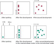
Figure 1: 2D TLC on a single plate. Top TLC plate: a single layer (silica, C18 or cellulose) developed in two directions with different mobile phases, 90° to one another, with drying between developments. Bottom TLC plate: a dual layer (C18 and silica, adjacent to one another) developed in two different modes to improve separation of complex mixtures.
Note in each case, the components never need be removed from the plate before the second development is begun, saving time and potential loss. After development, other procedures and documentation as described below can be continued.
The ease of application of 2D TLC is one example of an advantage of TLC compared to HPLC. It is much more difficult and costly to accomplish a successful 2D HPLC method. 2D HPLC often requires the collection of sample fractions or complex column switching. The biggest problem is often a mismatch of the mobile phase used in one mode compared to the second mode. If the mobile phases are immiscible, then drying and reconstituting each sample fraction is usually required before introduction into the second column. This is never a problem with 2D TLC because any initial mobile phase is evaporated from the plate before the second development is started.
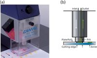
Figure 2: TLCâMS interface: (a) front of instrument and (b) schematic of extraction section. (Courtesy of Camag USA.)
Multiple development either manually or with Camag's Automatic Multiple Development (AMD 2) instrument can also greatly improve the resolution of complex mixtures. The typical mode of automatic multiple development uses a multiple-step mobile-phase gradient with successive runs that have decreasing strength (less polar for a silica gel layer) and increasing development distance, with vacuum drying of the plate between steps.
The development methods described above in this section involve migration of the mobile phase by capillary forces. There are now devices available for techniques that are categorized as forced-flow planar liquid chromatography (FFPLC) with which the mobile-phase migration is driven by other means. These include overpressured-layer chromatography (17), rotational planar chromatography (18) and pressurized planar electrochromatography (19).
Detection (Visualization) of Zones and Documentation of
Chromatograms: After drying the developed plate, compound zones are detected on the layer by their natural colour, natural fluorescence under 366nm UV light, quenching of fluorescence under 254nm UV light on an "F" layer containing a phosphor (termed an UV indicator), or as coloured, UV-absorbing or fluorescent zones after reaction with an appropriate chemical reagent (postchromatographic derivatization). Chromogenic and fluorogenic reagents are applied by spraying or dipping a layer or exposing it to reagent vapours (for example, iodine). Chemical detection reagents can be nondestructive universal (iodine), destructive universal (for example, charring with sulphuric acid) or selective (for example, ninhydrin for amino compounds). Most chemical derivatization reactions require heating of the plate for completion. A forced air oven (with a solid glass or metal plate onto which the TLC plate is placed for even heating) or a Camag plate heater can be used.
Effect directed analysis (EDA) is becoming more important in food, pharmaceutical and phytochemical analyses. In EDA, there is no attempt to analyse all compounds in a sample, but instead the focus is on just the relevant (for example, harmful or bioactive) compounds detected by a bioassay coupled to TLC. These compounds can be the target analyte as well as unknown metabolites, side products, process contaminants, adulteration products and residues. Bioassays used with TLC include microbiological assays (bioautography), bacterial assays (the Camag BioLuminizer bioluminescence detector utilizing Vibrio fischeri) and biochemical detections using enzymes, including immunoassays. TLC–EDA was reviewed by Morlock and Schwack (20).
If the chromatogram and its visualization produce visible results, it is possible to simply photocopy in black and white or colour, or scan the TLC plate to document it. However, a commercial system for photography is needed if documentation of a combination of coloured, fluorescent and quenched zones is required. Camag, Desaga and Analtech all offer photodocumentation systems.
Identification of Zones: Separated zones are initially identified based on the correspondence of R f values and detection characteristics between sample and standard zones on the same plate, but must be independently confirmed by other evidence, such as off-line and on-line coupling of TLC with various spectrometric methods. Scanning densitometers have a spectral mode that allows measurement of UV/vis spectra of zones directly on the plate and comparison with standards. In addition, peak purity can be examined by carrying out spectral studies at the peak start, middle and end.
One of the most important and active areas of current research is hyphenation of TLC with MS for identification or structure elucidation, which was reviewed by Morlock and Schwack (20,21). Many TLC–MS coupling approaches were covered in these reviews, but the elution headbased technique is the most interesting because of the commercial availability since mid-2009 of the Camag TLC–MS interface. Details of its use are available from Camag. The reviews by Morlock and Schwack also cover hyphenation of TLC with UV/vis, fluorescence, Fourier transform infrared (FT-IR) and surface-enhanced Raman spectrometry in various combinations to characterize samples as completely as possible.
Quantitative Analysis by Densitometry: Most reported quantitative analyses by TLC involve reflectance scanning in the absorbance or fluorescence mode using a Camag TLC Scanner 3, a Desaga CD-60 densitometer or a J&M diode array detector scanner. Standard zones are applied to a plate to create a calibration curve of peak area or height vs. weight through linear, nonlinear, polynomial or Michaelis–Menten regression, and weights of bracketed sample zones on the plate are interpolated from the curve. Videodensitometers with a visible or UV light source, camera and software for image capture and quantification are also available from Camag and Desaga, but these do not allow measurement at an optimal wavelength as the slit scanning densitometers with a monochromator do.
Stationary Phases
The chronology of the introduction of different TLC sorbents, plate formats and instruments was reviewed in an historical article (22). As in column chromatography, the chemistry and size of the particles as well as the thickness of the layer affects the speed and resolution of the separations that are possible. Silica gel is the predominant TLC layer because of its high selectivity for a large range of compounds with different functional groups. Supposedly, similar layers according to the label from different manufacturers (for example, the most widely used layer by far, silica gel 60 F254) with the same layer thickness can give quite different results, perhaps because of the unstated binder type and content, and should be comparatively evaluated before substitution of plates in a given method.
Binders: In addition to the chosen sorbent, precoated plates usually contain a binder to help the particles adhere to the support that serves as the backing for preparation of the layer. The most widely used commercial TLC and highperformance TLC (HPTLC) plates contain an organic polymeric binder, such as polymethacrylate at a concentration of 1–2%; these are harder, smoother, more durable and generally give better separations than G layers. The classical TLC plates designated as "G" for gypsum (calcium sulphate) binder are still available from EMD/Millipore, Macherey-Nagel and Analtech. The nature of the binder in a plate can affect separations and detection based on colour reactions.
Fluorescent Indicators: Layers containing an indicator that fluoresces when irradiated with 254- or 366-nm ultraviolet light are designated as "F" or "UV" layers. These layers are used to facilitate the detection of compounds that absorb at these wavelengths and give dark zones on a bright background (fluorescence quenching). F254 indicators can give green (zinc silicate) or blue (magnesium tungstate) fluorescence. F366 indicators can be an optical brightener, fluorescein or a rhodamine dye (23). Some precoated plates have both indicators to detect compounds that absorb at both wavelengths (F254+366 plates). Plates designated with an "s" have a UV indicator that is acid stable (F366s plates). Lux TLC plates (EMD/Millipore) and Adamant TLC and NanoSILHPTLC plates (Macherey-Nagel) have enhanced brightness because of increased levels of the F254 indicator.
TLC vs. HPTLC
The differences between TLC and HPTLC can be seen in Figure 3 and Table 1. The effect of the smaller particle size and the shortened development distances combine to give higher efficiency, shortened development times, increased sensitivity and a larger number of samples processed simultaneously.

Figure 3: Comparison of the separation of dansyl amino acids on (a) a classical TLC silica gel 60 plate and (b) an HPTLC silica gel 60 plate under identical conditions. Samples (bottom to top): N-α-dansyl-L-asparagine (lowest Rf), α-dansyl-L-arginine, dansyl-L-cysteic acid, N-dansyl-L-serine, dansyl-glycine and N,N-didansyl-L-tyrosine (highest Rf). Sample volume: TLC, 4 µL; HPTLC, 0.3 µL; mobile phase: ethyl acetateâmethanolâpropionic acid (22:10:3,v/v/v); migration distance: TLC, 10 cm; HPTLC, 5 cm; analysis time: TLC, 42 min; HPTLC, 13 min, 45 s; detection: UV at 366 nm. (Chromatograms courtesy of Merck KGaA, Darmstadt, Germany.)
TLC and HPTLC silica gel 60 plates with etched identification codes, spherical particle plates (not irregular silica gels as used in most standard layers) and thinner layers for special applications are also available. These can be checked out on the manufacturers' websites (EMD/Millipore, Macherey-Nagel and Analtech).
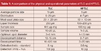
Table 1: A comparison of the physical and operational parameters of TLC and HPTLC.
Plates with Channels and Preadsorbent Zone
Kieselguhr (diatomaceous earth) and silica 50000 are small surface area, weak adsorbents that are used as the lower 2–4 cm inactive preadsorbent (also called concentration zone) in the manufacture of silica gel and C18 preadsorbent plates. Samples applied to the preadsorbent zone run with the mobile-phase front and reform into sharp, narrow, concentrated bands at the preadsorbent–analytical sorbent interface. This leads to efficient separations with minimum time and effort in manual application of samples, and possible sample cleanup by retention of interferences in the preadsorbent, which can be modified by dipping (then drying) into reagents that can capture unwanted impurities once spotted.
Channeled (laned) TLC and HPTLC plates have tracks scribed in the layer that are 9 mm wide with 1 mm between channels (19 useable channels across a 20-cm wide plate). Advantages include no possible cross-contamination of zones during development; exact location of zones during application, which facilitates alignment of a densitometer light beam for automated scanning; and easier removal of separated zones by scraping, before recovery by elution, without contaminating or damaging adjacent tracks.
Pretreatment of Plates
To remove extraneous materials that may be present because of manufacture, shipping or storage conditions, it is recommended to preclean plates before use by development to the top of the plate with methanol (24). The plate is then dried in a drying oven or plate heater for 30 min at 120 °C. If the plate is being used immediately, it will equilibrate with the laboratory relative humidity (which should be controlled to 40–60% and recorded regularly) during sample application. Plates that are prewashed do not need activation by heating unless they have been exposed to high humidity. More complete suggestions for initial treatment, prewashing, activation and conditioning of different types of glass- and foil-backed layers have been published (24).
Glass plates can be cut to a smaller size using a commercial glass cutter and broken with grozier pliers for better results (Figure 4). Scissors, a razor knife, razor blade or circular cutter (as used to cut fabric) can all be used to cut plastic or aluminum sheets. A straightedge should be used, and care must be taken to avoid damaging the sorbent layer and your fingers.

Figure 4: Cutting tools for scoring and breaking glass TLC plates: (a) pistol-grip glass cutters and (b) grozier pliers. These items are available from hobby or stained glass stores. Images from author’s collection (F.R.).
Silica Gel
Silica gel (silica) is by far the most frequently used layer material in TLC. The structure is held together by bonded silicon and oxygen (siloxane groups), and separations take place primarily because of differential migration of sample molecules caused by selective hydrogen bonding, dipole–dipole interaction and electrostatic interactions with surface silanol groups (Si-OH). The intensity of these adsorption forces depends on the number of effective silanol groups, the chemical nature of the sample molecules to be separated and the elution strength of the mobile phase.
Differences in the type and distribution of silanol groups for individual sorbents may result in selectivity differences, and separations may not be exactly the same for brands of silica gel layers from different manufacturers. Typical properties of silica gels suitable for planar chromatography are as follows (25): mean hydroxyl (silanol) group density, approximately 8 µmol/m2 (independent of silica gel type); mean pore diameter between 40 and 120 Å (4–12 nm); specific pore volume (vp ) between 0.5 and 1.2 mL/g; mean particle size and particle size distribution are shown in Table 1; and specific surface area (SBET ) between 400 and 800 m2 /g. Silica gel 60 has a 60-Å pore diameter and is the most commonly used type in TLC and HPTLC. Precoated plates used in a laboratory with 40–60% relative humidity and temperature of 20 °C will become equilibrated to a hydration level of 11–12% water, will not require preactivation unless earlier exposed to high humidity and will give consistent R f values within this moisture range.
Figure 5 shows a typical highresolution separation with symmetrical, narrow densitometer peaks obtained on a silica gel HPTLC plate.
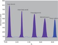
Figure 5: Densitogram of a neutral lipid standard mixture showing peaks of (left to right) cholesterol, oleic acid, triolein, methyl oleate and cholesteryl oleate, which serve as marker compounds for free sterols, free fatty acids, triacylglycerols, methyl esters and steryl esters, respectively. 4 µL of a solution containing 0.2 µg/µL solution (0.8 µg) of each compound was applied to a laned Analtech HPTLC silica gel preadsorbent plate, which was developed with petroleum etherâdiethyl etherâglacial acetic acid (80:20:1) in a vapour-saturated Camag HPTLC twin-trough chamber. The lipid bands were detected by spraying the plate with 5% ethanolic phosphomolybdic acid solution and heating at 110 °C for 10 min, and densitometry was carried out at 610 nm using a Camag Scanner 3 with winCATS software.
Silica Gel Bonded Phases Reversed-Phase Bonded Layers:
Reversed-phase TLC is used more now because the bonded phases eliminate previous attempts to use coated layers, which required mobile phases saturated with the coating agent, paraffin or silicon oils.
Similar to HPLC, alkylsiloxane-bonded silica gel 60 with dimethyl (RP-2 or C2), octyl (RP-8 or C-8) and octadecyl (RP-18 or C18) functional groups are most widely used for reversed-phase TLC of organic compounds (polar and nonpolar, homologous compounds and aromatics), weak acids and bases after ion suppression with buffered mobile phases and polar ionic compounds using ion-pair reagents. Layers from different companies with the same bonded group can have different percentages of carbon loading and give different separation results. The hydrophobic nature of the layer increases with both the chain length and the degree of loading of the groups. Alkylsiloxane-bonded layers with a high level of surface modification are incompatible with highly aqueous mobile phases and are used mainly for normal phase separations of low polarity compounds. Problems of wettability and lack of migration of mobile phases on reversed-phase plates with high proportions of water have been solved by adding 3% NaCl to the mobile phase to attain better wettability (C18 layers; Whatman/GE Healthcare) or preparing "water-wettable" layers (for example, RP-18W; EMD/Millipore) with less exhaustive surface bonding to retain a residual number of silanol groups. The latter layers with a low degree of surface coverage and more residual silanol groups exhibit partially hydrophilic, as well as hydrophobic character and can be used for reversed-phase TLC and normal-phase TLC with purely organic, aqueous–organic and purely aqueous mobile phases.
Chemically bonded phenyl layers are also classified as reversed phase; their separation properties have been reported (23) to be very similar to RP-2 although the aromatic diphenyl bonded groups (with their p-p interaction possibilities) would appear to be very different than ethyl groups. RP-2 layers function with a normal-phase mechanism when developed with purely organic mobile phases and a reversed-phase mechanism with aqueous–organic or purely aqueous mobile phases.
Reversed-Phase Bonded Layers for Enantiomer Separations: To make a chiral TLC plate it is necessary to coat or bond various chiral phases. These reagents are costly to make and any resulting plate would be too expensive for one use and disposal. Only one commercial layer is available under the name Chiralplate (MachereyNagel) for separation of enantiomers by the mechanism of ligand exchange. These consist of a glass plate coated with C18 bonded silica gel and impregnated with the Cu(II) complex of (2S, 4R, 2'RS)-N-(2'-hydroxydodecyl)-4-hydroxyproline as a chiral selector.
As an alternative, it is possible to do chiral separations on standard plates with the chiral selector (that is, β-cyclodextrin or vancomycin) added to the mobile phase or impregnated into the layer. Examples of such separations and layers used are discussed in two book chapters (26,27).
Hydrophilic Bonded Layers: Hydrophilic bonded silica gel containing cyano, amino or diol groups bonded through short-chain nonpolar spacers (a trimethylene chain [-(CH2)3-] in the case of NH2 and CN plates) are wetted by all solvents, including aqueous mobile phases, and exhibit multimodal mechanisms. Polarity varies as follows: unbonded silica > diolsilica > aminosilica > cyano-silica > reversed-phase materials (24).
In the future, these layers might be of use with hydrophilic interaction chromatography (HILIC) as this mode gains favour for analysing polar mixtures with polar mobile phases in HPLC and TLC.
Cyano bonded layers: Cyano layers can act as a normal or reversed phase, depending on the characteristics of the mobile phase, with properties similar to a low capacity silica gel and a short-chain alkylsiloxane bonded layer, respectively.
Amino bonded layers: Amino layers are used in normal-phase, reversed-phase and weak basic anion-exchange modes. In normal-phase TLC, compounds are retained on amino layers by hydrogen bonding using less polar mobile phases than for silica gel. Activity is less than silica gel, and selectivity is different.
A special feature of amino precoated layers is that many compounds (for example, carbohydrates, catecholamines and fruit acids [24]) can be detected as stable fluorescent zones by simple heating of the plate at 105–220 °C (thermochemical activation).
Sugars, sugar alcohols, barbiturates, steroids, carbohydrates, phenols and xanthin derivatives have been separated on amino layers in various aqueous and nonaqueous mobile phases (24).
Diol bonded layers: Diol plates have functional groups in the form of alcoholic hydroxyl residues and unmodified silica gel has active silanol groups. The vicinal diol groups are bonded to silica with a nonpolar alkyl ether spacer group. Diol layers can operate with normal-phase or reversed-phase TLC mechanisms, depending on the mobile phase and solutes.
Polar compounds show reasonable retention by hydrogen bond and dipole-type interactions in the former mode, and in the reversed-phase mode retention with polar solvent systems is low but higher than with amino layers. A study of mixed mechanisms on cyano, amino and diol layers was reported (24).
Nonsilica Sorbents
Cellulose: TLC cellulose consists of long chains of polymerized β-glucopyranose units connected at the 1–4 positions. Precoated TLC and HPTLC crystalline cellulose (Avicel, Analtech) layers (400–500 glucose units) and native fibrous cellulose TLC layers (40–200 glucose units) without binder are used for the separation of polar substances, such as amino acids and carbohydrates. The mechanism is normal-phase partition with sorbed water as the stationary phase, although adsorption effects cannot be excluded in cellulose separations. Zones are generally less compact and development times are much longer than on silica gel.
Modified Celluloses: Cellulose has been surface modified to produce reversed-phase (acetylated cellulose), basic anion exchange (polyethyleneimine [PEI], aminoethyl [AE], diethylaminoethyl [DEAE] and ECTEOLA) or acidic cation exchanger (cellulose phosphate [P] and carboxymethylcellulose [CM]) layers. Acetylated cellulose has been used mostly as a chiral layer for separation of enantiomers (26). The cellulose ion exchangers have open structures that can be penetrated by large hydrophilic molecules such as proteins, enzymes, nucleotides, nucleosides, nucleobases and nucleic acids.
Alumina: Alumina (aluminum oxide) is a polar adsorbent that is similar to silica gel in its general chromatographic properties, but it has an especially high adsorption affinity for carbon–carbon double bonds and better selectivity toward aromatic hydrocarbons and their derivatives. It is available in basic (pH 9–10), neutral (pH 7–8), and acid (pH 4–4.5) form. The most used TLC aluminas are alumina 60 (type E), 90 and 150 (type T). Other properties of alumina can be found in reference 28. Alumina is not yet available as an HPTLC plate.
Polyamide: Polyamide 6 (nylon 6; polymeric caprolactam) is a synthetic organic sorbent that shows high affinity and selectivity for polar compounds that can form hydrogen bonds with the surface amide and carbonyl groups. Reported applications of polyamide TLC include separations of flavonoid aglycones, flavonol glycosides, leaf flavonoids and phenols (24).
Kieselguhr: Kieselguhr is natural diatomaceous earth consisting mostly of SiO2. It has high porosity and is completely inactive, used as a support for impregnated reagents (for example, ethylenediaminetetraacetic acid for separation of tetracycline broad spectrum antibiotics) and as a preadsorbent zone. Macherey-Nagel manufactures precoated 0.25-mm kieselguhr layers with and without UV254 indicators.
ProteoChrom Plates:
ProteoChrom plates were especially developed by EMD/Millipore for fast and easy separations of peptide and protein digests. The silica gel 60 F254 plates are for one dimensional (1D) separations and the 10 × 10 cm microcrystalline cellulose plates are for 2D separations to produce peptide maps.
Ultrathin-Layer Chromatography Plates
A miniaturized planar chromatography system assembled from an ordinary consumer ink-jet printer for application of low-nanolitre volumes and a flat bed scanner for documentation and quantification was proposed for determinations on monolithic and nanostructured ultrathin-layer chromatography (UTLC) phases (29). The system was shown, using dye mixture test solutions, to outperform existing TLC instruments for analysis on miniaturized plates with analyses performed in minutes, numerous samples run in parallel at a reduced cost and very low sample and reagent volumes, by using a familiar computer interface with common office peripherals. Commercial glass plates (6 × 3.6 cm) coated with a 10 µm thick monolithic silica gel layer (EMD/Millipore) and glass backed plates (5 or 10 × 2 cm) coated by depositing SiO2 hexagonal spiral nanostructures at a deposition angle of 84° in a 10-µm layer were developed in a small TTC chamber (29). The goal is to eventually develop a fully online, automated system for rapid, routine analysis.
Miscellaneous Layers
Resin Ion-Exchange Layers: Polygram Ionex-25 (Macherey-Nagel) are polyester sheets coated with a 0.2mm mixed layer of silica, a polystyrenebased strong acid cation or strong base anion exchange resin and a binder. They are suited for separation of organic compounds with ionic groups, such as amino acids and inorganic ions.
Impregnated Layers: Layers have been impregnated with various buffers, chelating agents, metal ions or other compounds to aid resolution or detection of certain mixtures (impregnation reagents are tabulated in reference 12).
Analtech precoated silica gel plates are available already impregnated with potassium oxalate to facilitate resolution of polyphosphoinositides, magnesium acetate for phospholipids, 0.1 M NaOH for organometallics and acidic compounds and silver nitrate for argentation TLC. Analtech manufactures plates containing ammonium sulphate for detection of compounds as fluorescent or charred zones after heating (vapour-phase fluorescence or self-charring detection).
Macherey-Nagel sells silica gel G plates with ammonium sulphate for separation of surfactants and caffeineimpregnated plates for chargetransfer TLC, used in the separation of polycyclic aromatic hydrocarbons (PAHs).
The most common impregnation reagent is silver, which allows separation of organic compounds with unsaturated groups such as fatty acids and sterols through π-complexation.
Mixed-Phase Layers: Mixed-phase layers have been homemade with a particular ratio of sorbents for specific applications. In addition, MachereyNagel offers the following 0.25-mm precoated mixed layers: aluminum oxide G/acetylated cellulose for separation of PAHs; cellulose/silica for separation of preservatives; and kieselguhr/silica for carbohydrate, antioxidant and steroid separations. Analtech sells Avicel cellulose-DEAE cellulose (9 + 1) layers that have a lower ion-exchange capacity and higher mobility for negatively charged molecules than DEAE alone.
Preparative Layer Chromatography
Commercial precoated plates for preparative layer chromatography (PLC) are available from a number of manufacturers (25). These are mostly silica gel with particle size distributions of 5–40 µm, 5–17 µm or 10–12 µm; layer thickness 0.5–2 mm; organic, gypsum or no foreign binder; and with or without a fluorescent indicator. A variety of bulk materials are also available from manufacturers for homemade preparation of preparative layers.
Analtech offers a unique tapered layer with preadsorbent for capillaryflow preparative separations and precast TLC silica gel GF rotors with 1000–8000 µm nominal thickness for use with the company's Cyclograph centrifugal instrument (Figure 6) for PLC applications. It is possible to see the operation of the Cyclograph instrument in a YouTube video.
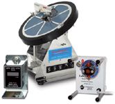
Figure 6: The Cyclograph centrifugal PLC instrument. (Courtesy of Analtech, Inc.)
HPTLC and TLC International Meetings
Interested readers should consider attending the next International HPTLC meeting, which will be held in two years. The last meeting (the 21st) was held in July 2011 in Basel, Switzerland. It was attended by 350 participants from 41 countries. More than 200 papers, posters and tutorials were presented. Information on past and future meetings can be found at www.hptlc.com.
Future Prospects for TLC
The following trends in TLC usage and research are likely to occur in TLC in the next few years:
- Increased use of silica-gel HPTLC vs. TLC layers and silica-gel bonded phases vs. plain silica gel for improved separations of certain mixtures.
- Increased use of chemometrics in TLC, for example, for mobilephase optimization through quantitative structure-retention relationship (QSRR) studies and prediction of retention using descriptors such as topological indices, and for the separation of overlapped densitometry peaks when sample constituents cannot be chromatographically resolved and enhancement of selectivity because of noise and background removal (30).
- Increased use of multidimensional planar chromatography (MD-PC) for separation of more complicated multicomponent mixtures by different combinations of 2D development, chromatographic plates with different properties, a variety of mobile-phase compositions, various forced-flow techniques and multiple development modes (31).
- Continued development of the miniaturized, automated system introduced by Morlock and colleagues (29) with newly prepared UTLC layers having nanoscale and monolithic structures (32).
- Increased development and application of hyphenated techniques, especially TLC–MS and TLC–EDA.
- Increased development of validated methods for industrial quality control (QC) and government regulation of synthetic drug formulations, herbal medicines and dietary supplements, maintaining these as the primary fields in TLC research.
- Increased use of horizontal chambers to double the number of samples analysed to drastically save time and money in chromatographic analyses. Morlock and Oellig (33) demonstrated the use of silica-gel TLC–densitometry with an HDC 2 chamber for identification and quantification of 25 water soluble dyes used as food additives; 12 min was required for 40 runs in parallel developed from both ends of a plate toward the centre (2 × 20 tracks) using 8 mL of mobile phase up to a migration distance of 50 mm. The total analysis time of all steps of the analysis was 60 min for 40 runs, for an overall time/run of 1.5 min and a solvent consumption of 200 µL. A sample throughput of 1000 samples/8 h day was reached by switching among working stations (application, development, and evaluation) in a 20 min interval, which tripled analysis throughput.
- Expanded application of existing manual–visual semiquantitative TLC screening methods (34) and transfer and validation of these, and additional new methods, to instrumental quantitative HPTLC–densitometry suitable for support of regulatory compliance actions (35) for mislabeled, substandard and counterfeit pharmaceutical products appearing worldwide, especially in underdeveloped countries.
Authors' note: As this article was being prepared, notice was sent by Hichrom Limited that it would be selling the Whatman HPLC columns (acquired from GE Healthcare). The Whatman/GE Healthcare TLC plates, however, were being discontinued as of midApril. Merck KGaA (EMD/Millipore in North America) has also discontinued the manufacture of the UTLC monolithic silica gel plates. Unfortunately, this leaves users of any discontinued TLC prepared plates with the task of finding replacement TLC products once their supplies have been depleted.
Fred Rabel, PhD, is president of his company, ChromHELP, LLC, giving seminars, courses and technical help in chromatography. He is on the editorial boards of the Journal of Liquid Chromatography and Related Technologies and Journal of Chromatographic Science. He has published many articles and chapters on various aspects of HPLC, TLC and column chromatography.
Joseph Sherma, PhD, joined the faculty of Lafayette College in 1958 after receiving a PhD degree in analytical chemistry from Rutgers University under the supervision of the renowned ion exchange chromatography expert Wm. Rieman III, and he is currently John D. and Frances H. Larkin Professor Emeritus of chemistry and continues to supervise undergraduate students in analytical method development and interdisciplinary analytical chemistryinvertebrate biology research.
"Column Watch" Editor Ronald E. Majors is a senior scientist at the Columns and Supplies Division, Agilent Technologies, Wilmington, Delaware, USA, and is a member of LCGC Europe's editorial advisory board. Direct correspondence about this column should be addressed to "Column Watch", LCGC Europe, 4A Bridgegate Pavilion, Chester Business Park, Chester Business Park, Wrexham Road, Chester, CH4 9QH, UK, or e-mail the editor, Alasdair Matheson, at amatheson@advanstar.com
References
(1) J. Sherma, J. AOAC Int., 95, in press (2012).
(2) G. Cudiamat, "Chromatography Market Profile," LCGC North Am., 27(12), 1030 (2009).
(3) Thin Layer Chromatography in Drug Analysis, L. Komsta, M. WaksmundzkaHajnos and J. Sherma, Eds. (CRC Press/Taylor & Francis Group, Boca Raton, Florida, in preparation to be published in 2013).
(4) J. Krzek and J. Sherma, J. AOAC Int. 93(3), 751–753 (2010).
(5) Thin Layer Chromatography in Phytochemistry, M. WaksmundzkaHajnos, J. Sherma and T. Kowalska, Eds. (CRC Press/Taylor & Francis Group, Boca Raton, Florida, 2008).
(6) E. Reich and J. Sherma, J. AOAC Int., 93(5), 1347–1348 (2010).
(7) K. Ferenczi-Fodor, B. Renger and Z. Vegh, J. Planar Chromatogr.-Mod. TLC 23(3), 173–179 (2010).
(8) H. Gao, L. Chen, G. Pan and C. Tu, LCGC North Am. 29(4), 348–357 (2011).
(9) G. Morlock and J. Sherma, J. AOAC Int. 92(3), 689–690 (2009).
(10) D.M. Khatri and P.J. Mehta, J. Planar Chromatogr.-Mod. TLC 24(5), 412–418 (2011).
(11) Handbook of Thin Layer Chromatography, 3rd Ed., J. Sherma and B. Fried, Eds. (Dekker, New York, New York, 2003).
(12) E. Reich and A. Schibli, High Performance Thin Layer Chromatography for the Analysis of Medicinal Plants (Thieme Medical Publishers, Inc., New York, New York, 2007).
(13) T.H. Dzido and T. Tuzimski in TLC in Phytochemistry, M. WaksmundzkaHajnos, J. Sherma and T. Kowalska, Eds. (CRC Press/Taylor & Francis Group, Boca Raton, Florida, 2008), pp. 119–174.
(14) B. Spangenberg in TLC in Phytochemistry, M. Waksmundzka-Hajnos, J. Sherma and T. Kowalska, Eds. (CRC Press/Taylor & Francis Group, Boca Raton, Florida, 2008), pp. 175–201.
(15) J. Sherma, J. AOAC Int. 93(3), 754–764 (2010).
(16) L.R. Snyder, J. Planar Chromatogr.-Mod. TLC 21(5), 315–323 (2008).
(17) E. Tyihak and E. Mincsovics, J. Planar Chromatogr.-Mod. TLC 23(6), 382–395 (2010).
(18) A.M. Moricz and H. Kalasz, J. Planar Chromatogr.-Mod. TLC 23(6), 415–419 (2010).
(19) A. Halka, P.W. Poocharz, A. Torbicz and T.H. Dzido, J. Planar Chromatogr.-Mod. TLC 23(6), 420–425 (2010).
(20) G. Morlock and W. Schwack, J. Chromatogr. A 1217, 6600–609 (2010).
(21) G. Morlock and W. Schwack, Trends Anal. Chem. 29(10), 1157–1171 (2010).
(22) J. Sherma and G. Morlock, J. Planar Chromatogr.-Mod. TLC 21(6), 471–477 (2008).
(23) P.E. Wall, Thin Layer Chromatography — A Modern Approach (Royal Society of Chemistry, Cambridge, UK, 2005).
(24) J. Sherma in Thin Layer Chromatography in Phytochemistry, M. WaksmundzkaHajnos, J. Sherma and T. Kowalska, Eds. (CRC Press/Taylor & Francis Group, Boca Raton, Florida, 2008), pp. 103–117.
(25) H.E. Hauck and M. Schulz in Preparative Layer Chromatography, T. Kowalska and J. Sherma, Eds. (CRC Press/Taylor & Francis Group, Boca Raton, Florida, 2006), pp. 41–60.
(26) J. Sherma in Thin Layer Chromatography in Chiral Separations and Analysis, T. Kowalska and J. Sherma, Eds. (CRC Press/Taylor & Francis Group, Boca Raton, Florida, 2007), pp. 43–64.
(27) K. Günther, P. Richter and K. Möller, in Chiral Separations, Methods and Protocols, G. Gübitz and M.G. Schmid, Eds. (Springer, New York, New York, 2004, Vol 243 of series Methods in Molecular Biology), pp. 29–59.
(28) H.E. Hauck and M. Mack in Handbook of Thin Layer Chromatography, 2nd Ed., J. Sherma and B. Fried, Eds. (CRC Press/Taylor & Francis Group, Boca Raton, Florida, 1996), pp. 101–128.
(29) G.E. Morlock, C. Oellig, L.W. Bezidenhout, M.J. Brett and W. Schwack, Anal. Chem. 82, 2940–2946 (2010).
(30) L.J. Komsta, J. AOAC Int. 95(3), (2012) DOI: 10.5740/jaoacint.SGE_Komsta_intro.
(31) T. Tuzimski in High Performance Thin Layer Chromatography (HPTLC), M. Srivastava, Ed. (Springer-Verlag, Heidelberg, Germany, 2011), pp. 247–310.
(32) A.M. Frolova and O.Y. Konovalova, J. Sep. Sci. 34(16–17), 2352–2361 (2011).
(33) G.E. Morlock and C. Oellig, J. AOAC Int. 92(3), 745–756 (2009).
(34) Global Pharma Health Fund E.V. Minilab TLC screening kit methods, http://www.gphf.org
(35) C. O'Sullivan and J. Sherma, Acta Chromatogr. DOI: 10.1556/AChrom.24.2012.2.2, in press.
Free Poster: NDSRI Risk Assessment and Trace-Level Analysis of N-Nitrosamines
April 25th 2025With increasing concern over genotoxic nitrosamine contaminants, regulatory bodies like the FDA and EMA have introduced strict guidelines following several high-profile drug recalls. This poster showcases a case study where LGC and Waters developed a UPLC/MS/MS method for quantifying trace levels of N-nitroso-sertraline in sertraline using Waters mass spectrometry and LGC reference standards.
New Guide: Characterising Impurity Standards – What Defines “Good Enough?”
April 25th 2025Impurity reference standards (IRSs) are essential for accurately identifying and quantifying impurities in pharmaceutical development and manufacturing. Yet, with limited regulatory guidance on how much characterisation is truly required for different applications, selecting the right standard can be challenging. To help, LGC has developed a new interactive multimedia guide, packed with expert insights to support your decision-making and give you greater confidence when choosing the right IRS for your specific needs.
Using the Carcinogenic Potency Categorisation Approach (CPCA) to Classify N-nitrosamine Impurities
April 25th 2025Learn how to manage nitrosamine impurities in pharmaceuticals with our free infographic. Discover how the CPCA approach establishes acceptable intake limits and guides the selection of NDSRI reference samples. Stay compliant and ensure safety with our ISO-accredited standards.

.png&w=3840&q=75)

.png&w=3840&q=75)


.png&w=3840&q=75)


.png&w=3840&q=75)
