Researchers Use Chip CE to Investigate Rhinovirus Replication
Researchers from Vienna University of Technology (TU Wien), Austria, have used chip capillary electrophoresis (CE) in combination with molecular beacons (MBs) to analyze the release of the RNA genome from a human rhinovirus.


Photo Credit: Laguna Design/Getty Images
Researchers from Vienna University of Technology (TU Wien), Austria, have used chip capillary electrophoresis (CE) in combination with molecular beacons (MBs) to analyze the release of the RNA genome from a human rhinovirus.1
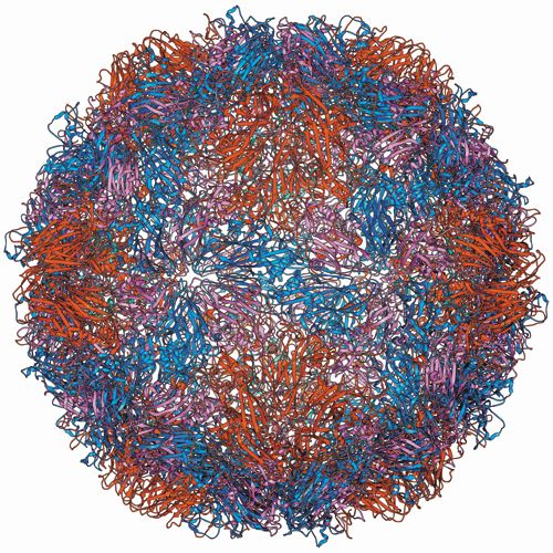
Human rhinoviruses (HRVs), the virus family responsible for the common cold, are problematic because of their proficiency to cause discomfort and also the significant challenges they present to researchers to analyze. Measuring only 30 nm in diameter,2 the non-enveloped, icosahedral particles are not easily visualized or characterized, with 160 serotypes identified to date. These factors have contributed to considerable knowledge gaps surrounding the basic functioning of these very common viruses.
“The exact mechanism of RNA release from a rhinovirus in vivo from inside the protective virus capsid and through the lipid membrane of endosomes is at the moment still not completely understood,” said Günter Allmaier, TU Wien, Austria.
This is an incredibly important step in the virus’s replication process and understanding it could lead to potential targets for medical interventions. Viruses have no reproduction capability of their own, instead they utilize a host cell’s own apparatus to replicate. In order to achieve this, they must introduce their own genetic material into the cell and part of this process involves that RNA passing through the viral capsid (shell).3 Preventing this transfer of RNA and therefore virus replication would eliminate the virus.
The challenge for researchers was to develop a methodology capable of analyzing the release of viral RNA from the protective capsid and open the process up to future more inâdepth research.
The method developed uses molecular beacons (MBs), which “are oligonucleotide probes modified by a fluorophore and a fluorescence quencher at their 3’ and 5’ ends. In the absence of a complementary DNA or RNA target sequence, the terminal nucleotides of a molecular beacon form a helix stem. This in turn leads to spatial proximity between the attached fluorophore and quencher resulting in only very low intensity fluorescence. However, in the presence of a respective target, the molecular beacon hybridizes to the target, the stem opens up and an increase in fluorescence is recorded,” explained Weiss and Allmaier.
Through this fast, sensitive, and easyâtoâuse lab-on-a-chip-based method, Allmaier and his team can specifically detect the genome release using fluorescence detection after the sample components have been separated by chip CE. Thereby successfully setting the stage for further investigation into the mode of release of the RNA genome of a human rhinovirus.
The TU Wien team also hope the method could be adapted to other areas, including the investigation of gene therapy delivery systems and RNA release control.
References
- V.U. Weiss, G. Allmaier et al., Anal. Bioanal. Chem.408(16), 4209–4217 (2016).
- V.U. Weiss et al., Anal. Chem.87, 8709–8717 (2015).
- R. Fuchs and D. Blaas, in Structure-based study of viral replication, (Singapore: World Scientific Publishing 2008) pp.1–42
Look out for the interview with Günter Allmaier on this research in the next issue of The Column.


.jpg")


Regulatory Deadlines and Supply Chain Challenges Take Center Stage in Nitrosamine Discussion
April 10th 2025During an LCGC International peer exchange, Aloka Srinivasan, Mayank Bhanti, and Amber Burch discussed the regulatory deadlines and supply chain challenges that come with nitrosamine analysis.

