
News|Articles|March 21, 2025
- March 2025
- Volume 21
- Issue 1
Influence of Concentration in Conventional GPC/SEC and Advanced Detection GPC/SEC
Author(s)Daniela Held
Sample concentration is a parameter that can influence the quality of gel permeation chromatography/size-exclusion chromatography (GPC/SEC) separations and the obtained results. Understanding this influence can help to support the development of reliable GPC/SEC methods.
Advertisement
Gel permeation chromatography/size‑exclusion chromatography (GPC/SEC) is a technique that separates macromolecules based on their hydrodynamic volume in solution. It is used for many different types of large molecules, ranging from synthetic polymers highly disperse in molecular weight to monodisperse proteins. Users of this technique typically require information such as molar masses and/or molar mass distribution as well as size and size distribution.
Sample concentration is always an important parameter in method development. From a detection perspective, a high sample concentration is beneficial to ensure a good signal to noise (S/N) for GPC/SEC detectors. However low concentrations are also favorable because this reduces the amount of sample required. In addition, higher concentrations may affect the separation in the pores of the GPC/SEC columns, which may result in physically meaningless variations of sample molar mass with the injected amount.
Understanding how the sample concentration affects separation, as well as the accuracy and reproducibility of the derived results, is crucial for establishing reliable GPC/SEC methods.
Concentration Influence on Separation
GPC/SEC is used to separate large molecules, sometimes with very high molar masses, which leads to specific requirements concerning sample preparation (1). Selecting the right concentration range is part of sample preparation and method optimization.
A sample concentration (or more precisely, injected mass) that is too high can negatively influence the chromatograms. In contrast to other liquid chromatography (LC) techniques, a concentration that is too high in GPC/SEC typically increases the elution volume and, even worse, changes the peak shape. Depending on the detection options used, such a change in elution volume and peak shape results in significant changes of the outcome.
The maximum applicable concentration depends on two sample characteristics: the sample’s molar mass and its dispersity. Therefore concentration recommendations are typically for molar mass ranges and differentiate between narrowly distributed/monodisperse and broadly distributed samples. Table 1 lists typical concentrations to start with depending on molar mass and dispersity, while Table 2 summarizes recommendations for injection volumes based on the number of columns used.
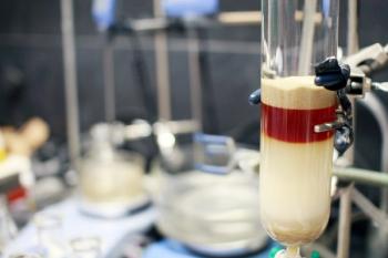
To get an idea of how the concentration affects the peak position and shape for different molar mass ranges, a mixture of two narrowly distributed reference materials were injected using different concentrations and refractive index (RI) detection. Figure 1 shows the overlay of the chromatograms obtained.
As expected, the peak increases with the sample concentration (detector linearity is rarely a problem in GPC/SEC), but with increasing concentration the peaks are shifted to higher elution volumes. This effect is more pronounced for the higher molar mass sample. It is also clearly visible that the peak shape changes with higher sample concentration. This is especially problematic when conventional column calibration would be used to determine the molar mass distribution results.
This does not mean that the concentrations given in Figure 1 will always be too high for any kind of sample in this molar mass region. For samples with a broader molar mass distribution, the concentration can be much higher than in Figure 1 (please refer to Table 1). An easy test to perform if the injected mass is too high is to inject a sample with different injection volumes. If there is no peak shift or deterioration, the concentration is most probably in the right region. If in doubt, testing different concentrations and injection volumes is recommended.
Sample Concentration Influence on Results Based on Advanced Detection vs. Column Calibration
Depending on the required results, a GPC/SEC system can consist of very different components. Instrumentation setups range from systems comprising just a single detector to multi-detection systems with three or more detectors in a detector train. A former Tips & Tricks installment discusses the requirements of typical GPC/SEC setups and the differences compared to other LC techniques (2). The differences between the GPC/SEC detector types are addressed in several installments (3).
In general, GPC/SEC detectors are classified into concentration detectors and molar mass sensitive detectors. The classification is based on their response to sample properties as shown in equation 1:
With
KDetector: detector constant;
kSample: sample specific constant; for example, refractive index dn/dc or extinction coefficient ε;
c: concentration;
x: coefficient for molar mass dependency
of the signal.
For a concentration detector, such as a refractive index (RI) detector, or a UV detector, x equals 0, resulting in a simplified equation 2:
For molar mass sensitive detectors, the coefficient x is typically higher than 0. For static light scattering detectors, x= 1; for viscosity detectors x is related to the Mark-Houwink exponent. All detector types show a dependence and response on the sample concentration, but the influence on the results will be very different depending on the detectors applied.
Concentration Influence When Working with Molar Mass Sensitive Detectors: Molar mass sensitive detectors, such as light scattering detectors, are very popular because they can measure the molar mass directly. However, light scattering is not parameter free, and concentration is one of the required inputs. A molar mass sensitive detector always requires knowledge of the concentration (or eluted mass), otherwise the desired result molar mass cannot be determined. The detailed influence of an incorrect concentration and other parameters on the accuracy of the molar mass result depends on the analysis options applied in GPC/SEC light scattering (4).
Typically, the error influence for the concentration is linear, meaning that 5% error in concentration will also result in 5% error in molar mass. On a positive note, the light scattering detector would be able to verify if a concentration that is too high is responsible for a broad peak, as shown in Figure 1 for the sample with the high molar mass (red trace). The molar mass obtained from the light scattering detector would stay constant over the elution volume, indicating a missing size/molar mass separation.
Concentration Influence When Working with Column Calibration: If only a concentration detector, such as a RI detector, is used, it is not possible to verify the separation on the column independently. To obtain molar mass results, a column calibration using several narrowly distributed reference materials or one or more broadly distributed reference materials needs to be established. However, if such a column calibration curve is used to determine the molar mass distribution, the sample concentration is not required as an input parameter. If the concentration is sufficiently low, so that the peak is neither shifted or distorted, it is irrelevant if the concentration is 1.5 mg/mL or 2 mg/mL because only ratios of the different molar masses contribute. On a positive note, the peak area can also serve as an indicator of concentration. Please note that this is also the reason why molar mass sensitive detectors are used in combination with concentration detectors. The concentration detector measures the concentration required by the advanced detector.
Summary
- A concentration that is too high in GPC/SEC will result in shifts to higher elution volume and distorted peak shapes.
- The higher the molar mass, the lower the concentration needs to be.
- Narrowly distributed samples require much lower concentrations than broadly distributed samples.
- If in the right concentration regimen, the accuracy of the concentration will affect results obtained with molar mass sensitive detectors while results obtained using column calibration will be unaffected.
References
- Reinhold, G. Tips & Tricks GPC/SEC: Sample Preparation. The Column 2011, 7 (15), 13–17.
- Held, D. Tips & Tricks GPC/SEC: What Are the Differences Between GPC, SEC, and GFC, and How Do You Get Started with the Technique? The Column 2018, 14 (10), 2–8.
- Held, D.; Kilz, P. Tips & Tricks GPC/SEC: Understanding Positive and Negative Detector Signals. The Column 2008, 17–20.
- Held D.; Kilz, P. Qualification of GPC/GFC/SEC Data and Results
in Quantification in LC and GC; Kromidas, S.; Kuss, J., Eds.;
Wiley VCH, 2009.
Articles in this issue
Newsletter
Join the global community of analytical scientists who trust LCGC for insights on the latest techniques, trends, and expert solutions in chromatography.
Advertisement
Related Content
Advertisement


Advertisement
Advertisement
Trending on LCGC International
1
Best of the Week: Profiling Endoenous Protein Complexes, Previewing Riva 2026
2
Restek Introduces GC Columns Featuring TriMax Technology for Trace-level Sensitivity
3
Riva Returns Alive and Kicking
4
DoE-Optimized GC–FID Method for Robust Terpene Profiling in Essential Oils
5